DuoScanTM Imaging
检测样品
检测项目
关联设备
共5种
下载方案
方案详情文
DuoScanTM成像技术是一个共焦成像模式,它可以利用高精度、超快速摆动的反射镜生成各种尺寸的大激光光斑,也可以从深紫外到近红外做纳米级步长的成像。 DuoScanTM具有多种成像功能: ﹡在大范围内迅速获取信息 ﹡不移动样品进行低至50nm步长的精细扫描 ﹡大光斑平均光谱
智能文字提取功能测试中
Page 2/4 Page 3/4 DuoScanTM Imaging The innovative DuoScanTM Imaging system extends the imaging capabilities of HORIBA Scientific’s Ramaninstruments from sub-micron to macro-scale mapping. Generating confocal images becomes faster,easier andmore flexible, from the deep UV to the IR. The Raman microscope has become a powerful analyticaltool for many applications including pharmaceuticals,polymers, semiconductors, forensics and life science.The new generation of Raman microscopes offers a non-destructive and non-contact method of sample analysis atthe micron level. More particularly, Raman imaging providesthe spatial distribution of the various molecular species withina heterogeneous sample, making it possible to produce fastand accurate 1 mm) obtained in macro-mapping mode. The colorscorrespond to the integrated intensity of the Si band (red)and of the respective G bands of the CNTs (green and blue). Once the CNT of interest has been located, a high-definitionimage ofthe CNT is then obtained by zooming in with DuoScanTMin step-by-step micro-mapping mode (inset). In that case, theapparent width of the CNT (366nm) is a convolution of the tubediameter with the laser spot size. This shows the superior spatialresolution achievable with a confocal microscope. Fig. 5: Macro-map and zoomed-in step-by-step map of a CNT obtained with DuoScanTM.(Sample courtesy of Dr Kalbac, Heyrovsky Institute, Czech Republic) Example 2: Component distribution on a pharmaceuticaltablet Macro mapping can be used to look at component distributionon very large pharmaceutical samples. In this example, a17.5 mm by 7 mm pellet was analyzed. As the DuoScanTMautomatically matches the image pixel size to the scannedarea, the full surface is covered, ensuring no spot is missed. As with the previous example, step-by-step mapping can. be performed on a region of interest to look at finer details(Figure 6). Fig. 6: Raman maps from a pharmaceutical tablet (A) Macro-mapping, 8240 macro-points, total time 400 s (full tablet) (B) Step-by-step image of a region of interest, 10000 points, total time 13 minutes (500x500 um) Web links: http://www.horiba.com/scientific/products/raman-spectroscopy/raman-imaging/duoscan/ http://www.horiba.com/scientific/products/raman-spectroscopy/raman-imaging/duoscan/duoscan-application-examples/ JOBIN YVON Technology info.sci@horiba.com www.horiba.com/scientific Scientific France:+33 (0)1 64 54 13 00Germany: +49 (0)89 4623 17-0Italy: +3925760 3050Japan: +81 (0)3 62064717Brazil::+55(0)11 5545 1514 From sub-micron to macro, from UV to IR, we've got your sample covered!JOBIN YVONTechnologyHORIBAExplore the futureAutomotive Test Systems Process & Environmental Medicall Semiconductor| Scientific HORIBAExplore the futureAutomotive Test Systems Process & Environmental Medical I Semiconductorr Scientific
关闭
-
1/4

-
2/4
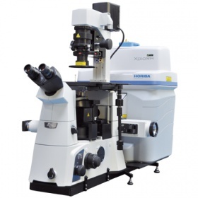
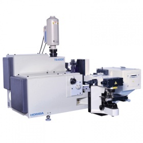
上海巨纳科技有限公司为您提供《DuoScanTM Imaging》,该方案主要用于中检测,参考标准《暂无》,《DuoScanTM Imaging》用到的仪器有HORIBAXploRA INV智能型全自动拉曼光谱仪、HORIBA HR Evolution高分辨拉曼光谱仪、HORIBACombiscopeXploRA原子力拉曼联用系统、Horiba XploRA INV 多功能拉曼及成像光谱仪、HORIBA T64000三级拉曼光谱仪。
我要纠错


 咨询
咨询