

方案详情文
智能文字提取功能测试中
A)NleQIFVKTLTG KTITLEVEPS DTIENVKAKI QDKEGIPPDQ QRLIFAGKQL EDGRTLSDYN IQKESTLHLV LRLRGG hydrolysis of diUb (PDB:4BOS). Note: K11 di- and triUb were prepared as reported Racemic X-ray Structures of K27-linked Ubiquitin Chains Preparedby Total Chemical Synthesis Man Panl*, Shuai Gaol#, Yong Zhengl#, Xiaodan Tan', Huan Lan', Xianglong Tan,Demeng Sun', Lining Lu', Tian Wang', Qingyun Zheng, Yichao Huang’, Jiawei Wang',LeiLiul* Tsinghua-Peking Center for Life Sciences, Ministry of Education Key Laboratory of Bioorganic PhosphorusChemistry and Chemical Biology, Department of Chemistry, Tsinghua University, Beijing 100084, China;StateKey Laboratory of Biomembrane and Membrane Biotechnology, Center for Structural Biology, School of LifeSciences, Tsinghua University, Beijing 100084, China Supplementary Information 1 . Ge n e r al I n f or m a ti o n . . . . S2 a.Materialsb.HPLC and FPLC c. Molecular biology and biochemistry d. Mass spectrometry e. Protein Expression and Purification 2. Experimental Section.. ..S5 a. Synthesis and characterization of peptides b. Chemical synthesis D-K27-linked di- and triubiquitin c. Protein folding d. Circular dichroism (CD) experiments e. X-ray crystal structure determination. f. MS/MS identification of specific isopeptide bond g. Cloning of deubiquitinase OTUD2 Mutants 3. Experimental figures. . S10 1. General Information a. Materials 2-Chlorotrityl resin and rink amide resin were purchased from Hecheng Technology(Tianjing,China). Fmoc-amino acids includingFmoc-Lys(Alloc)-OH and1-Hydroxy-7-azabenzotriazole (HOAt) were purchased from CS Bio, GL Biochem(Shanghai, China). Ethyl cyanoglyoxylate-2-oxime (oxyma) was purchased mAdamas-beta (Shanghai, China). N,N-Diisopropyl-carbodiimide (DIC),1,2-ethanedithiol,N,N-diisopropylethylamine (DIPEA), triisopropylsilane (TIPS) and4-mercaptophenylacetic acid (MPAA) were purchased from Ouhe Technology(Beijing, China). Dithiothreitol(DTT) was purchased from Aladdin (Shanghai, China).Acetonitrile (HPLC grade) was purchased from J.T. Baker (Phillipsburg, NJ,USA).Na2HPO4.12H2O, CH;OH, guanidine hydrochloride (Gn·HCl), Et2O, Fmoc-hydrazineand N,N-Dimethylformamide (DMF) were purchased from Sinopharm ChemicalReagent. Thioanisole and trifluoroacetic acid (TFA) (HPLC grade) were purchasedfrom J&K Scientific (Beijing, China). CH2Cl2 (DCM) and NaNO2 were purchasedfrom Beijing Chemical Works (Beijing, China). Glycyl1-(2,4-dimethoxyphenyl)-2-mercaptoethyl auxiliary andFmoc-N-(2,4-dimethoxybenzyl) glycine were purchased from Nantong PPTIDEBiotech ltd (Jiangsu, China). b.HPLC and FPLC Analytical RP-HPLC was run on a SHIMADZU (Prominence LC-20AT) instrumentusing an analytical column (Grace Vydac C18 and welch xb-c4, 250 mm × 4.6 mm, 5um particle size, flow rate 1.0 mL/min, rt). Analytical injections were monitored at214 nm and 254 nm. Semi-preparative HPLC was run on a SHIMADZU (ProminenceLC-20AT) instrument using a semi preparative column (Grace Vydac C18, 250× 25mm, 10 um particle size and welch xb-c4, 150 mm×21.2 mm and welch xb-c18, 250mm x21.2 mm, 5 um particle size, flow rate 7.0 mL/min). Solvent A was 0.1% TFAin acetonitrile, and solvent B was 0.1% TFA in Deionized Distilled Water. Bothsolvents were sonicated for 10 min before use. FPLC was run on a GE Healthcare (AKTA purifier 10 UPC-F920) instrument using a Superdex 75 column or Mono Q column. The injections were monitored at280 nm. All the buffers were filtered through 0.22 um filter paper and sonicated for 10min beforeuse. c. Molecular biology and biochemistry Primers were ordered from Biomed Biotech (Beijing) Co., Ltd. All enzymes wereordered from New England Biolabs (NEB). Bacterial cells were grown in LB(Luria-Bertani) broth or LB agar medium (Sigma). All pictures of protein gels weretaken on ChemDocXRS+ (Bio-Rad). For SDS-PAGE, samples were loaded onto12-15% SDS-PAGE gels were electrophoresed for 15 min at 100 V and 40 min at 200V. d. Mass spectrometry High-resolution ESI mass spectra were measured on Agilent 6210 Time of FlightMass Spectrometer. Normal ESI mass spectra were measured on a Bruker DaltonicsData Analysis 3.0 workstation. e. Protein Expression and Purification: Protteeiinn was texpressed from modified pGEX-4T-1vectors, which introduced aPreScission Protease cleavage site, and C terminal His6-tag. Protein was expressed inE. coli BL21 (DE3) cells that were grown to an OD600 of 0.8 at 37 ℃ and inducedwith 200 mM IPTG for 16-20 hr at 20 ℃. Large-scale protein expression wasperformed in 2L-8L LB medium supplemented with amp. antibiotic. Proteinpurifications were performed at 4 ℃. Cells were lysed by sonication in 40-160 mLlysis buffer (200 mM NaCl, 20 mM Tris [pH8], 5 mM DTT, 1mM PMSF), andcleared by centrifugation 16000 rpm for 30 min. The cleared lysate was incubatedwith 2 mL equilibrated Glutathione Sepharose 4B resin for 1 hr, and subsequentlywashed with 500 mL buffer A (20 mM Tris [pH8], 5 mM DTT) plus 500 mM NaCl,and 200 mL buffer A plus 50 mM NaCl. The GST tag was cleaved on the resin with50 ug GST-tagged PreScission protease overnight. Cleaved protein was eluted withbuffer A plus 50 mM NaCl to a final volume of 15 mL and subjected to anion exchange chromatography (MonoQ, GE Healthcare) with a NaCl gradient from 50 to500 mM. Concentrated proteins were pooled and subjected ttogel filtration(Superdex75) in buffer A plus 200 mM NaCl. Proteins were concentrated to 2-20mg/mL using a 10 kDa MW cut-off concentrator and flash-frozen in liquid nitrogen. g. General protocol of hydrazide-based native chemical ligation. The hydrazide peptide (1 umol, 1 eq) was dissolved in 1 mL ligation buffer (6 MGn-HCl, 200 mM NaH2PO4, pH 3.0) pre-cooled in an ice bath (-10 ℃). Then 10 uLof 1 M NaNO2 (10 umol, 10 eq) was added dropwise and the reaction was incubatedfor 30 min at -10℃ to fully convent the hydrazide to the acyl azide. Next MPAA (6.8mg, 40 umol, 40 eq) was added and the pH was adjusted to 5.0 for 5 min to generatethe thioester peptide. Finally, the N-terminal Cys peptide (1 umol, 1 eq) was addedand the pH was adjusted to 6.5 to initiate standard native chemical ligation. Thereaction was monitored by analytical RP-HPLC. 2. Experimental Section a. Synthesis and characterization of peptides Table S1 sequence of synthetic peptide Peptide segment Peptide sequence Acm-Cys*-Gly-CONHNH2(1) C(Acm)GKQLEDGRTLSDYNIQKESTLHLVLRLRGCO- NHNH Metj-K27aG-Phe45-CONHNH2(2) MQIFVKTLTGKTITLEVEPSDTIENVK(aG)AKIQDKEGIPPD QQRLIF-CONHNH2 Met'-Phe+s-CONHNH(5) MQIFVKTLTGKTITLEVEPSDTIENVKAKIQDKEGIPPDQQR LIF-CONHNH, Cys46-Gly76-CONH2(7) CGKQLEDGRTLSDYNIQKESTLHLV LRLRGG-CONH2 Cys-Gly-CONH(7*) CGKQLEDGRTLSDYNIQKESTLHLVL-CONH2 The synthesis of peptide 46-76-NH2 (7) and 46-71-NH2(7*) were conducted on rinkamide resin with estimated loading of 0.48 mmol/g. The rest segment coupling wasprocessed on the other resin named Fmoc-NHNH, based 2-Chlorotrityl resin withestimated loading of 0.5 mmol/g. Before amino acid coupling, the 2-chlorotrityl resinshould be pretreated to obtain the C-terminal hydrazide. 4 eq Fmoc-NH-NH2 and 8 eqDIPEA were added to the resin and stirred for 2 h at room temperature, which wasturned into Fmoc-NH-NH-2-trityl resin. During the11-45-K27aG-NH-NH2 peptide synthesis, Lys27was chargedasFmoc-Lys(Alloc)-OH instead of Fmoc-Lys(Boc)-OH for the specific protection. Afterepeptidesynthesiswas completed,the resin was first incubated with[(Ph3)P]4Pd/PhSiH to remove the Alloc protecting group and then coupled withglycine auxiliary in DIC/HOAt system. The subsequent procedure was the same asdescribed above. All peptide segments can be obtained in hundred milligrams quantityand purified by preparative reverse-phase HPLC from each 0.25 mmol synthesis scalein 3 days. b. Chemical synthesis D-K27-linked di- and triubiquitin The overall route for chemical synthesis D-K27-linked di- and triubiquitin wasbasically the same as L-type chains with several changes. To minimize the cost ofD-type amino acids, auxiliary-group assisted ligation was not used because of itsrelatively low efficiency. We chose cysteine instead of glycine auxiliary, which couldbe desulfurizated to generate Ala to mimick Gly. We also mutated Metl to Nle, because Met could be oxidized during SPPS. After the synthesis of D-type peptidesegments, followed by subsequent chemical ligations, we obtained D-k27-diubquitin(Figure S3) and D-k27-triubiquitin (Figure S4). c. Protein folding Lyophilized K27-linked ubiquitin chains were dissolved in buffer (100 mM Na2HPO4,6 M Gn-HCl; 150 mM NaCl;pH 3) at a protein concentration of 2 mg/mL, and thentransferred to a dialysis membrane and dialyzed overnight against ubiquitin foldingbuffer (20 mM Tris, 150 mM NaCl, pH 7.5) with gradually decreased urea(8M-0M).Then diluted protein solution was concentrated using an Amicon Ultra-15 10-kDaMWCO centrifugal filter device (Millipore) and high-speed centrifuged at 14000 rpmprior to loading onto a pre-equilibrated superdex 75 column (GE Life Science) usingan AKTA FPLC system. The elution buffer consists of 50mM NaCl and 20mM Tris atpH 7.5 and the flow rate was 0.4 mL/min. Fractions containing pure K27-linkedubiquitin chains were concentrated to about 5 mg/mL and frozen at -80℃ forstorage. d. Circular dichroism (CD) experiments Circular dichroism experiments were tested at 298K using Applied PhotophysicsPistar 元-180 CD spectrometer in the center of biomedical analysis, TsinghuaUniversity. The concentrations of K27-linked diubiquitin, K27-linked triubiquitin,D-K27-linked diubiquitin, D-K27-linked diubiquitin were 0.1-0.2 mg/mL, and thethickness of CD cuvette was 1mm. The wavelength range was of 190-260 nm and thestep was 1 nm. Each sample was measured three times. e. X-ray crystal structure determination. For the crystal structure of K27-linked diUb, we carried out molecular replacement inspace group C2. The molecular replacement solution packed well with 1/2 L-typediUb molecular and 1/2 D-type diUb molecular in the asymmetric unit. With the usingof PHENIX in structure refinement, Rwork and Rfree of the model decreased to 23%and 29%. For the crystal structure of K27-linked triUb, we carried out molecular replacement inspace group H3. The molecular replacement solution packed well with 1/3 L-typediUb molecule and 1/3 D-type diUb molecule in the asymmetric unit. With the usingof PHENIX in structure refinement, Rwork and Rfree of the model decreased to 25%and 30%. Moreover, the Rfree values for the crystal structure of K27-linked di- and triUbcannot fully demonstrate the quality of the data especially at these resolutions (1.55 Aand 2.1 A). Therefore we tried lower symmetry space group P1, which is reportedhelpfully in reducing in Rfree. However, this only has little effect on reducing theRfree of both structures. The map ofK27-linked diUb cannot be resolved in phasemolecular replacement with CCP4 in space group P1 (Rfree up to 55%), and the Rfreeof crystal structure of K27-linked triUb in space group P1 reduced to 29%. We alsotried higher completeness (92.5%) at the cost of resolution. The Rfree of bothstructures still cannot be reduced (data not shown). f. MS/MS identification of specific isopeptide bond Gel bands of proteins with catalase activity were excised for in-gel digestion, andproteins were identified by mass spectrometry as previously described. Briefly,proteins were disulfide reduced with 25 mM dithiothreitol (DTT) and alkylated with55 mM iodoacetamide.IIn-gel digestion was performedusing sequencinggrade-modified trypsin in 50 mM ammonium bicarbonate at 37 ℃ overnight. Thepeptides were extracted twice with 1% trifluoroacetic acid in 50% acetonitrileaqueous solution for 30 min. The peptide extractss were then centrifuged in aSpeedVac to reduce the volume. For LC-MS/MS analysis, peptides were separated by a 60 min gradient elution at aflow rate 0.250 pL/min with a Thermo-Dionex Ultimate 3000 HPLC system, whichwas directly interfaced with a Thermo LTQ-Orbitrap Velos pro mass spectrometer.The analytical column was a homemade fused silica capillary column (75 um ID,150 mm length; Upchurch, Oak Harbor, WA) packed with C-18 resin (300 A, 5 um;Varian, Lexington, MA). Mobile phase A consisted of 0.1% formic acid, and mobile phase B consisted of 100% acetonitrile and 0.1% formic acid. An LTQ-Orbitrap massspectrometer was operated in the data-dependent acquisition mode using Xcalibur2.0.7 software and there is a single full-scan mass spectrum in the Orbitrap (400-1800m/z, 30,000 resolution) followed by 20 data-dependent MS/MS scans in an ion trap at35% normalized collision energy (CID). MS/MS spectra from each LC-MS/MS run were searched against the ubiquitindatabase using Proteome Discoverer (Version 1.4) searching algorithm. The searchcriteria were as follows: full tryptic specificity was required; two missed cleavageswere allowed; carbamidomethylation was set as fixed modification; oxidation (M)were set as variable modifications; precursor ion mass tolerance was 10 ppm for allMS acquired in the Orbitrap mass analyzer; and fragment ion mass tolerance was0.8 Da for all MS2 spectra acquired in the LTQ. High confidence score filter (FDR<1%) was used to select the “hit” peptides. The corresponding MS/MS spectra weremanually collected²3. g. Cloning of deubiquitinase OTUD2 Mutants cDNA of full-length human OTUD2 was synthesized by Genescript and amplified by PCR using 2 primers: ‘CGCCATATGTTTGGTCCGGCTAAAG'&‘CCGCTCGAGTCAGACTTCACCAAAATTC'.‘OTU+ZnF'Domain ofOTUD2(132-348) &OTU'Domain of OTUD2(132-314) was also amplified by PCRusing corresponding primers. Then they are all cloned into modified pGEX-4T-1vectors, which introduces a PreScission Protease cleavage site, via NdeI and XhoIdigestion and ligation. OTUD2-MutS2 (I292Q, V295Q) was generated by two roundmutagenesis, first with primer F-GCTCTAATGATGACCAAGTGCTGGTTCAGG&R-CCTGAACCAGCACTTGGTCATCATTAGAGC, then withF-CCTGAACCAGCACTTGGTCATCATTAGAGC & R-GTTCCAGTGCCTGTTGCAGCACGATGTCAT. Mass observed 24967 Da and minus 24473 Da which means that the first five amino acids were missing duringovernight cleavage. OTUD2-MutS1 (I201Q) was generated by mutagenesis withprimer F-TTATTCTGAAGCACAACTGGGCAAAACC& R- GGTTTTGCCCAGTTGTGCTTCAGAATAA. h.SDS-PAGE analysis of K27-linked Ub chains. We noticedanunusualelectrophoretibcehavior of K27-linkedtriUb inn ttheSDS-PAGE of DUB essay: there are two unexpected bands with molecular weightabove triUb (25 kDa) (Fig. 4B). SDS-PAGE analysis of synthetic K27-linked triUbalone also showed the same phenomenon (Fig. S8A-B), while the linear triUb andsynthetic K27-linked diUb both showed only one band. Further MS/MS identificationof these two additional bands exhibited that both two bands were both identified asubiquitin and K27-linked Gly-Gly modificationwas alsoo verified (Fig. S8B,Supplementary Fig. 9-10), which ruled out the possibility of impurity introduced intoK27-linked triUb during synthesis process. During later attempts to explain theformation of two higher bands, we are surprised to find that the sample preparationprocess for the SDS-PAGE can result in the occurrence of the two bands (Fig. S8B): Itis likely that different heating time of SDS-PAGE loading sample would make thesynthetic K27-linked triUb aggregate in different level. Indeed, we did observe thetwo additional bands increased when extending heating time (Fig.S8C). 3. Experimental figures A +4H +4H1+ 1-45-27AG-NH-NH. B 1346.2 883.2 acm-46-75-NH-NH, +5H* +5H* +3H* 1077. +6H*+7706.8 1177.2 +6H* 588.8 897.8 +3H* 1794.6 0 0 Time(min) Figure S1. Characterization of synthetic peptide segments. a) Analytical HPLC chromatograms (a=214 nm) of purifiedsegment 2 and ESI-MS (observed 5380.8Da, calculated 5381.0Da). B) AnalyticalHPLC chromatograms (A=214 nm) of purifiedsegment 1 and ESI-MS (observed 3528.8 Da, calculated 3528.9 Da). C) Analytical HPLC chromatograms ()=214 nm) ofpurified segment 5 and ESI-MS (observed 5127.9Da, calculated 5127.6 Da). D) Analytical HPLC chromatograms (=214 nm)of purified segment 7 and ESI-MS (observed 3499.5 Da, calculated 3499.9 Da). E) Analytical HPLC chromatograms (A=214nm) of purified product 46-71-NH2 and ESI-MS (observed 2960.3Da, calculated 2960.1Da). (Note: HPLC conditions: a lineargradient of 20-60% acetonitrile (containing 0.1% TFA) in water (containing 0.1% TFA) over 35 min) 0 Time(min) Figure S2. Characterization of synthetic peptide fragments. a) Analytical HPLC chromatograms (A=214 nm) of purifiedfragment 3 and ESI-MS (observed 8878.9 Da, calculated 8878.5Da.). B) Analytical HPLC chromatograms (= 214 nm) ofpurified fragment 4 and ESI-MS (observed 8610.9Da, calculated 8610.7Da.). C) Analytical HPLC chromatograms (=214 nm)of purified fragment 6 and ESI-MS (observed 13706.8 Da, calculated 13706.8Da). D) Analytical HPLC chromatograms (A=214nm) of purified fragment 9 and ESI-MS (observed 8681.4Da, calculated 8682.7Da). E) Analytical HPLC chromatograms (a=214 nm) of purified fragment 10 and ESI-MS (observed 12079.0Da, calculated 12079.8Da). (Note: HPLC conditions: a lineargradient of 20-60% acetonitrile (containing 0.1% TFA) in water (containing 0.1% TFA) over 35 min) Figure S3. Chemical synthesis of D-K27-linked diubiquitin (8D). A) The amino acid sequence of D-K27-linked diubiquitinwith Gly76 of distal ubiquitin mutated to Ala. B) Synthetic route. C) Analytical HPLC chromatogram (A=214 nm) of isolatedproduct 8D. Inset: Observed ESI-MS of the main peak and deconvoluted electrospray ionization mass spectrum of 8D (observedmass = 17088 Da, calculated mass=17088.7 Da, average isotopes). D) Gel filtration chromatography chromatogram (1=280 nm)of product 8D. Figure S4. Chemical synthesis of D-K27-linked triubiquitin (11D). A) The amino acid sequence of D-K27-linked triubiquitinwith Gly76 of distal and middle ubiquitin mutated to Ala. B) Synthetic route. C) Analytical HPLC chromatogram (A=214 nm)of isolated product 11D. Inset: Observed ESI-MS of the main peak and deconvoluted electrospray ionization mass spectrum of11D (observed mass =25631 Da, calculated mass=25631.5 Da, average isotopes). D) Gel filtration chromatographychromatogram (A=280 nm) of product 11D. Table S2. Data collection and structure refinement statistics Data collection K27-linked diUb K27-linked triUb K27-linked triUb spacegroup c2 R3 P1 44.828 63.411 52.844 90.000 51.420 51.420 137.74590.000 51.75851.749 54.977 90.02 cell parameters 109.162 90.000 90.000 120.000 118.15119.95 Resolution(A) 1.55 2.1 Rmerge 0.064(0.339) 0.112(0.721) 0.11(0.89) I/oI 13.20(2.63) 7.01(0.74) 8.64(0.63) Completeness (%) 78.7(45.4) 85.8(28.1) 87.2(60.9) Redundancy 3.0(2.5) 2.5(1.2) 1.7(1.4) Refinement Resolution(A) 1.554 2.104 1.987 No.reflections 13688 10189 22698 Rwork / Rfree 0.2339/0.2946 0.2477/0.3044 0.2302/0.2971 No. atoms Protein 1212 1164 3406 Ligand/ion 8 0 Water 106 71 331 B-factors Protein 22.88 37.395 33.4 Ligand/ion C 0 0 Water 30.93 37.729 36.88 R.m.s. deviations Bond lengths (A) 0.012 0.008 0.017 Bond angles (°) 1.46 0.931 2.309 Figure S5. MS/MS identification of K27-linked diubiquitin. The MS/MS sequencing data verifies the specific formation ofthe Lys27 linked isopeptide bond with a false discovery rate <1%. Figure S6. Electron density maps K27-linked diubiquitin. A Di-UB-K27 with OTUD2" Tri-UB-K27 with OTUD2 0 min 5 min 30min 0 min 5 min 30min Ub3 DUB Ub2 Ub C D Di-UB-K11 with OTUD2WT Di-UB-K11 with OTUD2S1 Di-UB-K11 with OTUD2S2Marker(0 min 5 min 30 min 0 min 5 min30 min 00 min 5 min 30 min LUb E F Tri-UB-K11 with OTUD2WT Tri-UB-K11 with OTUD2S1 Tri-UB-K11 with OTUD2S2 Tri-UB-K27 with OTUD2WT Tri-UB-K27 with OTUD2S1 Tri-UB-K27 with OTUD2 11 0 min 5 min 30 min 0 min 5 min 30 min 0 min 5 min 30 min 0 min 55 min 30 min 0 min 5 min 30 min 0 min 5 min 30 mir Ub3 DUB DUB Ub3 Ub2 Ub2 Ub Ub K27-diUb with OTUD2 K27-triUb with OTUD2 G) H) Figure S7. Mutagenesis DUB assays with K27-linked Ub chains. Assays comparing isolated catalytic domains of WT OTUD2(OTUD2WT, aa 132-348), S1 site mutant (OTUD2S1, aa 132-348, I201Q) and S2 site mutant (OTUD2S2, aa 132-348, I292Q,V295Q). A-B)DUB essay of K11, 27 di- and triUb towards WT OTUD2. C-D) DUB essay of K11, 27 di-towards WT, S1 andS2 sites mutant OTUD2. E-F) DUB essay of K11, 27 tri- towards WT, S1 and S2 sites mutant OTUD2. G) DUB essay of K27 di-and triUb towards WT, S1 and S2 sites mutant OTUD2 at time point of 50min. G) Distal Ub position in OTUD2 during the Figure S8 SDS-PAGE analysis of K27-linked Ub chains. A) Tryptic MS/MS spectra confirmed the Lys27-linked isopeptidebond formation of the up two bands in K27-linked triUb SDS-PAGE marked with blue dash box, respectively. B) SDS-PAGEidentification of K27-linked triUb (K27-triUb), K27-linked diUb (K27-diUb), M0-linked triUb (Linear-diUb) and newlysynthesized K27-linked triUb (K27-triUb). C) SDS-PAGE for K27-linked triubiquitin with loading sample boiling time ladderat 95℃. Figure S9. MS/MS identification of up band in SDS-PAGE OF K27-linked triubiquitin. The MS/MS sequencing dataverifies the specific formation of the Lys27 linked isopeptide bond with a false discovery rate <1%. Figure S10. MS/MS identification of middle band in SDS-PAGE OF K27-linked triubiquitin. The MS/MS sequencingdata verifies the specific formation of the K27 linked isopeptide bond with a false discovery rate <1%. Reference 1.Okamoto, R.; Mandal, K.; Sawaya, M. R.; Kajihara, Y.; Yeates, T. O.; Kent, S. B. Angew.Chem. Int. Ed.2014,53,5194-8. 2. Weeks, S.D.; Grasty K.C.; Hernandez-Cuebas L.; Loll P.J. Proteins 2009,77,753-759. 3. Li, X.; Tao, J.; Han, J.; Hu, X.; Chen, Y.; Deng, H.; Zhang, G.; Hu, X.; Mi, K. Protein Cell2014,5,182-185. 4. Bremm,A.; Freund, S. M.; Komander, D. Nat. Struct. Mol. Biol. 2010,17,939. S s 清华大学刘磊教授团队用CEM LIBERTY BLUE全自动微波多肽合成仪合成了长达228个氨基酸的多肽,是目前世界上最大的合成结晶纯蛋白,做出如此世界级的突破性成绩!祝贺他们! 文章发表在最新一期的美国化学学会杂志上,摘要如下: Quasi-Racemic X-ray Structures of K27-Linked Ubiquitin Chains Prepared by Total Chemical SynthesisMan Pan†, Shuai Gao†, Yong Zheng†, Xiaodan Tan†, Huan Lan†, Xianglong Tan†, Demeng Sun†, Lining Lu†, Tian Wang†, Qingyun Zheng†, Yichao Huang†, Jiawei Wang‡ and Lei Liu*†*lliu@mail.tsinghua.edu.cn†Tsinghua-Peking Center for Life Sciences, Ministry of Education Key Laboratory of Bioorganic Phosphorus Chemistry and Chemical Biology, Department of Chemistry, Tsinghua University, Beijing 100084, China‡State Key Laboratory of Biomembrane and Membrane Biotechnology, Center for Structural Biology, School of Life Sciences, Tsinghua University, Beijing 100084, ChinaJournal of the American Chemical SocietyPublication Date (Web): June 6, 2016DOI: 10.1021/jacs.6b04031Quasi-racemic crystallography has been used to determine the X-ray structures of K27-linked ubiquitin (Ub) chains prepared through total chemical synthesis. Crystal structures of K27-linked di- and tri-ubiquitins reveal that the isopeptide linkages are confined in a unique buried conformation, which provides the molecular basis for the distinctive function of K27 linkage compared to the other seven Ub chains. K27-linked di- and triUb were found to adopt different structural conformations in the crystals, one being symmetric whereas the other triangular. Furthermore, bioactivity experiments showed that the ovarian tumor family de-ubiquitinase 2 significantly favors K27-linked triUb than K27-linked diUb. K27-linked triUb represents the so-far largest chemically synthesized protein (228 amino acids) that has been crystallized to afford a high-resolution X-ray structure.
关闭-
1/19
-
2/19
还剩17页未读,是否继续阅读?
继续免费阅读全文产品配置单
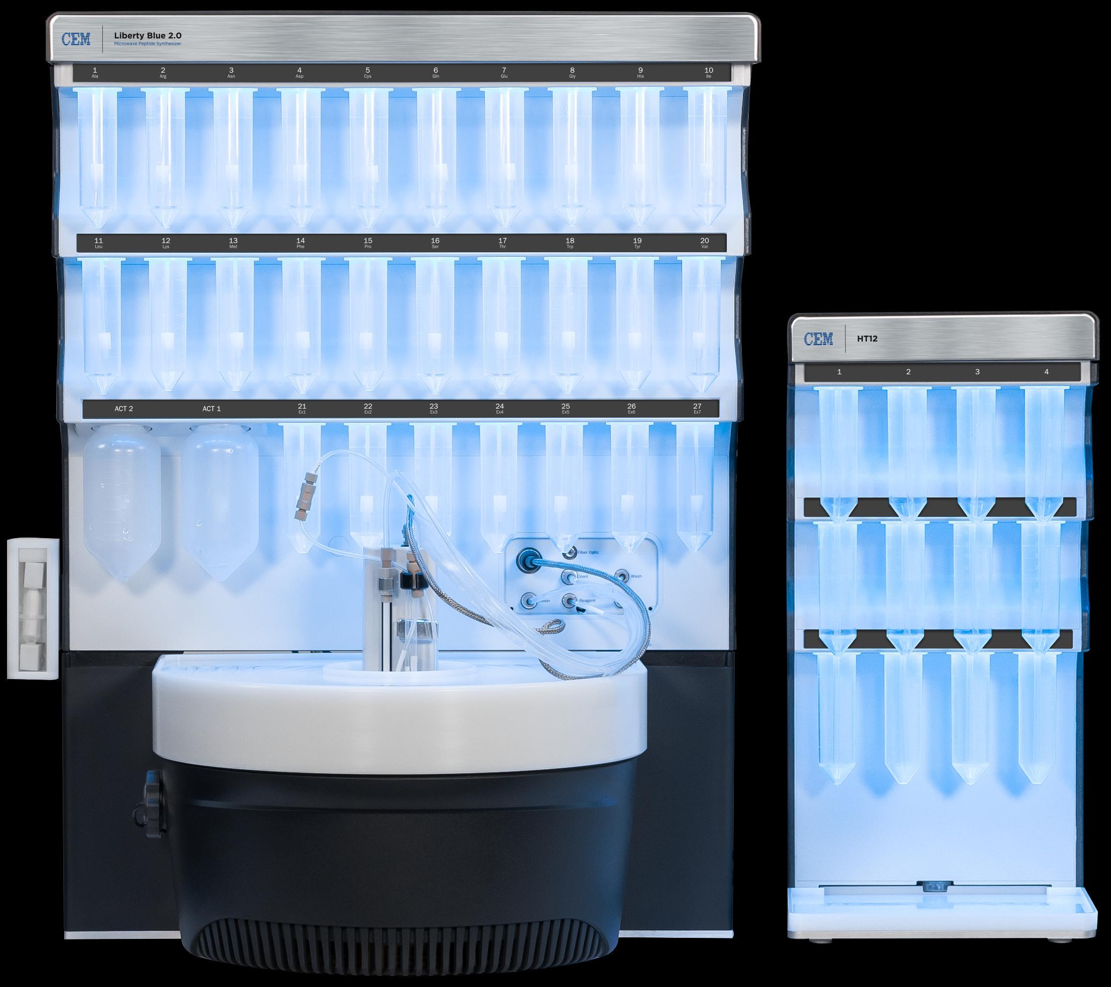
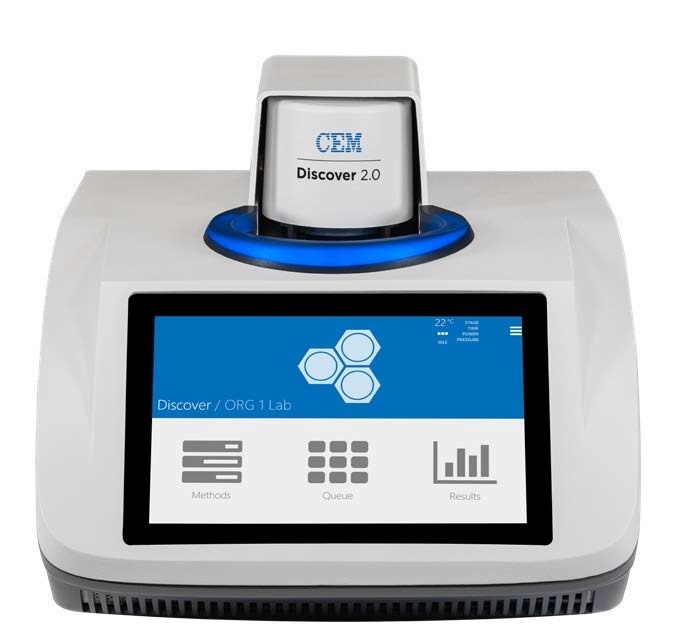
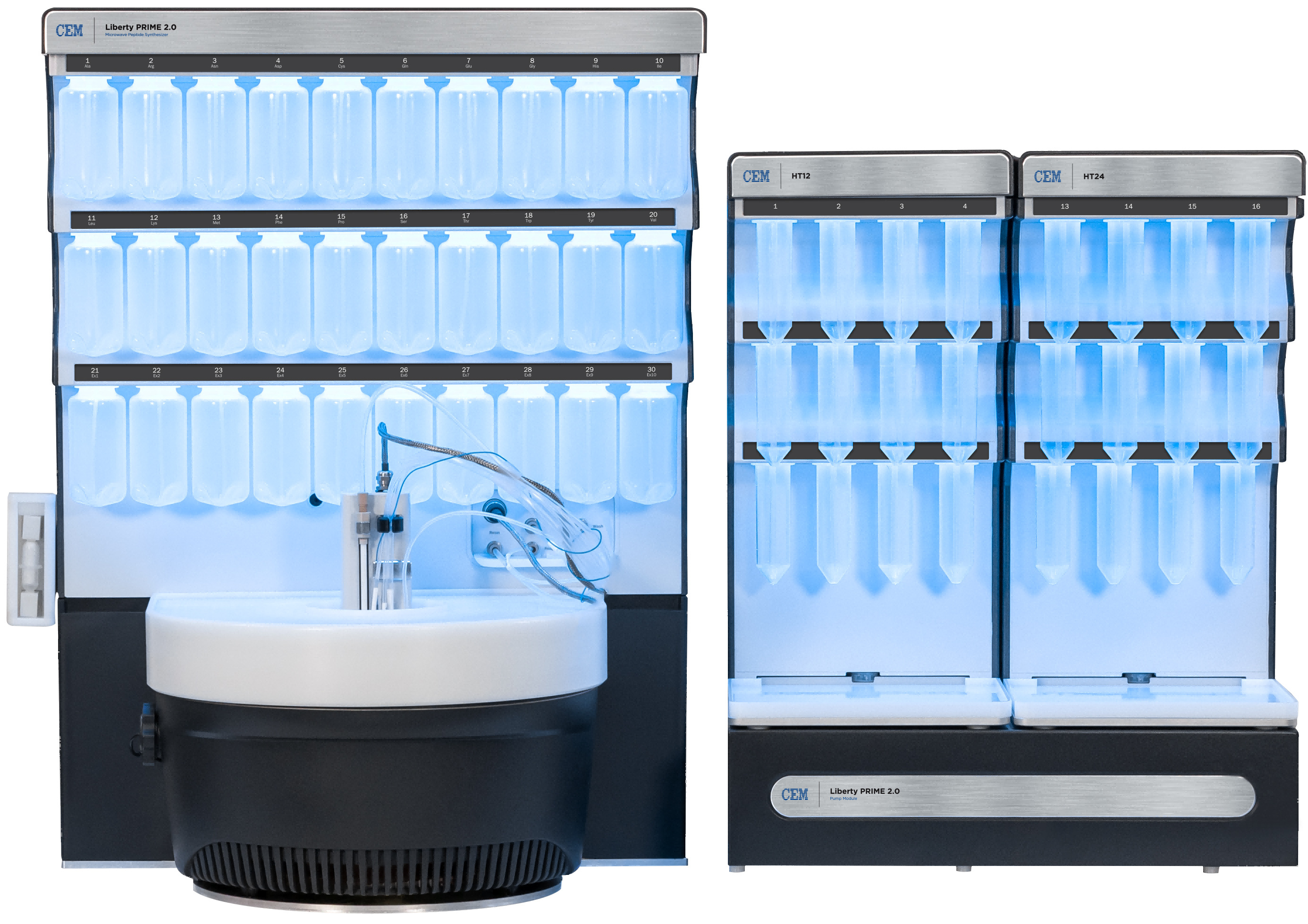
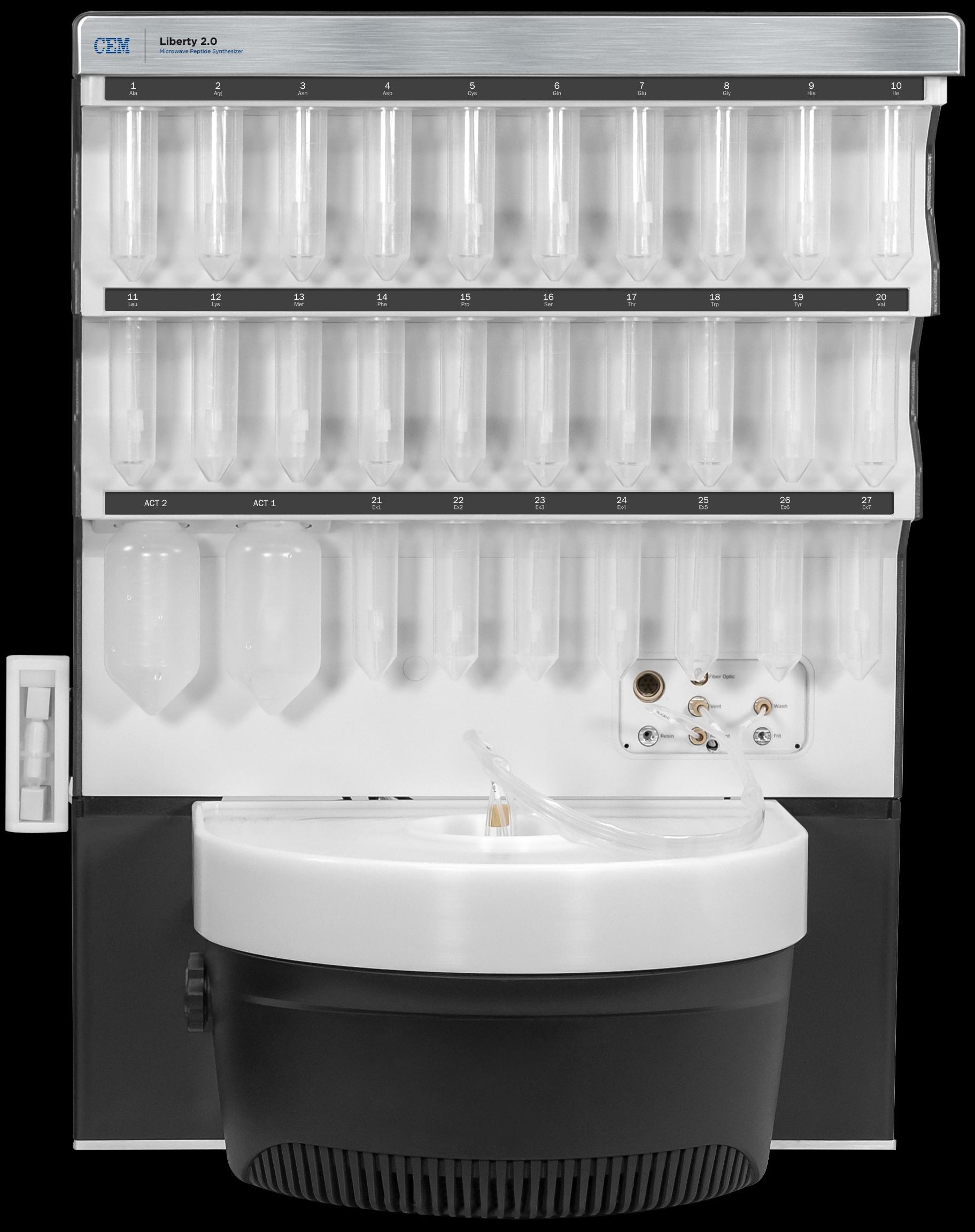
培安有限公司为您提供《多肽中合成检测方案(多肽合成仪)》,该方案主要用于化药新药研发中化合物发现检测,参考标准《暂无》,《多肽中合成检测方案(多肽合成仪)》用到的仪器有CEM LibertyBlue 2.0 全自动微波多肽合成仪、CEM Discover 2.0 300mL腔体环形聚焦单模微波合成仪。
我要纠错
推荐专场
相关方案
 咨询
咨询