

方案详情文
智能文字提取功能测试中
Analytical Biochemistry 555 (2018) 22-25Contents lists available at ScienceDirectAnalytical Biochemistry Y. Zhang et al.Analytical Biochemistry 555 (2018) 22-25 journal homepage: www.elsevier.com/locate/yabio Identification of adeno-associated virus capsid proteins using ZipChip CE/MS Yun Zhang*, Yan Wang, Zoran Sosic, Li Zang, Svetlana Bergelson, Wei ZhangAnalytical Development, Biogen Inc, Cambridge, MA, USA ARTICLEINFO A B S TR ACT Keywords:ZipChip CE/MSIntact mass analysisGene therapyAdeno-associated virusSerotypeIdentity test A simple and rapid identity test of adeno-associated virus (AAV) serotypes is important for supporting the AAVgene therapy development, as it relates to its efficacy and safety. The current mass spectrometry-based identitytests require extensive sample preparation steps, relatively large sample quantities and long analysis time.Herein, we describe a simple and novel microfluidic ZipChip CE/MS method used to characterize AAV capsidproteins. The three capsid proteins of AAV2 were separated and identified within 4 min using 5 nL of sampledirectly from a polysorbate-containing formulation buffer. This rapid method can be suitable to confirm AAVserotype identity. Recombinant adeno-associated viruses (AAVs) are popular genetransfer vectors in clinical gene therapy field because of their lack ofpathogenicity, low immunogenicity, and persistent gene expression invarious tissue types. AAVs are capable of infecting both dividing andnon-dividing cells and providing continuous, long-term expression oftherapeutic genes [1,2]. Currently, AAVs have been used for over 150ongoing clinical trials to target different genetic diseases [3]. AAV is asmall virus with single-stranded DNA genome enclosed in a capsid shell[1], which can be used to deliver DNA to target cells [4]. A specific AAVserotype preferentially transduces specific cell types or organs sinceeach AAV serotype has different tissue tropism, transduction efficiency,and antigenic reactivity. There are 13 human and non-human primateAAV serotypes and more than 150 genotypes have been identified[1,5-7]. In addition, AAV serotype has a direct effect on efficacy andsafety of clinical AAV gene therapies [8]. An AAV capsid contains a total of sixty copies of three capsid viralproteins (VPs) including VP1 (~87kDa), VP2 (~73kDa), and VP3(~61kDa) at an approximate population ratio of 1:1:10, respectively,which are arranged into an icosahedral structure to form the capsidshell [8,9]. The three VPs share a common C-terminal sequence. Theentire sequence of VP3 is contained within VP2, and the VP2 sequenceis contained within VP1[9]. In general, the AAVs share high homologyof amino acid sequences among the VPs. In addition, it was reportedthat single amino acid changes in the capsid proteins can improve transduction with less immunogenicity [10]. Thus, methods which canconfirm the AAV serotype and detect small differences in sequence,such as single-residue variations, are highly desirable. Mass spectrometry (MS) has become an increasingly powerful toolto characterize VPs and identify AAV serotypes [11,12].In 2009, a MSpeptide mapping-based capsid identity test was introduced. Thismethod can distinguish different AAV serotypes based on the massdifferences of unique fragments from proteolytic digestion of differentAAV serotypes. It can also detect single-amino-acid mutations com-pared to the wild-type AAV. However, this method requires serotype-specific protease and relatively complicated sample preparation stepsincluding running a denaturing gel, in-geldigestion, LC-MS/MS ana-lysis, and database search [12].Recently, Jin et al. introduced a directLC/MS intact protein analysis method to characterize VPs in a variety ofAAV serotypes [11]. This approach takes 4 h including sample pre-paration, LC/MS and data analysis, which is at least 6-fold faster thanthe protein digestion method. However, sample preparation includingpre-concentration step and buffer exchange are still required due to lowprotein concentration. High application potential of microchip and capillary electrophor-esis in separation, analysis, and characterization of proteins has beenrecognized [13-15]. Recently, a microfluidic ZipChip CE/MS approachhas been reported to characterize C-terminal variants of intact mAbmolecule, N-glycosylation profile monitoring, glycoform analysis andtiter determination directly from mAb cell culture supernatant withminimum sample preparation, and fast analysis time [16-20]. Here, we ( A bbreviations: CE, c apillary electrophoresis; MS, mass spectrometry; A A V, ad e no-associated virus; VP s , capsid viral proteins; LC , liquid chromatography; AGTC , Appli e d GeneticTechnologies Corporation; R F , r a dio f r equency; D R P, DNase I-r e sistant v e ctor particles; A c., Acetylation; Exp., experimental; Theo., theoretical ) ( * Corresponding author. ) ( E-mail address: y un . z h a ng @b i o g en . c o m ( Y . Zhang). ) ( h ttps :// doi . o r g /1 0. 1 016/ j.a b . 2018 . 06.006 ) ( R eceived 15 March 2 0 18; Received in r e vised form 7 June 2 0 18; A c cepted 7 June 2018 ) describe a simple, rapid, and fully-automated microfluidic ZipChip CE/MS method for identifying VPs from AAV samples to support the re-combinant AAV gene therapy development. The objectives of this workwere to distinguish wild-type AAV2 and an AAV2 mutant with threetyrosine (Y)-to-phenylalanine (F) mutations directly from polysorbate20-containing formulation buffer samples. Following ZipChip separa-tion of three VPs of either wild-type AAV2 or an AAV2 mutant, both N-and C-terminal sequences were identified based on the measured ac-curate masses of the expected protein sequences, thus demonstrating anacceptable specificity of this analysis. Materials and methods Materials A wild-type AAV2 (AAV2) and an AAV2 mutant (AAV2tYF) [21]samples containing 0.014% m/m polysorbate 20 were provided byApplied Genetic Technologies Corporation (AGTC). ZipChip HR(cat#810-00140) and ZipChip Peptides Kit (cat#810-00167) includingZipChip BGE peptides (pH 2.4) and peptide diluent were purchasedfrom 908 Devices (Boston, MA). ZipChip CE/MS The ZipChip CE ion source and autosampler were purchased from908 Devices (Boston, MA) and installed following the vendor's in-struction manual. The device was positioned in front of the inlet of aThermo Q Exactive HF mass spectrometer (Thermo, San Jose, CA). AnHR ZipChip with 22 cm long separation channel was used for sampleseparation analysis. 25 uL of either 3.11E12 DRP/mL (DRP: DNase I-resistant vector particles) AAV2 or 4.90E12 DRP/mL AAV2tYF wasdiluted 5 times with Peptide Kit sample diluent and then the dilutedAAV2 or AAV2tYF was transferred to an autosampler vial. 40 uL ofdiluted AAV2 or AAV2tYF was loaded to ZipChip sample well by theautosampler. The setting of mass spectrometer was mass resolution15000, in-source CID 0 eV, 1 micro scan, AGC target 1E6, maximuminject time 50, spray voltage 0kV, capillary temperature at 200℃, S-lens radio frequency (RF) level 50, Aux gas heater temperature 0C,sheath gas flow rate 2, Aux gas flow rate 0 with data acquired over amass range of m/z 800-2500. Data acquisition was accomplishedthrough Xcalibur (Thermo Scientific) tune page, which was triggered byZipChip software used to control the ZipChip output. The settings of CEwere: injection load time 30 s at 2 Pa (5nL), analysis run time 10 min,pressure assist start time 0.5 min, and field strength 500V/cm.Deconvolution of the mass spectra was performed using the deconvo-lution algorithm in the Biopharma Finder 2.0 software (Thermo, SanJose, CA). Results and discussion clearly without the interference from polysorbate 20. The separation ofthe analytes is based on differences in electrophoretic mobility, which isa function of the charge and size of the analytes. Positively chargedanalytes migrate toward the ESI orifice in the electric field, whileneutral and negatively charged components move back out of the se-paration channel to waste. Thus, the neutral detergent polysorbate 20can be separated from sample to avoid the detergent ion suppressioneffect on MS detection. As shown in the deconvoluted mass spectra ofthe peaks at 3.35 min, 3.38 min, and 3.42 min (Fig.1E, F, and 1G), thedetected masses for the three peaks are 81856 Da, 66488Da, and59973 Da, respectively, which agree well with the expected masses ofacetylated VP1 (amino acid sequence 2-735 with a 42 Da mass shiftindicating a single N-terminal acetylation), VP2 (amino acid sequence139-735), and acetylated VP3 (amino acid sequence 204-735 with a42 Da mass shift indicating a single N-terminal acetylation), respec-tively. Mass errors for the three VP peaks are less than 20 ppm (TableS1). The peaks corresponding to the salts in the sample matrix weredetected in a migration time window between 1.30 min and 3.20 min.In addition, each analysis consumed 5 nL of sample, which is over10000 times less than the amount previously used for RP-LC/MS intactmass analysis [11]. A rapid CE/MS analysis also showed superior se-paration of VP1 and VP2 compared to the RP-LC/MS intact massmethod [11]. Ac: acetylation. Exp.: experimental; Theo.: theoretical In addition to the wild-type AAV2, we further analyzed an AAV2mutant with three amino acid mutations to evaluate the specificity ofthis analysis. Total ion electropherogram of AAV2tYF is shown inFig. 2A. The total analysis time is 10 min and the separation of the threeVPs was also completed in less than 4 min, with all protein peaks de-tected in a migration time window between 3.60 min and 3.90 min andwith an average peak width at base of 3 s (the inset of Fig. 2A). Fig.2B,C, and 2D show the mass spectra for the peaks with the migration timesof 3.68 min, 3.71 min, and 3.79 min, respectively. The charge dis-tributions for the three VPs were also observed clearly without the in-terference from polysorbate 20. As shown in the deconvoluted massspectra of the peaks at 3.68 min, 3.71 min, and 3.79 min (Fig. 2E, F, and2G), the detected masses for the three peaks are 81807 Da, 66436 Da,and 59924 Da, respectively, which agree well with the expected massesof acetylated VP1 (amino acid sequence 2-735 with a 42 Da mass shiftindicating a single N-terminal acetylation), VP2 (amino acid sequence139-735), and acetylated VP3 (amino acid sequence 204-735 with a42 Da mass shift indicating a single N-terminal acetylation), respec-tively. Mass errors for the three VP peaks are equal or less than 60 ppm(Table S1), which is within the acceptable assay variability. Thus,ZipChip CE/MS intact mass method can distinguish wild-type AAV2and an AAV2 mutant that differ in only three amino acids. Ac: acetylation Exp.: experimental; Theo.: theoretical In summary, we report a novel, rapid, and fully-automated ZipChipCE/MS method to separate and measure the accurate masses of threeVPs in support of AAV serotype identification. The wild-type AAV2 andan AAV2 mutant that differ in three amino acids have been successfullyanalyzed and corresponding masses for VPs were accurately de-termined. This fast analysis approach allows direct analysis of diluted,low concentration samples from polysorbate-containing formulationbuffers as well as small material consumption, which is often importantconsideration due to potential limited sample availability. Taken to-gether, this type of analysis could be used as a rapid screening methodto confirm AAV serotype identity or monitor VP heterogeneity to sup-port the recombinant AAV gene therapy development. 100 NL:1.17E8 VP3 1007A 3.79 NL: 3.66E8 Conflicts of interest Appendix ASupplementary data The authors declare no competing interests. Fig. 1. (A) Total ion electropherogram ofAAV2 VPs with the migration time rangefrom 0.1 min to 5.0 min; the migration timerange from 3.20 min to 3.60 min is shown inthe inset. Mass spectra of AAV2 VPs sepa-rated in Fig. 1A: (B) peak at 3.35 min, (C)peak at 3.38 min, and (D) peak at 3.42 min.Deconvoluted mass spectra of AAV2 VPsseparated in Fig.1A: (E) peak at 3.35 min,(F) peak at 3.38 min, and (G) peak at3.42 min. Fig. 2. (A) Total ion electropherogram ofAAV2tYF VPs with the migration time rangefrom 0.1 min to 5.0 min; the migration timerange from 3.60 min to 3.90 min is shown inthe inset. Mass spectra of AAV2tYF VPs se-parated in Fig. 2A: (B) peak at 3.68 min, (C)peak at 3.71 min,and (D) peak at 3.79 min.Deconvoluted mass spectra of AAV2tYF VPsseparated in Fig. 2A:(E) peak at 3.68 min,(F) peak at 3.71 min, and (G) peak at3.79 min. Acknowledgement This research did not receive any specific grant from fundingagencies in the public, commercial, or not-for-profit sectors. We thank AGTC for kindly providing AAV2tYF and wild-type AAV2samples. Supplementary data related to this article can be found at http://dx.doi.org/10.1016/j.ab.2018.06.006. References [1] E. Hastie, R.J. Samulski, Adeno-associated virus at 50: a golden anniversary ofdiscovery, research, and gene therapy success—a personal perspective, Hum. GeneTher. 26 (2015) 257-265. ( [ 2 ] L . L isowski, S .S. T a y , I. E . A l e x a nd e r, Ad eno-ass o ciated v i r u s ser o types fo r g en e t herap e utic s, C u rr . Opi n . Pharmac o l . 24 ( 20 15)59 - 67. ) ( [3] A . B en ne tt , S . P a t el, M . M i et z s c h , A. Jo s e , B. L i n s- A u st i n , J . C. Y u, B . Bothner, R . M cKen n a , M. A g b a n d je- M c K enna, T h e r m al s t a bi li t y a s a s e t ermi nan t of AAV s er o t yp e i de n t i t y, Mol . Th er. Methods Cl i n . D e v . 6 ( 2017 ) 171- 1 8 2. ) ( [4 ] M . F . N a so , B . T o m k o wi c z, W.L . Pe r r y , W . R . S tr ohl, Ad eno - a ssoc ia te d vi r us ( A A V) as a vecto r for g ene t herapy , BioDrugs 3 1 ( 201 7) 3 17 - 3 34. ) ( [5] G . G a o , L . H . Va nd enb e rghe , J .M. W ilson, Ne w re c o mbinant se r o ty p e s o f AA V e1 v e cto r s , Cu r r . G e n e Ther . 5 ( 2005) 285-297. ) ( [6 ] L . L i so w s k i , A.P . Dane, K. Chu, Y . Zh a n g , S .C. C u n ni n g ham, E . M . W i ls o n , S . N y g aar d , M . G r o m pe , I . E . A l e xan d er , M . A . K a y , S e l e c tio n an d e v a l u a t i on o fc l inic a ll y re l ev a n t AAV variant s i n a x e nog raf t l ive r m o del, N a t u r e 5 06 ( 2 014) 3 82- 3 86. ) ( [ 7 ] M . M i e tz s ch, F . B r oeck e r, A . R e i nh a r dt, P. H . S e e b erger , R. H e i l bro n n , Diff e r ential a d e no - ass ociat ed v i r u s sero t y pe - s pe c i fi c i n te r a c t i on pa t te r n s wi t h s y nt he t ic h e - p arin s a nd othe r gly c an s, J . Vir o l . 88 (201 4 ) 2991 - 3 003 . ) ( [8 ]R . J. S a m ul s ki , N . M uzyc zka , A A V - me d ia t ed ge n e t h e r ap y for res e arc h an d the r - a peu t i c p urposes , A nnu. R ev .Vi r o l . 1 ( 2 01 4) 4 2 7-451. ) ( [9 ] ] K . Van V l i e t , V . Blouin, M . A g band j e - M cKe n na,R . O. Snyder , P r o teo l ytic mappin g of t he adeno-associate d v i rus c a psid , Mo l. Ther . 1 4 (2006) 809 -8 21. ) ( [10] C . L i, N. D i p rimio , D.E . B o w l e s , M .L . H i rsch, P. E. Monah a n, A . A s ok a n , ) ( J . R abinowit z , M . A g b a ndje-Mc K enna, R.J.S a m ul ski, S i ng l e a mi no acid m odi f ica- t i o n of ad eno- a sso cia t ed v i r u s c a p s id c hang es t r a nsdu c t ion a nd hum o ral immu n e p ro f ile s , J . V i r o l . 8 6 ( 201 2) 7 7 52 - 7 7 59. ) ( [ 1 1] X . J i n, L . L i u , S . N ass , C . O ' R i o rd a n , E. P a s t o r , X .K . Z h a n g, Di re ct li q u id chr o m a- t o g ra p hy/ ma s s spect ro m et ry an a ly s i s f o r c o m plete c h a r acterizat i on of r e c o m bin a nt a de n o-a s s o cia ted v i rus c a p s id pr ot ei ns , Hu m . G e ne Th er. M et h . 28(20 1 7) 2 55-267. ) K. Van Vliet, Y.Mohiuddin, S. McClung, V. Blouin, F. Rolling, P. Moullier, ( [12 ] M . Ag b a ndje-M c K enna, R . O . S n yd er, Ade n o-asso c iate d vir u s cap s id ser oty pe ide n - 十 . 1 1 t i f ic a t i on : a nal yti c al me thod s d e v e lo p me n t a nd a p pl i c a t i on, J . V i r o l . M e t ho d s 15 9 ( 2 009) 1 6 7-1 7 7. ) ( [ 13] S . S te p a n ova, V. K asicka, A na l ys i s of p ro t e i n s a n d pe p tide s by el e ctromig r a ti o n m et h od s i n m icr o ch i p s , J. S epar. S c i . 40 (2 0 1 7 ) 2 2 8-2 50 . ) [14] M. Dawod, N.E. Arvin, R.T. Kennedy, Recent advances in protein analysis by ca-pillary and microchip electrophoresis, Analyst (Cambridge,U.K.) 142 (2017)1847-1866. [15] S. Stepanova, V. Kasicka, Recent developments and applications of capillary andmicrochip electrophoresis in proteomic and peptidomic analyses, J. Separ. Sci. 39(2016)198-211. [16]J.S. Mellors, V. Gorbounov, R.S. Ramsey, J.M. Ramsey, Fully integrated glass mi-crofluidic device for performing high-efficiency capillary electrophoresis and elec-trospray ionization mass spectrometry, Anal. Chem. 80 (2008) 6881-6887. [17]Y. Wang, P. Feng, Z. Sosic, L. Zang, Monitoring glycosylation profile and proteintiter in cell culture samples using ZipChip CE-MS, J. Anal. Bioanal.Tech. 8(2017)1-8. [18]E.A. Redman, N.G. Batz, J.S. Mellors, J.M. Ramsey, Integrated microfluidic capil-lary electrophoresis-electrospray ionization devices with online MS detection forthe separation and characterization of intact monoclonal antibody variants, Anal.Chem. 87 (2015) 2264-2272. [19]K. Khatri, J.A. Klein, J.R. Haserick, D.R. Leon, C.E. Costello, M.E. McComb, J. Zaia,Microfluidic capillary electrophoresis-Mass spectrometry for analysis of mono-pIsaccharides, oligosaccharides, and glycopeptides, Anal. Chem. 89 (2017)6645-6655. ( [20]A . G. C hambers, J .S . M e l lors , W .H. H e nle y , J.M . R ams e y, M on ol i t h i c in t eg r at i o n o f t wo-dim e nsional l i q u i d chr omat o g r a ph y -capi l l a ry e l e c tr o p hores i s a n d e l e c t r o - s pray i o n i z a t ion on a mi c r o fl u i dic d e v i c e , A n al . C hem . 8 3 ( 2 01 1 ) 8 42 - 849. ) ( [21]G . -J . Ye , E . B u d zy n ski, P . S onn e nta g, T . M . No rk, P . E. M i l ler, A . K. Sh ar m a, J . N . Ve r H o ev e, L. M . S m ith, T . Ar ndt, R . C alcedo , C . Ga s k i n , P . M . R o bi n son , D .R. K n o p, W .W . H a usw i rt h , J .D . C h ul a y , S afe t y an d bi o dis t rib u tion e v al uat i on i n c y n om olg u s m aca q ues of rAAV2 t YF- P R1.7 - hCNGB3, a r eco m bin a n t A A V v ec tor for treatment of 5 , a c h romat o psi a , H um . G e n e T h e r . C l i n. D e v. 2 7 ( 2 0 1 6) 37 - 4 8. ) 重组腺相关病毒 (AAVs, adeno-associated viruses)由于其致病性差、免疫原性低、在多种组织中持续表达等特点,成为临床基因治疗领域的热门基因转移载(gene transfer vectors)。AAV 能够感染分裂细胞和非分裂细胞,并提供治疗基因的持续、长期表达。目前,AAV已被用于150多个正在进行的针对不同遗传疾病的临床试验。AAV 是一种小病毒,单链DNA基因组被包裹在衣壳中,可用于将DNA传递给靶细胞。特定的 AAV 血清型 serotype 优先传递特定的细胞类型或器官,因为每个AAV血清型具有不同的组织取向性、转导效率和抗原反应性。有13个人类和非人类灵长类AAV血清型和150多个基因型已经确定。此外,AAV 血清型 serotype 直接影响临床AAV基因治疗的有效性和安全性。 AAV 衣壳共含有三种病毒蛋白 (VPs,viral proteins) 的 60 份拷贝,三种病毒蛋白包括VP1 (∼87 kDa)、VP2 (∼73 kDa)、VP3 (∼61 kDa),其种群比例约为1:1:10,排列成一个二十面体结构,形成衣壳。这病毒蛋白共享一个共同的C端序列,整个 VP3 序列包含在 VP2 中,VP2 序列包含在 VP1 中。AAVs 与 VPs 之间的氨基酸序列有很高的同源性。此外,据报道,在衣壳蛋白中即使是单个氨基酸的变化,也会改善免疫原性较低的转导。因此,目前对能够确定AAV血清型和检测序列上的微小差异如单残留变异的方法具有高度需求。 2009年研究人员提出了一种基于质谱肽定位的衣壳同一性检测方法(J. Virol. Methods, 2009,159:167–177)。该方法能根据不同 AAV 血清型的蛋白水解产物的质量差异来区分不同的 AAV 血清型。与野生型AAV相比,它还能检测到单氨基酸突变。然而,这种方法需要血清特异性蛋白酶和相对复杂的样品制备步骤,包括进行变性凝胶、凝胶内消化、LC-MS/MS分析和数据库检索。最近,金等人介绍了一种直接的 LC/MS 完整蛋白质分析方法来表征各种AAV血清型中的VPs(Hum. Gene Ther. Meth., 2017, 28: 255–267)。该方法包括样品制备、LC/MS和数据分析4小时,比蛋白质消化法至少快6倍。然而,由于蛋白质浓度较低,样品的制备还需要进行包括预浓缩和缓冲液交换的步骤。 微流控芯片和毛细管电泳在蛋白质的分离、分析和表征方面具有巨大的应用潜力。ZipChip 是美国908Devices 公司开发出的全新的基于微流控芯片的毛细管分离平台,通过与高分辨质谱仪的联用,可以实现生物蛋白大分子的快速、高效的分离。近期的多篇文章报道了ZipChip CE-MS 用于表征完整抗体分子以及抗体电荷变异体、N-糖基化谱监测、糖型分析和滴度测定等应用。本解决方案提供了一种简单、快速、全自动的微流控芯片CE/MS方法,用于从AAV样本中识别VPs,以支持重组AAV基因治疗的发展。以野生型 AAV2 和含有三个酪氨酸(Y)到苯丙氨酸(F)突变的AAV2突变体为测试样品,所有的仪器为908Devices 公司的ZipChip 离子源结合Thermo Fisher 公司的Q Exactive HF质谱仪,如图1所示,采用的芯片为908Devices公司ZipChip HR 芯片和背景缓冲溶液为ZipChip Peptide 试剂盒。 采用ZipChip CE-MS区分样品中的变异体,成功地分析了野生型AAV2和AAV2突变体在三个氨基酸上的差异,并准确地确定了VPs的相应质量,如图2所示。根据所测到的准确的蛋白序列,对N端和C端序列进行了鉴定,进一步验证了该方案的可靠性。ZipChip分离是基于电泳迁移率的差异,电泳迁移率取决于分析物的电荷和尺寸。带正电荷的分析物在电场作用下进入微流控毛细管分离通道并沿着通道向ESI 喷雾端迁移,而中性和带负电的成分从分离通道回流至废液池。因此,ZipChip的另一独特优势是可以将中性洗涤剂聚山梨酸盐polysorbate 20从样品中分离出来,以避免洗涤剂对MS检测的离子抑制影响。 这种快速分析方法允许直接分析稀释的、低浓度的样品,能够从含有多山梨酸盐的配方缓冲液中直接分离,并且消耗的样品量非常少(1-20纳升),这些都是潜在的样品受限而需要考虑的重要因素。综合考虑,这种基于微流控技术的芯片毛细管电泳-质谱联用技术(ZipChip CE-MS)可作为AAV血清型鉴定或病毒蛋白异质性监测的快速筛选方法,以支持重组AAV基因治疗的发展。
关闭-
1/4
-
2/4
还剩2页未读,是否继续阅读?
继续免费阅读全文产品配置单


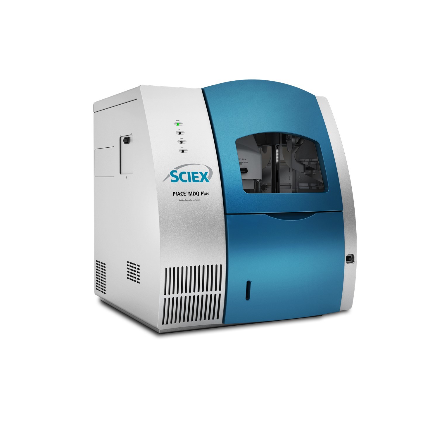
908 Devices为您提供《腺相关病毒中变异体检测方案(毛细管电泳仪)》,该方案主要用于其他中变异体检测,参考标准《暂无》,《腺相关病毒中变异体检测方案(毛细管电泳仪)》用到的仪器有908Devices ZipChip AS 毛细管电泳质谱联用系统 (自动进样款)、ZipChip 肽段分析试剂盒。
我要纠错
相关方案
 咨询
咨询