

方案详情文
智能文字提取功能测试中
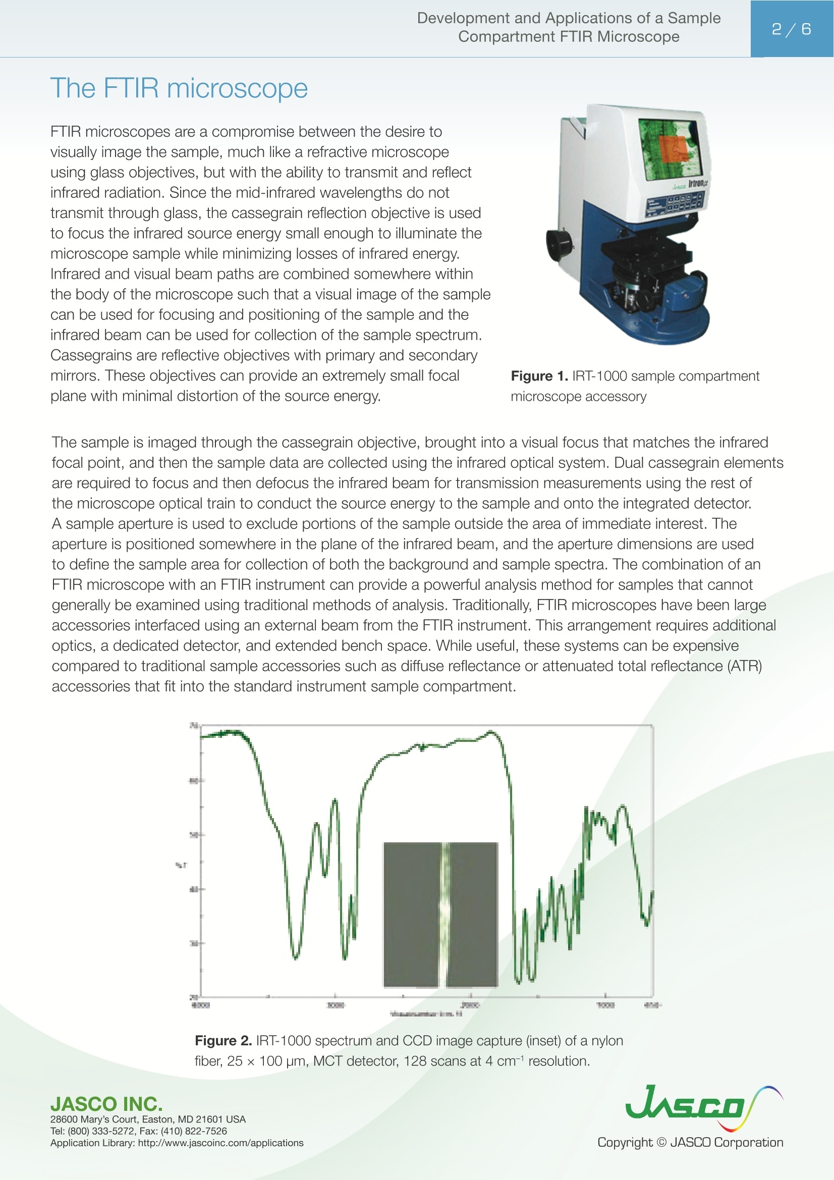
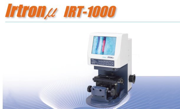
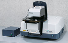
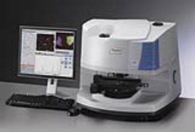
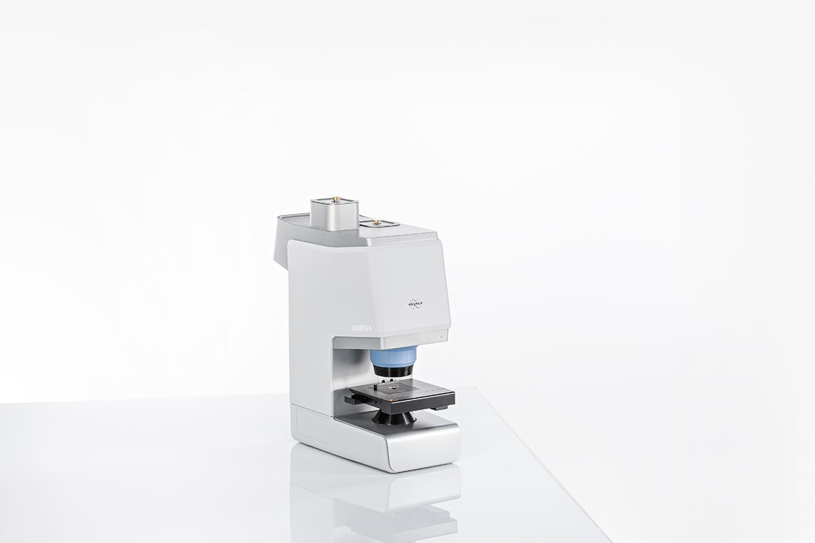
20世纪60年代中期FTIR光谱仪的引入激发了人们对红外显微镜的兴趣,因为干涉仪的吞吐量增加了,并且能够进行多次扫描以提高信噪比。红外显微镜系统最初由Digilab和Spectra-Tech在20世纪70年代末和80年代初引入。IRLAN由Spectra Tech于1983年推出,作为FTIR显微镜配件,可与各种制造商的仪器连接。由于能够以透射或反射模式从小至15μm的样品中收集红外光谱,红外显微镜配件成为从250μm或更小的样品区域收集红外光谱的首选工具。FTIR显微镜有许多应用,包括聚合物和纤维分析、药物和材料分析问题、取证、半导体、生物化学和化学分析应用。对于几乎任何可以用传统红外方法扫描的样品,FTIR显微镜都可以用来收集样品碎片的红外光谱。红外显微镜的空间分辨率主要由用于分析的红外波长的衍射极限决定,通常为12–20μm。显微镜中的孔径系统用于控制样品光束的空间尺寸,并且该孔径不能小于聚焦到样品上的红外波长。本应用说明通过实验示例说明了:IRT-1000是一款真正的FTIR显微镜配件。显微镜能够很容易地设置在仪器样品室中,无需对准,可以使用标准DTGS检测器或液氮冷却的MCT检测器。IRT-1000能够收集许多不同类型样品的透射率、反射率或ATR光谱,提供了一系列性能不受影响的功能。Application Note Development and Applications of a SampleCompartment FTIR Microscope2/6 Development and Applications of a Sample Compartment FTIR Microscope Since the early to mid-1940's, scientists using infrared spectroscopy have been trying to obtain spectral data from ever smaller samples. Starting with micro-KBr pellets and then beam condensers and micro-MIR (multiple internal reflectance) accessories, the ability to obtain a spectrum from an extremely small sample has been attempted on a regular basis. I ntroduction The introduction of FTIR spectrometers i n t he mid-1960's sti m ulated the interest i n infrared microscopy due to the increased through p ut of the interferometer and the ability to perform mult i ple scans to increase the signal-to-noise rat i o.Infrared microscope systems were ini t ially i ntroduced in the l ate 1970's and early 1980’s by Digi l ab and Spectra-Tech.The IRPlan was i ntroduced in 1983 by Spectra-Tech as an FTIR mi c roscope accessory that could be interfa c ed with instruments from various manufacturers. With the abi l ity to collect infrared spectra from samples as small as 15um in either transmission or reflection mode, the infrared microscope accessory became the preferred tool for t he collection of infrared spectra from sample areas of 250 pm or less. JASCO's I RT -5000/7000 F T/I R Microscope Series There are numerous applications for FTIR microscopy, including polymer and f iber analysis, pharmaceutical and mate r ials analysis problems, forensics, semiconductors, biochemistry, and chemical analysis applicat i ons.For almost a n y sample that can be scanned with a traditional i nfrared method, the FTIR microscope can be used to col l ect an inf r ared spectrum from a fragment of a sample. The spatial resolution of an infrared microscope is pri m ar i ly determined by the diffraction l imit of the i nfrared wavelengths used for analysis, usually 12-20 pm. The aperture system i n the microscope is used to control the spatial dimensions of the sample beam, and this aperture cann o t be smaller than the i nfrared wavelengths to be focused onto the sample. FTIR microscopes are a compromise between the desi r e to visually i mage the sample, much l i ke a refractive microscope using glass objectives, but wi t h the abi l i t y to transmit and ref l ect infrared radiation. Since the mid-infrared wavelengths do not transmit through glass, the cassegrain reflection objective is used to focus t he infrared source energy small enough to illuminate the microscope sample while minimiz i ng losses of i nfrared energy.Infrared and visual beam paths are combined somewhere within the body of the microscope such that a visual image of the sample can be used for focusing and posit i oning of t he sample and t he infrared beam can be used for collection of the sample spect r um.Cassegrains are reflect i ve objec t ives with primary and secondary mirrors. These objec ti ves can provide an extremely small focal plane with minimal distortion of the source energy. Figure 1. IRT -1000 sample compartment mi c rosc o pe accessory The sample is imaged t hrough t he cassegrain objective, brought i n to a visual focus that matches the infrared focal point , and then the sample data are collected using the infrared optical system. Dual cassegrain elements are required to foc u s and then defocus the infrared beam for transmission measurements using the rest of the microscope optical train to conduct the source energy to the sample and onto the integrated detector.A sample aper t ure is used to exclude portions of the sample outside the area of i mmediate interest . The aperture is positioned somewhere in the plane of the i nfrared beam, and the aperture dimensions are used to define the sample area for collection of both t he background and sample spectra. The combination of an FTIR microscope with an FTIR instrument can provide a powerful analysis method for samples that cannot ge n erally be examined using traditional methods of analysis. Traditionally, FTIR microscopes have been large accessories i nterfaced using an external beam f rom the FTIR instrument. This arrangement requires additional optics, a dedicated detector, and extended bench space. While useful , these systems can be expensive compared to traditional sample accessories such as diffuse reflectance or attenuated total reflectance (ATR) accessories that fi t into the standard i ns t rument sample compart m ent. Figure 2. IRT -1000 s p ec t rum and CCD i mage capt u re (i n set) of a nylon fiber, 25×100 pm, MCT detector, 128 scans at 4 cm-1resolutio n . The IRT -1000 (F i gure 1) (Jasco, Easton, MD) is an FTIR microscope accessory that f its i nto the i nstrument sample compartment of the FT/IR-4000 or FT /IR-6000 series i nstruments (Jasco). The IRT -1000 i n tegrates a manual stage, charge couple device (CCD) video camera, and LCD video screen i n an i nterchangeable accessory that weighs no more than 15 lb and ut i l i zes the standard i nstrument detector(s) for data collect i o n .The microscope can collect sample data with ei t her the standard deuterated t ri glycine sulfate (DTGS) or an optional liquid nitrogen cooled mercury cadmium telluride (MCT) detector for collection of infrared spectra.The microscope accessory can be used to collect spectral data f rom 15,000 to 250 cm-1, depending on the instrument config u rat i on. Figure 3. Pol ye ster f iber (inset -CCD vid e o capture ), 4 cm -1r e sol ut ion;comparison o f i n st ru me nt de t ector performa n ce. Figure 4. DTGS and MC T dete c tor scans of a PTFE f il m sample, 128scans, 4 cm-1 resol u tion, 100×100 pm aperture. Wi t h the accessory, samples as smal l as 20 um can be visualized, and spectral data can be col l ected us i ng either t ransmission or reflec t ion mode. The addition of an ATR objective, such as diamond, germanium (Ge),or zinc selenide (ZnSe), allows the collect i on of ATR spectra from samples, minimizing the requiremen t for extensive sample preparation. The IRT-1000 can be i ntegrated with the i nstrument power supply a n d PC software, or can be operated in stand-alone mode using a separate power supply and front panel controls for the co l lec t ion of either background or sample spectra, while additional controls are used to set the si z e of the samp l e aperture or Aperture Through Observat i on System (ATOS). This sample aperture system, which is illum i nated by an integrated red LED, can be set as small as 20×20 pm or as large as 800 ×800 pm. Figure 5. DTGS detect o r scans of a pol y mer la mina t e sample: Polyet h ylene (blue) and nylon (red) 128 sc a ns, 4 cmresolu t ion. Figure 6. GeO, crystal sample, 256 scans, 4 cm- resolut i on, DTGS detector . Figure 2 illustrates a spectrum of a si m ple nylon fiber tha t was collected using the IRT-1000 microscope accessory. The f iber was f l attened and mounted across an open aperture on the standard sample holder for t he microscope. The sample f iber was visualized us i ng the visible system, the ATOS apertu r e was set to exclude areas other t han t he fiber, t he f iber was moved away f rom the aperture, an d t he backg r ound of an open area was collected. The fiber was t hen moved i nto t he aperture area, and the sample spectrum of the fiber itself was collected. Figure 3 demonstrates a comparison between t he MCT and DTGS detector performance for a polyester fiber sample. The sample was f lattened and mounted over an open aper t ure on the standard sample holder.The ATOS aperture was set to 35 ×80 pm and the data were collected wi t h the two different detectors. As illustrated in t he f igure, the DTGS detector is capable of providi n g si m ilar quali t y data for the same sample, but does require a greater number o f scans, and t ime, to obtain the same quality data as the MCT detector. For samples that contain f unctional groups that absorb outside the range of a standard MCT detector , the DTGS detector can prov i de an advantage for t hese measurements given that the DTGS detector has a greater spectral range down to 400 cm-1. Figure 4 illustrates a comparison between t he spectra of a thin film sample of poly(tetrafluoro-ethylene) (PTFE) sample collected using the two different detectors. T he DTGS detector was able to obta i n the peaks associated wit h the C - F bending modes below 550 cm -1, while the MCT detector spectral range is limited to approx. 600 cm-1. Whi l e i t is possible to specify an MCT detector with a greater spectral ra n ge, t here is a sacri f ice in sensitivity performance with this detector and often it i s elected to obtain a m edium-band or narrow-band MCT with the attendant rest r icted spectral range. Figure 5 demonstrates the spectra collected f rom the cross-section of a polymer laminate sample. The spectrum of the polypropylene layer was collected using the ATOS aper t ure set to 20×700 pm; t he nylon layer was larger and the aperture was set to 50×700 um for the spectrum collection. The DTGS detector was used to collect t he spectra of the polymer layers. The spectra demonstrate the ability of the ATOS aperture to exclude the portions of the nylon layers that are on either side of t he polyethylene layer in the laminate sample. When the FTIR is configured with a Csl beamsplitter, t he spectral range of the instrument can be extended into the far-IR region approaching 200 cm-1. Figure 6 illustrates the spec t rum o f a germanium oxide crystal t hat has several absorption bands below 600 cm-1. The GeO, crystal was supported on a polyethylene sheet, the band at 725 cm-1due to the polyethylene. The data were collected with a standard DTGS detector , which can obtain data f r om organometallic samples, well beyond t he range of even a wide-band MCT detector used in standard FTIR microscope systems. To obtain reflectance spectra, t he top cassegrain optic is used; i n frared sou r ce energy i s directed t hrough one-hal f of the cassegrain, and the other half used to collect the reflected energy and direct i t onto the detector.The sample aperture is set as desired for the sample, a background spectrum collected from a reference mirror, and the desired sample area co l lected as a sample spectrum and ratioed versus the background spectrum of t he reference m irror. In F i gure 7, the reflectance spec t rum of a soda can l ining is displayed; the spectrum was collected with the MCT detector and an aperture set t ing of 100 ×100 pm was used. The resul t s of a library search provided a similar spec t rum that was a two-part epoxy compound. Figure 7. Refl e ctance sc a n of beverage can l ini n g, 128 scans, 4 cm-1resol u t i on. Figure 8. ZnSe AT R ob j ective spectrum of layer i n a po l ymer laminate sample, 256 scans,MCT detector , 250×250 um. The addition of an ATR objective provides the capability to collect sample spectra with a minimum of sample preparation. F i gure 8 demonstrates a spectrum of a polymer laminate sample collected using a ZnSe in t erchangeable ATR object i ve a n d the MCT detector. The ac t ive sample contact area for the ATR objective was 250 pm; the poly(ethylene terephthalate) sample was much la r ger than the active area of the ATR objecti v e. Conclusion The IRT-1000 is an FTIR microscope accessory that truly is an accessory. Capable of being easily set into the instrument sample compartment, the microscope requires no alignment and can utilize either the standard DTGS detector or a liquid nitrogen cooled MCT detector. With the ability to collect transmittance, reflectance,or ATR spectra o f many different types of samples, the IRT -1000 offers a range of capabilities without compromise i n performance . Applicatio n Library: http://www.j ascoinc .com/appl i c a t i ons
关闭-
1/6
-
2/6
还剩4页未读,是否继续阅读?
继续免费阅读全文产品配置单
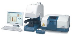
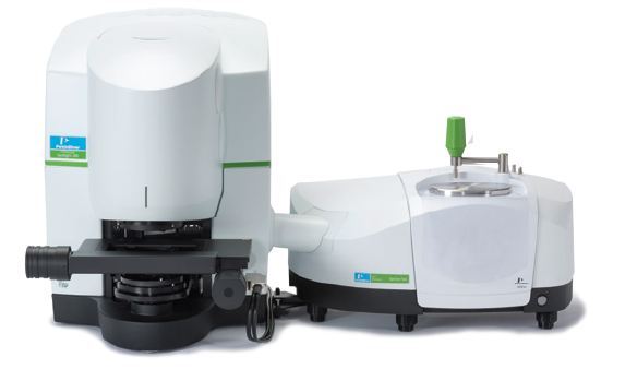
佳士科商贸有限公司为您提供《样品舱FTIR显微镜综述》,该方案主要用于其他中FTIR显微镜、透射率、反射率、ATR光谱检测,参考标准《暂无》,《样品舱FTIR显微镜综述》用到的仪器有jasco,小型红外显微镜,jasco IRT-1000、JASCO傅立叶变换红外光谱仪FT/IR-6000、JASCO红外显微镜IRT-7200。
我要纠错
推荐专场
其它光谱仪
更多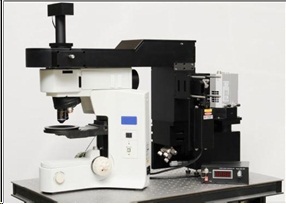
相关方案
 咨询
咨询