【PalmSens4电化学应用】基于还原氧化石墨烯和分子印迹共聚物的电化学传感器,用于测定环丙沙星的聚苯胺−聚邻苯二胺
检测样品 环境水(除海水)
检测项目 综合

 铜牌会员
29 篇解决方案
铜牌会员
29 篇解决方案
方案详情文
智能文字提取功能测试中
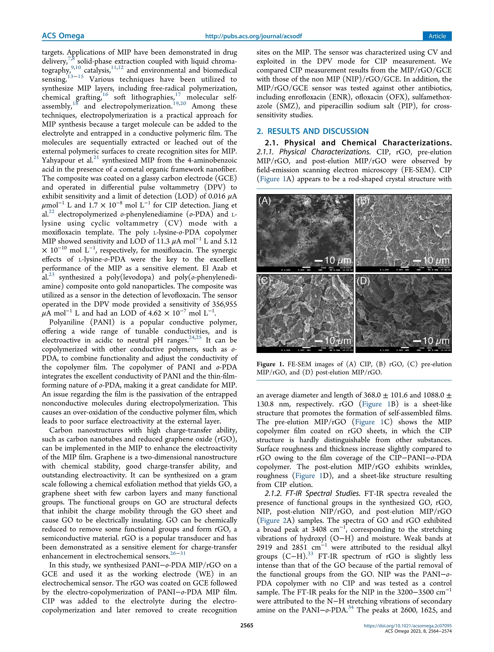
水中抗生素的污染是病原体产生抗生素耐药性(ABR)的主要原因,危害全球人类健康和粮食安全。环丙沙星(CIP)是一种合成氟喹诺酮(FQ)抗生素,据报道,在某些地区,其在地表水中的浓度超过了生态毒理学预测的无作用浓度。本研究使用具有CIP识别位点的聚苯胺(PANI)和聚邻苯二胺(o-PDA)的电聚合分子印迹聚合物(MIP)制备了CIP传感器。将MIP涂覆在还原氧化石墨烯(rGO)修饰的玻碳电极(rGO/GCE)上,并在差分脉冲伏安法(DPV)模式下进行CIP检测。该传感器在1.0×10^-9至5.0×10^-7 mol/L CIP范围内表现出优异的响应,传感器检测限和灵敏度分别为5.28×10^-11 mol/L和5.78μA mol/L。该传感器对CIP的灵敏度是其他测试抗生素的1.5倍,包括恩诺沙星(ENR)、氧氟沙星(OFX)、磺胺甲恶唑(SMZ)和哌拉西林钠盐(PIP)。还研究了传感器装置的再现性和可重复使用性。--摘自ACS Omega 2023, 8, 2564-2574, https://doi.org/10.1021/acsomega.2c07095cc①③ ArticleACS Omegahttp://pubs.acs.org/journal/acsodf Electrochemical Sensor Based on a Composite of ReducedGraphene Oxide and Molecularly Imprinted Copolymer of Polyaniline−Poly(o-phenylenediamine) for Ciprofloxacin Determination: Fabrication, Characterization, and Performance Evaluation Jedsada Chuiprasert, Sira Srinives, Narin Boontanon, Chongrak Polprasert, Nudjarin Ramungul, Napat Lertthanaphol, Apisit Karawek, and Suwanna Kitpati Boontanon* Cite This: ACS Omega 2023, 8, 2564−2574 Read Online ACCESS Metrics & More Article Recommendations s*ıSupporting Information ABSTRACT: Contaminationofantibioticsin waterisa major cause of antibiotic resistance (ABR) in pathogens that endangers human health and food security worldwide. Ciprofloxacin (CIP) is a synthetic fluoroquinolone (FQ) antibiotic and is reportedly present in surface water at a concentration exceeding the ecotoxicological predicted no-efefct concentration in some areas. This studyfabricateda CIP sensor usinganelectropolymerized molecularly imprinted polymer (MIP) of polyaniline (PANI) and poly(o-phenylenediamine) (o-PDA) with CIP recognition sites. The MIP was coated on a reduced graphene oxide (rGO)-modifeid glassy carbon electrode (rGO/GCE) and operated under a difefrential pulse voltammetry (DPV) mode for CIP detection. The sensor exhibited an excellent response from 1.0× 10⁻⁹ to 5.0 CIP template × 10−7molL−1CIP, showing a sensor detection limit and sensitivity of 5.28× 10⁻¹¹molL−1 and 5.78 uAmol−1L, respectively. The sensor’s sensitivity for CIP was 1.5 times higher than that of the other tested antibiotics, including enrofloxacin (ENR), ofloxacin (OFX), sulfamethoxazole (SMZ), and piperacillin sodium salt (PIP). The reproducibility and reusability of the sensor devices were also studied. 1. INTRODUCTION Ciprofloxacin (CIP) is a synthetic second-generation fluo-roquinolone (FQ) antibiotic used to prevent and treat infectious bacterial diseases. It is a widely used medicine for humans and veterinary use, providing excellent bactericidal responses against gram-negative and gram-positive pathogens. Generally, more than 75% of the consumed CIP is unmetabolizedandexcretedintotheenvironment, polluting surface water, groundwater, anddrinking water.1,2Several reports reveal the presence ofng L−1tong L−1 CIP in wastewater at water treatment facilities, which contributes to antibiotic resistance (ABR) in pathogens and leads to difcfulit-to-treat or even untreatable infections. The issue of ABR has become a global concern that requires urgent action to reduce the impact and control the expansion of resistance.3 Several analytical methods were employed to determine CIP in water,including high-performance liquid chromatography (HPLC),4fluorescencespectrophotometry,5atomic absorp-tion spectrometry, and capillaryelectrophoresis.6 Although thesetechniquesarestandardizedandreliable,theyrequire costly instruments, are time-consuming, and are impractical for on-site monitoring. Therefore,a mobilesensingdevicethat provides quick concentration measurements at a reasonable priceisessential. An electrochemical sensorisa monitoring device that relies on redox reactions to yield sensing responses to a target analyte. Such a device can be used as an alternative to conventional techniques for detecting CIP with a quick response time, the potential to be installed in a compact area, and excellent mobility for on-site measurements. The molecularlyimprinted polymer (MIP) is a technique that carries specifi sites for a target molecule using the molecule as the template. It has recently received signifciant attention owing to its high selectivityand sensitivity toward Received: November 3, 2022Accepted: December 19, 2022 Published: January 3, 2023 targets. Applications of MIP have been demonstrated in drug delivery,7,8 solid-phase extraction coupled with liquid chroma-tography,9,10 catalysis,11,12 and environmental and biomedical sensing.13−15 Various techniques have been utilized to synthesize MIP layers, including free-radical polymerization, chemical grafting,16 soft lithographies,17 molecular self-assembly,18 and electropolymerization.19,20 Among these techniques,electropolymerizationisapracticalapproachfor MIP synthesis because a target molecule can be added to the electrolyte and entrapped in a conductive polymeric flim. The molecules are sequentially extracted or leached out of the external polymeric surfaces to create recognition sites for MIP. Yahyapour et al.21 synthesized MIP from the 4-aminobenzoic acid in the presence of a cometal organic framework nanofiber. The composite was coated on a glassy carbon electrode (GCE) and operated in difefrential pulse voltammetry (DPV) to exhibit sensitivity and a limit of detection (LOD) of 0.016 pA umol−1L and 1.7× 10⁻⁸mol L−1 for CIP detection. Jiang et al.22 electropolymerized o-phenylenediamine (o-PDA) and L-lysine using cyclic voltammetry (CV) mode with a moxifloxacin template. The poly L-lysine-o-PDA copolymer MIP showed sensitivity and LOD of 11.3 uAmol−1L and 5.12× 10⁻¹⁰ mol L−1, respectively, for moxifloxacin. The synergic efefcts of L-lysine-o-PDA were the key to the excellent performance ofthe MIP as a sensitive element. El Azab et al.23synthesized a poly(levodopa) and poly(o-phenylenedi-amine) composite onto gold nanoparticles. The composite was utilized as a sensor in the detection of levofloxacin. The sensor operated in the DPV mode provided a sensitivity of 356,955uAmol−1L and had an LOD of 4.62× 10⁻⁷mol L−1. Polyaniline (PANI) is a popular conductive polymer, ofefring a wide range of tunable conductivities, and is electroactive in acidic to neutralpH ranges.24,25It can be copolymerized with other conductive polymers, such as o-PDA, to combine functionality and adjust the conductivity of the copolymer flim. The copolymer of PANI and o-PDA integrates the excellent conductivity of PANI and the thin-flim-forming nature of o-PDA,making it a great candidate for MIP. An issue regarding the flim is the passivation of the entrapped nonconductive moleculesduringelectropolymerization. This causes an over-oxidation of the conductive polymer flim,which leads to poor surface electroactivity at the external layer. Carbon nanostructures with high charge-transfer ability, such as carbon nanotubes and reduced graphene oxide (rGO), can be implemented in the MIP to enhance the electroactivity of the MIP flim. Graphene is a two-dimensional nanostructure with chemical stability, good charge-transfer ability, and outstandingelectroactivity.Itcanbesynthesizedonagram scale following a chemical exfoliation method that yields GO, a graphenesheet with few carbonlayersand many functional groups. The functionalgroupson GO are structuraldefects thatinhibitthecharge mobilitythroughthe GO sheetand cause GO to be electrically insulating. GO can be chemically reduced to remove some functional groups and form rGO, a semiconductive material. rGO is a popular transducer and has been demonstrated as a sensitive element for charge-transfer enhancement in electrochemical sensors.26−31 In this study, we synthesized PANI−o-PDAMIP/rGO on a GCE and used it as the working electrode (WE) in an electrochemical sensor. The rGOwas coated on GCE followed by the electro-copolymerizationof PANI−o-PDA MIP flim. CIP was added to the electrolyte during the electro-copolymerization and later removed to create recognition sites on the MIP. The sensor was characterized using CV and exploited in the DPV mode for CIP measurement. We compared CIPmeasurement results from the MIP/rGO/GCE with those of the non MIP (NIP)/rGO/GCE. In addition, the MIP/rGO/GCE sensor was testedagainstotherantibiotics, including enrofloxacin (ENR), ofloxacin (OFX), sulfamethox-azole (SMZ), and piperacillin sodium salt (PIP), for cross-sensitivity studies. 2. RESULTS ANDDISCUSSION 2.1. Physical and Chemical Characterizations.2.1.1. Physical Characterizations. CIP, rGO, pre-elution MIP/rGO, and post-elution MIP/rGO were observed by feild-emission scanningelectron microscopy(FE-SEM). CIP (Figure 1A) appears to be a rod-shaped crystal structure with Figure1. FE-SEM imagesof(A) CIP, (B) rGO, (C) pre-elution MIP/rGO, and (D) post-elution MIP/rGO. an average diameter and length of 368.0± 101.6 and 1088.0±130.8 nm, respectively. rGO (Figure 1B) is a sheet-like structure that promotes the formation of self-assembled flims. The pre-elution MIP/rGO (Figure 1C) shows the MIP copolymer flim coated on rGO sheets, in which the CIP structure is hardly distinguishable from other substances. Surface roughness and thickness increase slightly compared to rGO owing tothe flim coverage ofthe CIP−PANI−o-PDA copolymer. The post-elution MIP/rGO exhibits wrinkles, roughness (Figure1D), anda sheet-like structure resulting from CIP elution. 2.1.2. FT-IR Spectral Studies. FT-IR spectrarevealedthe presenceoffunctionalgroupsinthesynthesized GO, rGO, NIP, post-elution NIP/rGO, and post-elution MIP/rGO (Figure 2A) samples. The spectra of GO and rGO exhibited a broad peak at 3408 cm−1, corresponding to the stretching vibrations of hydroxyl (O−H) and moisture. Weak bands at 2919 and 2851cm−1 wereattributed to the residual alkyl groups(C−H).33 FT-IR spectrum ofrGO is slightly less intense than that of the GO because of the partial removal of thefunctionalgroupsfrom the GO. NIP was the PANI−o-PDA copolymer with no CIP and was tested as a control sample. The FT-IR peaks for the NIP in the 3200−3500 cm−1were attributed to the N−H stretching vibrations of secondary amine on the PANI−o-PDA.34 The peaks at 2600, 1625, and (C) Post-elution MIP/rGO Pre-elution MIPc NIP rGO GO 10 20 30 40 50 60 70 80 90 20 (degree) Figure 2. (A) FT-IR spectra of GO, rGO, NIP, post-elution NIP/rGO, and post-elution MIP/rGO; (B) Raman spectra of GO, rGO, NIP, pre-elution MIP, andpost-elution MIP/rGO; and(C) XRD spectra of GO, rGO, NIP, pre-elution MIP, and post-elution MIP/rGO. 1598 cm−1 corresponded to O−H of the carboxyl group, C=C vibrations of quinoid, and C=C vibrations of benzenoid, respectively. The absorption band at 1497cm−1could be ascribed to the phenazine group,35 whereas the peak at 756cm−1 was associated withthe substituted phenyl ring. The post-elution NIP/rGO and post-elution MIP/rGO samples provided a similar IR pattern. The peaks at 3424, 2919, 2851, and 2600 cm−1 corresponded to N−H,C−H,C−H, and O−H groups, respectively,and were contributedbythefunctional groups on the copolymer and rGO. The copolymer (NIP and MIP) of the NIP/rGO and MIP/rGO provided peak signals at 1625, 1598, 1497, and 756 cm−1, corresponding to the C=C vibrations of quinoid, C=C vibrations of benzenoid, phenazine, and phenyl ring, respectively. 2.1.3. Raman and X-ray Difrfaction (XRD) Analysis. Raman spectroscopy is an efefctive tool for characterizing rGO because it provides information on the disordered amorphous structure and ordered graphitic structure ratio (ID/IG). For GO, rGO, and post-elution MIP/rGO, the IG and ID bands were present at 1340 and 1589 cm−1. The ID/IG for GO, rGO, and post-elution MIP/rGOwas 0.98, 0.91, and 0.94, respectively (Figure 2B), revealing an equivalent level of crystallinity. For NIP, the spectral peak at 1360 and 1514 cm−1corresponded to the C−N•+ stretching of delocalized polaronic charge carriers and the N−H bending deformation of protonated amines.36For the pre-elution MIP, the peaks at 1618 and 1379 cm−1 corresponded to the C−C stretching of an aromatic ring and a combination of CH2wagging and C−C stretching. The Raman spectra confrim that the composite contains copolymer and graphene; both phases maintain their functionalities and crystallinities. The crystallographic structures of GO, rGO, NIP, pre-elution MIP, and post-elution MIP/rGO were studied using XRD (Figure 2C).ForGO, the pattern showed only one broad peak at 20 = 12.3°, corresponding to the (001) plane of the graphene structure.37For rGO, two broad peaks were observed at 14° and 24°, corresponding to the (001) and (002) planes of rGO. For NIP, the XRD patterns at 14.6°, 22.1°, 25.3°, and 33.3° were observed,suggestingthepresenceof PANI and poly(o-PDA).38−40 Forpre-elution MIP, the XRD patterns were observed at 6.3°, 9.4°, and 18.9°, indicating the presence of the copolymer PANI and poly(o-PDA). Notably, the copolymer pattern was shifted from itsinitialposition,and other small peaks were observed. The shift can be attributed to the presence of CIP in the copolymer matrix.41For the post-elution MIP/rGO, signals from the rGO pattern overlapped with the copolymer, resulting in only one broad peak at 15.1°. 2.2. Electrochemical Characterization. Electrochemical currents from CV analysis were used to investigate the electroactivity of the modifeid electrodes in a background mediator of [Fe(CN)6]3−/4−. The anodic and cathodic peaks of the bare GCEwere observed at 0.26 and 0.11 V (Figure 3A). The height of the anodic peak was extracted as the response Figure 3.Electroactivity of the bare GCE (A), rGO/GCE (B), post-elution rGO/GCE (C), post-elution NIP/rGO/GCE (D), post-elution MIP/rGO/GCE (E), and in[Fe(CN)6]3−/4− mediator evaluated by CV at scan rate of 50 mV s−1 (n= 5). (A) Map Sum Spectrum Wt% C71.3O F 26.9 0000 Powered by Tru-Qe ZnRRROI PZn P (B) Map Sum Spectrum Wt% c69.5 27.3 Powered by Tru-Q@ S (CuZCu Figure 4.EDS pattern of the pre-elution MIP/rGO (A) and post-elution MIP/rGO (B); changes in Ip corresponding to different template removal eluents used in the preparation of MIP/rGO/GCE (C). (Ip). TheIpvalues for the bare GCE and rGO/GCE were 38.27 and 58.08 respectively, indicating a signifciant enhancement in charge-transfer ability owing to the incorpo-ration of rGO (Figure 3B). The post-elution rGO/GCE (Figure 3C) exhibited an Ip value of 56.92 uA, indicating no efefct from the elution process on the electroactivity of rGO/GCE. For NIP/rGO/GCE (Figure 3D),Ip was60.79slightly higher than that of rGO/GCE and post-elution rGO/GCE.We believe that NIP, a copolymer flim of PANI−o-PDA, can provide an extra surface area for the electrode or a reduction in the double layer of the electrode/copolymer interfaces. The post-elution MIP/rGO/GCE (Figure 3E) produced thehighest Ip of77.77 uA, which is attributed to an additional surface area from wrinkles and a sheet-like copolymer of the MIP.We determined the electroactive area of the electrode followingthe Randles−Sevciekequation (eq 1):32,42 where Ip denotes the anodic peak current (A),A denotes the electroactive area(cm2), Ddenotesthe diffusioncoefcfieint (4.0 ×10⁻⁶cm2s−1),n denotes the number of electrons participating in the redox reaction (n= 1), v denotes the scan rate (V s−1), and C denotes the concentration of the probe molecule (5.0 × 10⁻³ mol L−1 [Fe(CN)6]3−/4−). The electroactive areas of bare GCE, rGO/GCE, post-elution rGO/GCE, post-elution NIP/rGO/GCE, and post-elution MIP/rGO/GCE were 0.07, 0.09,0.09,0.10,and0.14cm2, respectively. 2.3. Variation in MIP/rGO/GCE Synthesis Conditions.2.3.1. Efefct of the SuspensionMedium of rGO.The rGO can be suspended in water and a polar aprotic solvent such as dimethylformamide (DMF). The efefct of the suspension medium was studied by suspending rGO in either ultrapure water (UPW) or DMF at a concentration of 1.0 mgmL−1. The suspension was drop-coated onto GCE. The electroactivity of rGO/GCEwas investigated in the CVmode (Figure S1a). The rGO (UPW)/GCE and rGO (DMF)/GCE provided Ip values of 54.56 and 48.42 pA, respectively. The GO (UPW)/GCE was studied as a control sample and yielded an Ip of 1.25 uA. Based on the Ip results, rGO suspension in UPWmediumwas used for subsequent experiments. 2.3.2. Efefct of pH in Electropolymerization. The pH of electrolytes plays a critical role in the electropolymerization of PANI and poly(o-PDA). Because the ANI and o-PDA are electroactive in the pH range of 1−4, the experiments were performed accordingly. The MIP was electropolymerized on rGO/GCE from the electrolytes in the pH range of 1−4 to obtain the MIP/rGO/GCE. The MIP/rGO/GCE was characterized using a CV to receive the Ip responses. The Ip of MIP/rGO/GCE (Figure S1b) slightly increased from 55.46to 59.43 and 60.71uA as thepH increased from 1 to 3. Furthermore, the Ip decreased to 55.87 uAwhen the pH of the electrolyte reached beyond 3.5. The results support the hypothesis that the copolymer is only well electropolymerized within the potential pH range. The dependence of MIP electropolymerization on the pH of electrolytes results from a surface cavity, oxidative state, and surface structure of the copolymer flim. Therefore, a medium solution (pH 3.0) was selected for the electropolymerization of the MIP. 2.3.3.EfefctofCIPConcentration. CIP wasused as a template for MIP synthesis. The concentration of CIP in the electrolytes can signifciantly afefct the number of cavities on MIP. To investigate the efefct of template concentration, MIP was polymerized on rGO/GCE at difefrent CIP concen-trations, rangingfrom 1.0 ×10⁻³to10.0 ×10−3 mol L−1(Figure S1c) (pH of electrolytes = 3). At CIP concentrations of 1.0× 10⁻³ and 2.5× 10⁻³mol L−1, the Ip values were 60.02and 60.29 uA, respectively. The highest Ip of 63.71 uA was obtained for MIP/rGO synthesized at a CIP concentration of 5.0× 10⁻³mol L−1. As the CIP concentration increased to 7.5× 10⁻³ and 10× 10−3mol L−1, the Ip value decreased to 59.61and 57.66 uA, respectively. This observation can be explained since CIP is lesselectroactivethantherGO and MIP, and excessive CIP coverage on MIP passivatestheelectroactive area and reduces the Ip value. 2.3.4. Efefct of the Monomer Concentration. The interaction between the monomersand CIP is essential to form aMIP flim.During the electropolymerization, the ANI to o-PDA ratio varied from 2:1 to 1:1, 1:2, 1:3, 1:4, and 1:5. As the ANI to o-PDA ratio decreased from 2:1 to 1:1, Ip increased from 56.88 to 59.88 pA (Figure S1d). This result explains the higher electroactivity of PANI than that of poly(o-PDA), which leads to a thicker copolymer flim and lower Ip value. A decrease in Ip value (52.26 uA) occurred as the ratio increased to 1:2. The Ip value decreased to 52.62, 48.16, and 48.94 uA as the ratio decreased from 1:2 to 1:3, 1:4, and 1:5, respectively. This lowering ofIp was attributedto the incorporation of poly(o-PDA) in the MIP/rGO, which reducedthe electro-activity of the copolymer flim.TheANI:o-PDA ratio of 1:1 was selected for the electropolymerization of the MIP. 2.3.5. Efefct of the Number of CV Cycles and Scan Rate. In the electropolymerization of PANI−o-PDA copolymer, the number of cycles for the CV scan is crucial for the characteristics of the flim. Electropolymerization was per-formed with cycle numbers ranging from 5 to 30 (Figure S1e). The flimwas employed asWE and studied for electroactivity in [Fe(CN)6]3−/4− mediator. The number ofcycles inelectro-polymerization process controls the thickness and electro-activity of the external layer of the MIP flim.33As the number rose from 5 to 10 and 15, Ip increased from 58.50 to 59.65 and 64.10uA. Thisincreasing trend ceased asIpdecreased to 61.07, 57.15, and 50.57 corresponding to the cyclic number of 20, 25, and 30, respectively. To investigate the efefct ofthescanrateonelectropolymerization, MIP was electro-polymerized in CVmode at difefrent scan rates in the 30−100mV s−1window.The Ipwas insensitive to the scan rate changes at 30, 40, 50, and 60 mV s−1, a relatively low scan rate range (Figure S1f), yieldingIpvaluesof64.23,64.10, 64.50,and 64.20 uA, respectively. However, the Ip rose more intensely at a high scan rate change of 70 to 80 mV s−1, yielding Ip values of 65.52 and 66.52 uA, respectively. A decrease in Ipwas observed as the scan rate increased to 100 mV s−1 owing to insufcfieint monomer diffusion to the WE and over-oxidation of the external layer of the copolymer flim. Thus, for the MIP synthesis, the number of CV cycles and the scan rate were set at 15 and 80 mV s−1, respectively. 2.3.6. Template Removal Eluents. Removal of CIP from MIP was required to create CIP recognition sites on the MIP flim. The removal process can be studied using EDS spectra comparing binding energy signals from the pre-eluted and post-eluted MIP/rGO/GCE. The signal from fluorine (F) revealed the presence of the CIP (Figure 4A). The CIP removal was performed by submerging MIP/rGO/GCE in a 1.0molL−1HCl solution for 180 s. TheF signalwas spotted at 0.70 eV for the pre-elution MIP/rGO and disappeared for the post-elution MIP/rGO (Figure 4B). The disappearance of the F signal indicates theabsence of CIP on the MIP after the removal step. In this study, water, ethanol, water−ethanol mixture (1:3, v:v), 1.0 mol L−1 NaOH, 1.0 mol L−1 NaOH solution−ethanol mixture (1:3, v:v), 1.0 mol L−1HCl, and 1.0mol L−1HCl solution−ethanol mixture (1:3, v:v) were tested as eluting solutions(eluents) for template removal(Figure 4C). Water, ethanol, and the water−ethanol mixture provided MIPwith Ip values of 65.87, 65.41, and 66.87 uA, respectively. The 1.0 mol L−1 NaOH yielded MIP with thelowestIpof 29.77uA. However, MIP eluted with1.0 mol L−1 NaOH solution−ethanol mixture, 1.0 mol L−1HCl, and 1.0 mol L−1HCl solution−ethanol mixtureprovidedIpvaluesof 69.87,71.60,and65.49 pA, respectively.Inthiscase,thesolution interacts with copolymericchains and causes thechains to release CIP. Difefrences in MIP electroactivities correspond to difefrent eluents used in the CIP removal and correlate with the interactions between the copolymeric chains and the eluents. 2.3.7. Time for CIP Removal. Elution time is an essential factor for CIP removal and MIP synthesis. Figure S1g shows changes in the Ip as the elution time varied from 30 to 300 s. The Ip response plateaued quickly in the 70−72.55 uA range as the elution time rose from 30 to 150 s. The Ip reached the maximum value of 76.42 uA at the elution time of 180 s and reduced to 67.44 and 66.59 uA as the elution time rose to 240and 300 s. 2.4. Optimization of Conditions for CIP Determina-tion. 2.4.1. Efefct of the Incubation pH on the CIPDetection Performance.For CIP sensing, the MIP/rGO/GCE electrode should rebind with CIP before the CIP determination via the DPV technique. In this study, the CIP solution was prepared by diluting a stock solution of CIP in 0.05 mol L−1 phosphate bufefr solution (PBS) at pH values ranging from 6.0 to 8.0, creating 1.0× 10⁻⁹mol L−1 of CIP. The electrode was further used in the electrochemical cell and operated in the DPVmode to obtain a responding current. Results were reported in the form of current variation (AI), in whichAIwas defined as I0−Ic,asubtractionofbaselinecurrent(I0)andcorresponding current (Ic) (Figure S2a). The AI increased as the pH increased from 5.0 to 8.0 and was unafefcted within a narrow pH range (6.5−7.0). Notably, although MIP/rGO/GCE was introduced into the incubation medium at difefrent pH values, it was operated in a DPVmode in the medium of pH 6.5.43−45 2.4.2. Incubation Time.The efefcts of CIP incubation time on the response of the sensor were investigated by introducing MIP/rGO/GCE to 1.0× 10⁻⁹molL−1CIP solution at pH 6.5. The time was varied in the range of 1−20 min. As the time increased from 1 to 3, 5, and 7 min, the current variation (△I =I0− Ic) increased from 37.65 to 49.46, 76.54, and 109.74 pA, respectively (Figure S2b). TheAI plateaued as the rebinding time lasted longer than 8.0 min. TheAI values were 110.37,121.37, 116.18, 120.17, and 119.64, corresponding to the rebinding time of 10, 13, 15, 18, and 20 min, respectively. The Figure 6.Performance of MIP/rGO/GCE sensor: (A)DPV of different CIP concentrations. Inset: plot ofAI vs (a−k) logarithmic of 1.0× 10⁻⁹ to 5.0× 10⁻⁷molL−1CIP (AI= difference in peak current in absence and presence of the CIP) and (B) sensor selectivity upon introduction to 1.0×10⁻⁶mol L−1 of CIP, ENR, OFX, SMZ, and PIP in PBS at pH 6.5. Table 1. Comparison of the MIP Analytical Techniques for the Detection of CIP electrode detection method linear range (umol L−1) LOD (umol L−1) sensitivity (uA umol−1L) references MIP microcantilever dynamic 1.5−150.9 0.8 2.6 Hz pg−1 47 Ch-AuMIP/GCE DPV 1−100 0.21 −47.6× 10⁶ 48 MIPs@SiO2−FITC fluorescent 0.004−0.25 0.004 49 MMWCNTs@MIP/CPE DPV 0.005−0.85 0.0017 22.766 50 MIP/PGE SWV 0.001−1000 0.000076 100× 106 51 MIP/rGO/GCE DPV 0.001−0.5 0.00005 5.78× 10⁶ this study results state that adsorption sites on the MIP reach equilibrium at the CIP concentration of1.0 ×10−9 mol L−1after the incubation time of 7.0 min. An optimal incubation time of 7.0min was used for this study. 2.5. Electrochemical Characteristics of MIP/rGO/GCE. The redox characteristics of the MIP/rGO/GCE sensor were analyzed using CV analysis. The CV curves were acquired in a [Fe(CN)6]3−/4−mediator at scan rates ranging from 10 to 150mV s−1 (Figure 5A). Anodic and cathodic (Ipc) currents were monitored from the peaks of a CV curve and reported correspondingtothescanrate(v1/2)(Figure5B). The CV curves of the peak currents of the reduction and oxidation of [Fe(CN)6]3−/4− were plotted against the square roots of the scan rates (Ipa vs v1/2 and Ipc vs v1/2). The anodic and cathodic currents increased linearly with an increase in the scan rate. The correlations were:Ipa(uA) = 3.19v1/2 + 11.48(R2 =0.9900) and Ipc (uA)=−4.04 v1/2− 14.65 (R2= 0.9814). The distinctive peaksofIpaandIpcindicated favorable electron transfer between MIP/rGO/GCE and [Fe(CN)6]3−/4− redox species. The characteristicof the MIP/rGO/GCE resembles that of a diffusion-controlled electrochemical reaction model, in which the sensing signal is directly proportional to the diffusion rate of the target analyte. 2.6. Analytical Performances toward CIP Detection. The MIP/rGO/GCE sensor was operated in the DPV mode and introduced to CIP at difefrent concentrations. The normalized peak current decreased as the CIP concentration increased (Figure 6A). The relation between the normalized current ( I) and CIP concentration was linear over two dynamic ranges of the MIP/rGO/GCE sensor. The frist range was at 1.0 × 10⁻⁹ to 1.0 × 10⁻⁸ mol L−1 with a regression equation of△I (uA)= 33.27 logCCIP (M) + 342.86 with anR2of 0.9613. The second range was at 2.5× 10⁻⁸ to 5.0× 10⁻⁷mol L−1with a regression equation of△I (uA)= 5.78 logCCIP(M) + 121.96 and a correlation coefcfieint (R2) of 0.8835. An LOD was determined within a linear correlation range, followingtheequation LOD = 3.3 SD/S, whereSD is the standard deviation of the intercept and S is the slope of the calibration curve.46The sensitivity of 5.78 pAmol−1L (n= 5) was obtained from the calibration plot ofAI and logCCIP. The LODwas 5.28× 10⁻¹¹mol L−1, indicating that the sensor can detect the CIP of 5.28× 10⁻¹¹mol L−1with 95% confidence. We compared our results with other MIP reports as summarized in Table 1 and other electrochemical methods in Table S1. Okan et al. developed MIP nanoparticles to modify a micromechanical cantilever sensor for CIP detection. The MIPmicrocantilever provided a sensitivity of 2.6 Hz pg−1with a LOD of 0.8 umol L−1 in the linear range of 1.5−150.9umol L−1.47 Surya et al. reported uses of chitosan-gold nanoparticle/MIP (Ch-AuMIP) to modify GCE for CIP detection. In the DPV mode, the sensor exhibited a LOD of 0.21 umol L−1 and sensitivity of −47.6 × 10⁶ °A umol−1 L within 1 to 100 pmolL−1 of the linear response range.48Wu et al. synthesized MIP on fluorescein isothiocyanate-modifeid silica (MIPs@SiO2-FITC). The detection was operated via the fluorescence techniques to obtain the LOD of 0.004 umol L−1and a linear range of 0.004−0.25 umol L−1.49 Bagheri et al. combined iron nanoparticles and MWCNT with MIP to synthesize magnetic MWCNT. The MWCNT−MIP compo-site was coated on the GCE.The electrode exhibited a LOD of 0.0017 umol L−1, a sensitivity of 22.766 uA umol−1 L, and a linearrangeof0.005−0.85 umol L−1.50 Yan et al. modifeid pencil graphite electrodes (PGE) with pyrrole and o-PDA MIP. In a square wave voltammetry (SWV) mode, the MIP/PGE provided a LOD of 0.000076 umol L−1 and a sensitivity of 100× 106 uA umol−1L in the linear range of 0.001−1000umolL−1.51 In this work, we synthesized the copolymer flim of PANI and poly(o-PDA) MIP on rGO/GCE using the electropolymerization process. The electrode was operated in the DPV mode, yielding a LOD of 0.00005 mol L−1,a sensitivity of 5.78× 10⁶ uA umol−1 L, and a linear range of 0.001−0.5 umol L−1. 2.7. Reproducibility and Repeatability. Five difefrent MIP/rGO/GCE sensors (n= 5) were prepared independently under identical conditions and tested for the net current responsesto1.0 ×10−9 mol L−1 CIP. The responses were collectedandcomparedtodeterminethereproducibilityof sensor fabrication. The responses from the fvie sensors showed a relative standard deviation (RSD) of 4.9%, indicating good reproducibility of the fabrication process. The repeatability of the sensor was determined by averaging sensing responses fvie times from the same sensor to the same concentration of CIP (1.0× 10⁻⁹molL−1) (Figure S3).The sensor revealed an RSD of 0.5%, indicating good repeatability in accurate readouts without pretreatment. 2.8. Selectivity of MIP/rGO/GCE. The selectivity of the MIP/rGO/GCE sensor was examined by comparing the DPV response (AI) to CIP in the presence of other interferences. The selected interferences in aquatic environments include antibiotic pollutants, such as ENR, OFX, SMZ, and PIP. The MIP/rGO/GCE sensors were fabricated and exposed to ENR, OFX, SMZ, and PIP at the same concentration of 1.0× 10⁻⁶mol L−1. Figure6B displays thecurrent intensity responses corresponding to CIP,ENR,OFX, SMZ, andPIP, inwhich the sensing response values to CIP are 1.5, 1.5, 1.6, and 1.9 times higher than those of ENR, OFX, SMZ, and PIP, respectively. The NIP-rGO/GCE sensor was tested as a control, revealing nosensingselectivityforthe CIP comparedto ENR, OFX, SMZ, and PIP.The imprinting factor (a) and selectivity factor (B) were computed using eqs 2 and 3: Both factors provide information regarding the sensitivity of the sensor toward a target analyte as calculated in Table S2. The a suggests the superiority of MIP over NIP,whereas the p focusesontherelativesensitivityofthetargetanalyteover other interferences. The a for CIP, ENR,OFX, SMZ, and PIP are 1.7, 1.1, 1.0, 1.1, and 1.1, respectively. The CIP is signifciantly greater than the other antibiotics, indicating a high MIP/rGO/GCE sensitivity toward the targetanalyte (CIP). The β for CIP,ENR,OFX, SMZ, and PIP are 1.0, 1.6, 1.6, 1.6, and 1.4,indicating thatthesensoris more sensitiveto CIP than other interference. 2.9. Determination of CIP in Actual Water Samples. TheMIP/rGO/GCE sensorwas designed to function as a real-time sensor for CIP detection. We investigated the perform-ance of the sensor against tap and pond water samples. The diluted water solution (actual water: PBS; 1:5; v:v) was transferred to the electrochemical cell and spiked with difefrent concentrations of CIP. Recovery tests were performed using DPV to determine CIP concentrations in water. The CIP concentration data from the sensor were compared with the actualconcentrationsinthe watersample (TableS3). The precisionofthesensor was determined usingrecovery(%), which is defined as the obtained data divided by the actual CIP concentration andmultiplied by 100 (Table S3).For tap water, the recovery (%) of 84.2, 87.8, 87.1, and 87.8 corresponded to the CIP spike of 2.5, 5.0, 7.5,and 10.0 mol L−1. The RSD remained less than 5% for all the data. For the pond water, the recovery (%) of 124.8, 123.4, 122.1, and 118.4 corresponded to the CIP spike of 2.5, 5.0, 7.5, and 10.0 mol L−1. The RSD was slightly higher than that of the tap water measurements and lower than 7.5% for all the data. Notably, the sensing data exhibited~15% reduction for tap water and~20% increment for pond water. This result requires further tests and analyses for water compositions and thus is part of our future experiments. 3. CONCLUSIONS The MIP/rGO/GCE-based electrochemical sensor was synthesized by electropolymerization in the presence of a CIPmolecular template. The copolymer flim of PANI−o-PDA functions as the MIP with recognition sites for the CIP. Subsequently, an MIP/rGO composite was fabricated to promote carrier mobility. The sensor was operated in the DPV mode and exhibitedhighsensitivityandselectivity for CIP with good reproducibility and repeatability. Furthermore, key factors for the synthesis of MIP and CIP measurements werestudied. The MIP/rGO/GCE sensor exhibited a high recovery rate, indicating a substantial potential to be used in realistic environments. Our future studies will focus on analyzing components in water samples, including surface water, groundwater, and tap water. 4. EXPERIMENTAL SECTION 4.1. Chemicals and Reagents.Graphite falkes (10 mesh,99.9%) were purchased from Alfa Aesar (Tewksbury, USA). Sulfuricacid(H2SO4,analyticalgrade,98%) was purchased from RCI Labscan Ltd.(Rongmuang, Thailand). Potassium permanganate (KMnO4, 99.0%) and L-ascorbic acid (C6H8O6,99.0%) were purchased from Ajax Finechem Pty Ltd. (Seven Hills, Australia). Sodium nitrate(NaNO3, Fluka Chemika,99%), hydrogen peroxide (H2O2, Mercks, 30%), CIP (HPLC grade), ENR (HPLC grade), OFX (HPLC grade), SMZ (HPLC grade), and PIP (HPLC grade) were purchased from Sigma-Aldrich (St. Louis, USA) and used as received. Aniline (ANI, ACS reagent), o-PDA (HPLC grade), potassium ferricyanide (K3Fe(CN)6, ACS reagent), potassium ferrocya-nide(K4Fe(CN)6·3H2O, ACS reagent),potassium chloride (KCl, ReagentPlus),and N,N-DMF (C3H7NO, anhydrous) were obtained from Sigma-Aldrich (St. Louis, USA). Hydro-chloric acid (HCl, ACS reagent, 37.0%), sodium hydroxide (NaOH, ACS reagent), and ethyl alcohol(C2H5OH, ACS reagent) were purchased from Merck (Darmstadt, Germany). The PBS (0.05 mol L−1) was prepared by mixing solutions of potassium dihydrogen phosphate(KH2PO4,reagent grade) and dipotassium hydrogen phosphate (K2HPO4, reagent grade), which were purchased from Merck (Darmstadt, Germany). A stock solution of 0.1 mol L−1CIPwas prepared by dissolving CIP in a 0.1 N HCl solution. All the aqueous solutions were preparedusing UPW (Millipore water, USA,18.2 MQ cm−1). 4.2. Synthesis of Reduced Graphene Oxide. rGO was supplied by the Nanocomposite Engineering (NanoCEN) Laboratory, Department of Chemical Engineering, Mahidol University. GO powder was synthesized following the chemical exfoliation method presented in a previous study.37Briefyl, we mixed 2.0 g of graphite falkes with 1.0 g of NaNO3 in 50 mL of H2SO4. The mixture was stirred in an ice bath at 0 °C for 2 h while 7.3 g of KMnO4was added. The mixture was transferred from thebathandstirredat30 °C for 2.5h. The viscous mixture was mixed with 55.0 mL of DI water and 7.0 mL of H2O2toterminatetheoxidationreaction. The mixture was flitered using a vacuum flitration apparatus to obtain a brownish GO slurry. The GO was rinsedsequentially with 3% (v/v) HCl and DI water until the pH 7 supernatant was achieved. GO powder was dried in a vacuum oven at 60 °C for 24 h. For a synthesis of rGO, GO suspension (1.0 mg mL−1) was prepared in an aqueous solution of 10 mg mL−1C6H8O6. The mixture was stirred for 24 h to reduce GO to rGO. The rGO powder was obtained using a vacuum flitration apparatus, rinsed with DI water,anddriedat 100°C for24h. The powder was kept in a desiccator for future use. 4.3. Preparation of the rGO/GCE. The GCE (3 mm diameter carbon disk) was sequentially polished with 0.3, 0.1, and 0.05 um alumina powder on polishing pads (CH Instruments, Inc.). It was rinsed with 1:1 HNO3/H2O (v/v), ethyl alcohol, and UPW, dried under a nitrogen stream, and heated to 60 °C for 10 min. The rGO suspension was prepared by suspending rGO (1.0 mg mL−1) in UPW with assistance from ultrasonication. The rGO suspension of 5.0 uL was dropped on the GCE and further dried in an oven (Memmert, Schwabach, Germany) at 60 °C to create a uniform rGO flim (1.42 ug mm−2). 4.4. Fabrication of MIP/GCE and NIP/GCE. The rGO/GCEwas employed in an electrochemical cell as a WE along with a counter electrode (CE, CHI115, CH Instruments Inc., Austin, USA) and reference electrode (RE, CHI111, CH Instruments Inc., USA). For the electropolymerization of MIP, the electrolyte was 1.0× 10⁻³ mol L−1 ANI, 1.0× 10⁻³ mol L−1 o-PDA, and 5.0× 10⁻³ mol L−1 CIP as a template in a background medium of HCl solution(pH 3). The electro-polymerization was performed inacyclicvoltammogram. A potential window of 0.0 to 0.7 Vwas applied to WE at 80 mV s−1 scan rate for 15 cycles (Figure S4a). The MIP-modifeid electrode was eluted by immersion in 1.0 mol L−1 HCl for 3min, creating imprinted cavities on the MIP. MIP/rGO/GCE was thoroughly rinsed with UPW and stored in a desiccator for future use. In addition, NIP/rGO/GCE was fabricated following a similar procedure with no CIP during the electropolymerization. The CV curve obtained from the electropolymerization is illustrated in Figure S4b. 4.5. Material Characterizations.The physical, chemical, and crystallographic properties were analyzed using FE-SEM (JSM 7610FPlus, JEOLLtd., Tokyo, Japan) equipped with an energy-dispersive X-ray spectrometer, FT-IR (Nicolet iS50spectrometer, Thermo Fisher Scientifci, Madison, USA), Raman spectroscopy (Jobin Yvon XploRA Plus, HORIBA, Kyoto, Japan), and XRD (D2 PHASER, Bruker, Bremen, Germany). For FE-SEM analysis, CIP, rGO, pre-elution MIP/rGO, and post-elution MIP/rGO were depositedinpowder form on an adhesive carbon tape. The samples were dried in a vacuum oven sputter-coated with Pt (JEC-3000FC, JEOL, Tokyo, Japan) at 20 mA for 25 s for better visualization. CIP was sprinkled onto the grid and sputter-coated with Pt flim. FE-SEM images were acquired using a 5.0 keV electron beam. FT-IR spectra were achieved over 4000−400 cm−1wavelength range, 4 cm−1 resolution, and 64 scanning cycles per spectrum using the KBr-disc technique. The solid powder was diluted in KBr diluent and pressed using a Perkin Elmer hydraulic device at an applied pressure of fvie tons. Raman spectra were created in the 200−2000 cm−1 range using Raman lasers of 532 nM at 0.70 mW and 785nm at 7.18 mW. For XRD analysis,the spectra were recorded from a 5−90° (20) window with a Cu-K X-ray radiation source. 4.6. Electrochemical Procedures. The electrochemical studies were performed on a PalmSens4 Potentiostat/Galvanostat electrochemical system (PalmSens BV, Utrecht, The Netherlands) using a PSTrace 5.9 electrochemistry software PC interface. The sensitive element on the GCE was used as the WE, consisting of silver/silver chloride (Ag/AgCl)RE and platinum (Pt) wire CE.For the CV analysis, the sensor was scanned within a potential window of−0.2 to 0.6 V at a scan rate of 50 mV s−1. An aqueous solution of 5.0× 10⁻³mol L−1 [Fe(CN)6]3−/4− (1:1) and 0.1 mol L−1KCl was used as a medium. For the DPV experiment, the WE was applied with a step potential (Estep) of 5.0 mV, pulse potential (Epulse) of 60.0 mV, pulse time (tpulse) of 0.02 s, and at a scan rate of 50mV s−1. High-purity nitrogen gas was used for degassing the electrolyte medium for 10 min before the experiments. For the statistical analysis,we used regression analysis to determine key parameters for the sensor at the 95% confidence level. Precision of electrochemical analysis was quantifeid where n = 5.The error bars of the results represent the SD of the mean by measurement. 4.7. Preparation of Actual Water Samples.Samples from the tap and pond water (13.796512N, 100.326857E) were collected during the summer time (April 8, 2022, 9:00a.m.) at Mahidol University,Salaya campus, Thailand. The values of temperature (26.0 and 28.7 °C), pH (7.19± 0.17and8.11 ±0.06), conductivity (0.28 and0.88 mS cm−1), salinity (0 and 0 g L−1), and dissolved oxygen (2.0 and 0.5 mg L−1) were averaged from three measurements for the tap and pond water, respectively. All samples were flitered with a glass fiber membrane(GF/B, 1.0 um pore size, Whatman, U.K.) and diluted at a ratio of 1:5 in PBS (pH 6.5) to a total volume of 10.0 mL. CIP stock solutions were diluted to CIP-containing water samples with concentrations of 2.5, 5.0, 7.5, and 10.0 nM. ASSOCIATED CONTENT s*ı Supporting Information The Supporting Information is available free of charge at https://pubs.acs.org/doi/10.1021/acsomega.2c07095. It covers results from variation in synthesis parameter of the MIP/rGO/GCE; conditionsfor CIP sensing; CV from electropolymerizationof MIP and NIP; compar-ison ofthesensor with otherelectrochemical sensors; selectivityofthesensor;andsignalrecoveryinactual water samples of the MIP/rGO/GCE sensor (PDF) AUTHOR INFORMATION Corresponding Author Suwanna Kitpati Boontanon−Graduate Program in Environmental and Water Resources Engineering, Department of Civil and Environmental Engineering, Faculty of Engineering, Mahidol University, Nakhon Pathom 73170, Thailand; Graduate School of Global Environmental Studies, Kyoto University, Kyoto 606-8501, Japan; orcid.org/0000-0003-2118-4577; Email: suwanna.boo@mahidol.ac.th Authors Jedsada Chuiprasert−Graduate Program in Environmental and Water Resources Engineering, Department of Civil and Environmental Engineering, Faculty of Engineering, Mahidol University, Nakhon Pathom 73170, Thailand; orcid.org/0000-0003-4507-7127 Sira Srinives−Nanocomposite Engineering Laboratory (NanoCEN), Department of Chemical Engineering, Faculty of Engineering, Mahidol University, Nakhon Pathom 73170, Thailand; orcid.org/0000-0002-7890-1103 Narin Boontanon− Faculty of Environment and Resource Studies, Mahidol University, Nakhon Pathom 73170, Thailand Chongrak Polprasert−Department of Civil Engineering, Faculty of Engineering, Thammasat University, Pathum Thani 12120, Thailand Nudjarin Ramungul−National Metal and Materials Technology Center, National Science and Technology Development Agency, Pathum Thani 12120, Thailand Napat Lertthanaphol−Nanocomposite Engineering Laboratory (NanoCEN), Department of Chemical Engineering, Faculty of Engineering, Mahidol University, Nakhon Pathom 73170, Thailand Apisit Karawek−Nanocomposite Engineering Laboratory (NanoCEN), Department of Chemical Engineering, Faculty of Engineering, Mahidol University, Nakhon Pathom 73170, Thailand Complete contact information is available at: https://pubs.acs.org/10.1021/acsomega.2c07095 Author Contributions J.C.: conceptualization, data curation, formal analysis, visual-ization, methodology, and writing-original draft and editing; S.S.:supervision, methodology, resources, investigation,and writing—review andediting; N.B.: methodologyand super-vision; C.P.: conceptualization and supervision; N.R.: funding acquisition and project administration; N.L.: resources; A.K.:formal analysis; and S.K.B.: supervision, formal analysis, funding acquisition, validation, and writing-review and editing. Notes The authors declare no competing financial interest. ACKNOWLEDGMENTS J.C. is grateful for the full Ph.D. scholarship from the Thailand Research Fund through the Royal Golden Jubilee (RGJ) Ph.D. program (PHD/0051/2561) with the National Research Councilof Thailand (NRCT). The authorsthank the On-site Laboratory Initiative of the Graduate School of Global Environmental Studies, Kyoto University, fortheir financial support. We also acknowledge the Mahidol University Frontier Research Facility (MU-FRF) for instrumental support and the MU-FRF scientists Nawapol Udpuay, Dr.Suwilai Chavean-ghong, and Chawalit Takoon for their kind assistance in materialanalysisof Raman spectroscopy, XRD, andenergy-dispersive X-ray spectrometer-equipped FE-SEM. ABBREVIATIONS ABR, antibiotic resistance;ANI, aniline; CE, counter electrode; CIP, ciprofloxacin; CV, cyclic voltammetry; DMF, dimethyl-formamide; DPV, differential pulse voltammetry; ENR, enrofloxacin; FQ, fluoroquinolone; FT-IR, Fourier transform infrared; GCE, glassy carbon electrode; HPLC, high-perform-anceliquidchromatography; LOD, limit ofdetection; MIP, molecularly imprinted polymer; NIP, nonmolecular imprinted polymer; OFX, ofloxacin; o-PDA, o-phenylenediamine; PANI, polyaniline; PBS, phosphate bufefr solution; PGE, pencil graphite electrode; PIP, piperacillin sodium salt; RE, reference electrode; rGO, reduced graphene oxide; RSD, relative standard deviation; SD, standard deviation; SMZ, sulfamethox-azole; SWV, square wave voltammetry; UPW, ultrapure water; WE, working electrode REFERENCES (1) Zhang, X.; Lin, B.; Li, X.; Wang, X.; Huang, K.; Chen, Z. MOF-derived magneticallyrecoverable Z-scheme ZnFe2O4/Fe2O3perfo-rated nanotube for efficient photocatalytic ciprofloxacin removal. Chem. Eng. J. 2022, 430, No. 132728. (2) Yang, Y.-Y.; Zhao, J.-L.; Liu, Y.-S.; Liu, W.-R.; Zhang, Q.-Q.; Yao, L.; Hu, L.-X.; Zhang, J.-N.; Jiang, Y.-X.; Ying, G.-G. Pharmaceuticalsandpersonalcareproducts(PPCPs) andartificial sweeteners (ASs) in surface and ground waters and their application as indication of wastewater contamination. Sci. Total Environ. 2018,616−617, 816−823. (3) Carneiro, R. B.; Pozzi, E.; Corbi, J. J.; Zaiat, M. Ecotoxicity and antimicrobial inhibition assessment of effluent from an anaerobic bioreactor applied to the removal of sulfamethoxazole and ciprofloxacin antibiotics from domestic sewage.Water, Air, Soil Pollut.2021, 232, 1−13. (4) Sinthuchai, D.; Boontanon, S. K.; Boontanon, N.; Polprasert, C. Evaluation of removal efficiency of human antibiotics in wastewater treatment plants in Bangkok, Thailand.Water Sci. Technol. 2016, 73,182−191. (5) Sun, Y.; Dramou, P.; Song, Z.; Zheng, L.; Zhang, X.; Ni, X.; He, H. Lanthanide metal dopedorganic gel as ratiometric fluorescence probe for selective monitoring of ciprofloxacin. Microchem. J. 2022,179, No. 107476. (6) Bosma, R.; Devasagayam, J.; Singh, A.; Collier, C. M. Microchip capillary electrophoresis dairy device using fluorescence spectroscopy fordetectionofciprofloxacinin milksamples.Sci. Rep.2020,10,13548. (7) Shevchenko, K. G.; Garkushina, I. S.; Canfarotta, F.; Piletsky, S. A.; Barlev, N. A. Nano-molecularly imprinted polymers (nanoMIPs) as a novel approach to targeted drug delivery in nanomedicine.RSC Adv. 2022, 12, 3957−3968. (8) Han, S.; Yao, A.; Ding, Y.; Leng, Q.; Teng, F.; Zhao, L.; Sun, R.; Bu, H. A dual-template imprinted polymer based on amino-functionalized zirconium-based metal−organic framework for delivery of doxorubicinandphycocyanin with synergistic anticancer effect. Eur. Polym. J. 2022, 170, No. 111161. (9) Wang, G. N.; Yang, K.; Liu, H. Z.; Feng, M. X.; Wang, J. P. Molecularly imprinted polymer-based solid phase extraction com-bined high performance liquid chromatography for determination of fluoroquinolones in milk.Anal. Methods 2016, 8, 5511−5518. (10) de Oliveira, H. L.; da Silva Anacleto, S.; da Silva, A. T. M.; Pereira, A. C.; de Souza Borges, W.; Figueiredo, E. C.; Borges, K. B. Molecularly imprinted pipette-tip solid phase extraction for selective determination of fluoroquinolones in human urine using HPLC-DAD. J. Chromatogr., B 2016, 1033−1034, 27−39. (11) Li, X.; Chen, X.; Lv, Z.; Wang, B. Ultrahigh ciprofloxacin accumulation and visible-light photocatalytic degradation: Contribu-tion of metal organic frameworks carrier in magnetic surface molecularlyimprintedpolymers.J. ColloidInterfaceSci.2022,616,872−885. (12) Tang, M.; Wan, J.; Wang, Y.; Yan, Z.; Ma, Y.; Sun, J.; Ding, S. Developing a molecularly imprinted channels catalyst based on template effect for targeted removal of organic micropollutants from wastewaters.Chem. Eng. J. 2022, 445, No. 136755. (13) Wang, Y.; Cheng, J.; Liu, X.; Ding, F.; Zou, P.; Wang, X.; Zhao, Q.; Rao, H. C3N4 Nanosheets/metal−organicframework wrapped with molecularly imprintedpolymer sensor:fabrication,character-ization, and electrochemical detection of furosemide.ACS Sustainable Chem. Eng. 2018, 6, 16847−16858. (14) Sullivan, M. V.; Henderson, A.; Hand, R. A.; Turner, N. W. A molecularlyimprintedpolymer nanoparticle-based surface plasmon resonance sensor platform for antibiotic detection in river water and milk.Anal. Bioanal. Chem. 2022, 414, 3687−3696. (15) C orman, M. E.; Ozcelikay, G.; Cetinkaya, A.; Kaya, S. I.; Armutcu, C.; O zguir, E.; Uzun, L.; Ozkan,S. A. Metal-organic frameworksasan alternativesmart sensing platform fordesigning molecularlyimprintedelectrochemicalsensors. TrAC, Trends Anal. Chem. 2022, 150, No. 116573. (16) Zhang, Z.; Huang, L.; Sheng, S.; Jiang, C.; Wang, Y. MIL-125(Ti)-derived COOH functionalized TiO2 grafted molecularly imprinted polymersforphotoelectrochemicalsensing of ofloxacin. Sens. Actuators, B 2021, 343, No. 130119. (17) Singh, C.;Ali,M.A.;Reddy,V.; Singh, D.;Kim,C.G.; Sumana, G.; Malhotra, B. Biofunctionalized graphene oxide wrapped carbon nanotubes enabled microfluidic immunochip for bacterial cells detection. Sens. Actuators, B 2018, 255, 2495−2503. (18)Xing,W.;Yan,Y.;Wang,C.;Gao, J.;Yu,C.;Yan,Y.;Li,C.;Ma, Z.; Wu, Y. MOFs self-assembled molecularly imprinted membranes with photoinduced regeneration ability for long-lasting selective separation.Chem. Eng. J. 2022, 437, No. 135128. (19) Wang, Y.; Sun, X.; Cai, L.; Wang, H.; Zhang, B.; Fang, G.; Wang, S. A “signal on/off” biomimetic electrochemiluminescence sensor using titanium carbide nanodots as co-reaction accelerator for ultra-sensitive detection of ciprofloxacin.Anal.Chim.Acta 2022, 1206, No. 339690. (20) Abo-Elmagd,I. F.; Mahmoud, A. M.; Al-Ghobashy, M. A.; Nebsen, M.; El Sayed, N. S.; Nofal, S.; Soror, S. H.; Todd, R.; Elgebaly, S. A. Impedimetric sensors for Cyclocreatine phosphate determination in plasma based on Electropolymerized poly (o-phenylenediamine) molecularly imprinted polymers. ACS Omega 2021, 6, 31282−31291. (21) Yahyapour, M.; Ranjbar, M.; Mohadesi, A. Determination of ciprofloxacin drug with molecularly imprinted polymer/co-metal organicframeworknanofiber on modified glassy carbonelectrode (GCE). J. Mater. Sci.: Mater. Electron. 2021, 32, 3180−3190. (22) Jiang, Z.; Li, G.; Zhang, M. A novel sensor based on bifunctional monomer molecularly imprinted film at graphene modified glassy carbon electrode for detecting traces of moxifloxacin. RSC Adv. 2016, 6, 32915−32921. (23) El Azab, N. F.; Mahmoud, A. M.; Trabik, Y. A. Point-of-Care Diagnosticsfor Therapeutic Monitoringof Levofloxacinin Human Plasma Utilizing Electrochemical Sensor Mussel-Inspired Molecularly Imprinted Copolymer. J. Electroanal. Chem. 2022, 918, No. 116504. (24) Marmisollé, W. A.; Azzaroni, O. Recent developments in the layer-by-layer assembly of polyaniline and carbon nanomaterials for energystorageandsensingapplications. From syntheticaspectsto structural and functional characterization. Nanoscale 2016, 8, 9890−9918. (25) Fenoy, G. E.; Van der Schueren, B.; Scotto, J.; Boulmedais, F.; Ceolín, M. R.; Bégin-Colin, S.; Bégin, D.; Marmisollé, W. A.; Azzaroni, O. Layer-by-layer assemblyof ironoxide-decoratedfew-layer graphene/PANI: PSS composite films for high performance supercapacitors operating in neutral aqueous electrolytes.Electrochim. Acta 2018, 283, 1178−1187. (26) Mitra, R.; Saha, A. Engineering. Reduced graphene oxide based “turn-on”fluorescencesensorforhighlyreproducibleandsensitive detectionofsmall organicpollutants. ACS Sustainable Chem. Eng.2017, 5, 604−615. (27) Pham,T. S. H.; Mahon, P. J.; Lai, G.; Yu,A.Reduced graphene oxide nanocomposite modified electrodes for sensitive detection of ciprofloxacin.Electroanalysis 2018, 30, 2185−2194. (28) Fu, L.; Mao, S.; Chen, F.; Zhao, S.; Su, W.; Lai, G.; Yu, A.; Lin, C.-T.Graphene-based electrochemical sensors for antibiotic detection in water, food and soil: a scientometric analysis in CiteSpace (2011−2021).Chemosphere 2022, 297, No. 134127. (29) Goyal, A.; Sakata, T. Development of a Redox-Label-Doped Molecularly Imprinted Polymer on B-Cyclodextrin/Reduced Gra- phene Oxidefor Electrochemical DetectionofaStress Biomarker. ACS Omega 2022, 7, 33491−33499. (30) Pienutsa, N.; Yannawibut, K.; Phattharaphongmanee, J.; Thonganantakul, O.; Srinives, S. Titanium dioxide-graphene compo-site electrochemical sensor for detection of hexavalent chromium. Int. J. Miner., Metall. Mater. 2022, 29, 529−535. (31) Reddy, Y.V.M.; Shin, J. H.; Palakollu, V.N.; Sravani, B.; Choi, C.-H.; Park, K.; Kim, S.-K.; Madhavi, G.; Park, J. P.; Shetti, N. P. Strategies, advances, and challenges associated with the use of graphene-based nanocomposites for electrochemical biosensors.Adv. Colloid Interface Sci. 2022, 304, No. 102664. (32) Bard, A. J.; Faulkner, L. R.; White, H. S. Electrochemical methods: fundamentals and applications; John Wiley & Sons, 2022. (33) Liu, H.; Xie, M.; Pan, B.; Li, N.; Zhang, J.; Lu, M.; Luo, J.; Wang, H. In-situ intercalated pyrolytic graphene/serpentine hybrid as an efficient lubricant additive in paraffin oil. Colloids Surf., A 2022,652, No. 129929. (34) Gicevicius, M.; Kucinski, J.; Ramanaviciene, A.; Ramanavicius, A.Tuning the optical pH sensing properties of polyaniline-based layer byelectrochemical copolymerizationofaniline with o-phenylenedi-amine.Dyes Pigm. 2019, 170, 107457. (35) Al-Hussaini, A. S.; Elias, A. M.; El-Ghaffar, A.; Mahmoud, A. New poly (aniline-co-o-phenylenediamine)/kaolinite microcompo-sites for water decontamination. J. Polym. Environ. 2017, 25, 35−45.(36) Zhou, T.; Wu, S.; Cai, J.; Ruan, W. Rapid humidity sensors based on poly (o-phenylenediamine-co-aniline) spherical nano-particles.Polym. Bull. 2020, 77, 1095−1105. (37) Pienutsa, N.;Roongruangsree, P.; Seedokbuab, V.;Yannawibut, K.; Phatoomvijitwong, C.; Srinives, S. SnO2-graphene composite gas sensor for a room temperature detection of ethanol. Nanotechnology 2021, 32, 115502. (38) Ye, F.; Zhao, B.; Ran, R.; Shao, Z. A polyaniline-coated mechanochemically synthesizedtin oxide/graphenenanocomposite for high-power and high-energy lithium-ion batteries. J. Power Sources 2015, 290, 61−70. (39) Mosali, V. S. S.; Bowmaker, G.A.; Gerard, M.; Kilmartin, P.A.; Travas-Sejdic, J.; Zujovic, Z. D. Self-assembled centimetre-sized rods obtained in the oxidation of o-phenylenediamine and aniline. Polym. Int. 2015, 64, 1135−1141. (40) Lan, H.; Muslim, A.; Hojiahmat, M.; Wang, L.; Meng, Y.; Zhong, Q. Morphologyformation mechanism andelectrochemical performance of poly (o-phenylenediamine) based electrode materials. Synth. Met. 2021, 273, No. 116688. (41) Nugrahani, I.; Tjengal, B.; Gusdinar, T.; Horikawa, A.; Uekusa, H. A comprehensivestudyofanew 1.75hydrateofciprofloxacin salicylate: SCXRD structure determination, solid characterization, waterstability,solubility,anddissolutionstudy. Crystals2020,10,349. (42) Antilén, M.; Valencia, C.; Peralta, E.; Canales, C.; Espinosa-Bustos, C.; Escudey, M. Enrofloxacinbehaviorinpresenceofsoil extractedorganic matter:anelectrochemicalapproach. Electrochim. Acta 2017, 244, 104−111. (43) Yuphintharakun, N.; Nurerk, P.; Chullasat, K.; Kanatharana, P.; Davis, F.; Sooksawat, D.; Bunkoed, O. A nanocomposite optosensor containing carboxylic functionalized multiwall carbon nanotubes and quantum dots incorporated into a molecularly imprinted polymer for highly selective and sensitive detection of ciprofloxacin. Spectrochim. Acta, Part A 2018, 201, 382−391. (44) Feng, X.; Cheng, H.; Pan, Y.; Zheng, H. Development of glucose biosensors based on nanostructured graphene-conducting polyaniline composite.Biosens. Bioelectron. 2015, 70, 411−417. (45) Jalal, N. R.; Madrakian, T.; Afkhami, A.; Ghamsari, M. Polyethylenimine@ Fe3O4@ carbon nanotubes nanocomposite as a modifier in glassycarbonelectrodefor sensitive determinationof ciprofloxacin in biological samples. J. Electroanal. Chem. 2019, 833,281−289. (46) Beluomini, M. A.; da Silva, J. L.; Sedenho, G. C.; Stradiotto, N. R. D-mannitolsensorbasedon molecularlyimprintedpolymer on electrode modified with reduced graphene oxide decorated with gold nanoparticles.Talanta 2017, 165, 231−239. (47) Okan, M.; Sari, E.; Duman, M. Molecularly imprinted polymer based micromechanical cantilever sensor system for the selective determinationofciprofloxacin. Biosens. Bioelectron.2017,88,258−264. (48) Surya, S. G.; Khatoon, S.; Lahcen, A. A.; Nguyen, A. T.; Dzantiev, B. B.; Tarannum, N.; Salama, K. N. A chitosan gold nanoparticles molecularly imprinted polymer based ciprofloxacin sensor.RSC Adv. 2020, 10, 12823−12832. (49) Wu, C.; Cheng, R.; Wang, J.; Wang, Y.; Jing, X.; Chen, R.; Sun, L.; Yan, Y. Fluorescent molecularly imprinted nanoparticles for selective and rapid detection of ciprofloxacin in aquaculture water. J. Sep. Sci. 2018, 41, 3782−3790. (50) Bagheri, H.; Khoshsafar, H.; Amidi, S.; Ardakani, Y. H. Fabricationofanelectrochemical sensorbasedon magnetic multi-walled carbon nanotubes for the determination of ciprofloxacin.Anal. Methods 2016, 8, 3383−3390. (51) Yan,C.;Li, J.; Meng,T.;Liu,X.;Zhang, R.;Chen, Y.;Wang, G. Selective recognition of ciprofloxacin hydrochloride based on molecular imprinted sensor via electrochemical copolymerization of pyrroleando-phenylenediamine.Int.J. Electrochem. Sci. 2016,11,6466−6476. Recommended by ACS Antibacterial Potential of Novel Acetamide Derivatives of 2-Mercaptobenzothiazole: Synthesis and Docking Studies Ahmed Sadiq Sheikh, Babar Murtaza, et al. MARCH 09, 2023ACS OMEGA READC Experimental Investigation for Determining an Ideal Algal Biodiesel–Diesel Blend to Improve the Performance and Mitigate Emissions Using a Response Surface Methodology Elumalai Ramachandran, Prabhakar Sekar, et al. MARCH 06, 2023ACS OMEGA READC A Study to Predict Ignition Delay of an Engine Using Diesel and Biodiesel Fuel Based on the ANN and SVM Machine Learning Methods Nguyen Van Tuan, Ocktaeck Lim, et al. MARCH 07, 2023 ACS OMEGA READC Cytotoxic, Antidiabetic, and Antioxidant Study of Biogenically Improvised Elsholtzia blanda and Chitosan-Assisted Zinc Oxide Nanoparticles Athisa Roselyn Maheo, Mani Govindasamy, et al. MARCH 15, 2023 A C S O M E G A READC Get More Suggestions >
关闭-
1/11
-
2/11
还剩9页未读,是否继续阅读?
继续免费阅读全文产品配置单
雷迪美特中国有限公司为您提供《【PalmSens4电化学应用】基于还原氧化石墨烯和分子印迹共聚物的电化学传感器,用于测定环丙沙星的聚苯胺−聚邻苯二胺》,该方案主要用于环境水(除海水)中综合检测,参考标准《暂无》,《【PalmSens4电化学应用】基于还原氧化石墨烯和分子印迹共聚物的电化学传感器,用于测定环丙沙星的聚苯胺−聚邻苯二胺》用到的仪器有PalmSens4便携式电化学工作站。
我要纠错
推荐专场

相关方案
 咨询
咨询