


您还有0次抽奖机会
锤子
消耗积分 : 免积分
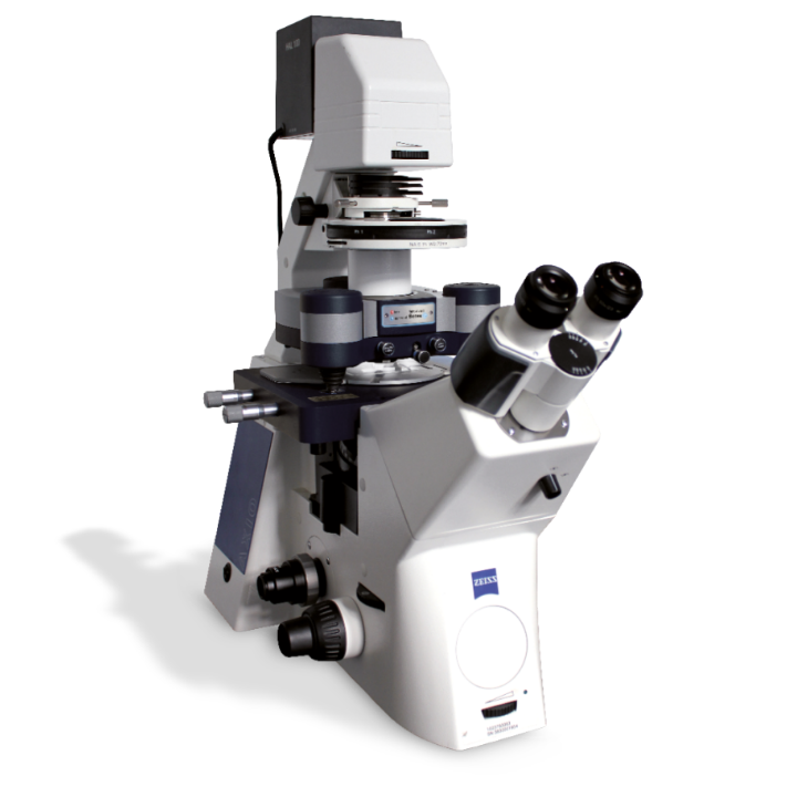
Fluorescence microscopy has become an indispensable tool in cell biology because it allows specifi c proteins to be visualized. Atomic force microscopy (AFM) is also becoming extensively used in the life sciences, but its development has largely followed an independent path and is used for somewhat different, but often complementary, purposes. Both methods can be applied to hydrated biological samples. Fluorescence microscopy is routinely used to image and track specifi cally labeled biological molecules in situ, illuminating a wide variety of cellular processes. In contrast, AFM is used for high spatial resolution imaging but also introduces the possibility of mechanically probing and manipulating samples without the need of complicated sample preparation. In this note, we briefl y describe the application of an AFM mounted on an inverted optical microscope to the characterization of the morphology of axons from cultured chick neurons using specifi c fl uorescent immunostaining and topographic imaging with AFM.
打开失败或需在电脑查看,请在电脑上的资料中心栏目,点击"我的下载"。建议使用手机自带浏览器。
