
产品详情
Anti-MCU antibody
种属反应性Human,Mouse,Rat
验证应用WB,ICC,IHC-P,FC
抗体类型兔多抗
免疫原Recombinant protein within human MCU aa 50-250.
偶联Non-conjugated
Anti-MCU antibody性能
形态Liquid
浓度1 mg/mL.
存放说明Store at +4℃ after thawing. Aliquot store at -20℃. Avoid repeated freeze / thaw cycles.
存储缓冲液1*PBS (pH7.4), 0.2% BSA, 50% Glycerol. Preservative: 0.05% Sodium Azide.
亚型IgG
纯化方式Protein affinity purified.
亚细胞定位Mitochondrion.
其它名称
moreAnti-MCU antibody应用
WB: 1:500-1:1,000
ICC: 1:50-1:200
IHC-P: 1:50 -1:200
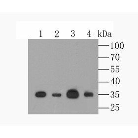
FC: 1:50-1:100Fig1: Western blot analysis of MCU on different lysates using anti-MCU antibody at 1/500 dilution.
Positive control:
Lane 1: A549
Lane 3: mouse spleen tissue
Lane 2: HL-60
Lane 4: mouse brain tissue
Fig2: ICC staining MCU in A431 cells (green). The nuclear counter stain is DAPI (blue). Cells were fixed in paraformaldehyde, permeabilised with 0.25% Triton X100/PBS.

Fig3: ICC staining MCU in A549 cells (green). The nuclear counter stain is DAPI (blue). Cells were fixed in paraformaldehyde, permeabilised with 0.25% Triton X100/PBS.

Fig4: ICC staining MCU in HT-29 cells (green). The nuclear counter stain is DAPI (blue). Cells were fixed in paraformaldehyde, permeabilised with 0.25% Triton X100/PBS.

Fig5: Immunohistochemical analysis of paraffin-embedded rat cerebellum tissue using anti-MCU antibody. Counter stained with hematoxylin.

Fig6: Immunohistochemical analysis of paraffin-embedded human lung cancer tissue using anti-MCU antibody. Counter stained with hematoxylin.

Fig7: Immunohistochemical analysis of paraffin-embedded mouse brain tissue using anti-MCU antibody. Counter stained with hematoxylin.

Fig8: Immunohistochemical analysis of paraffin-embedded human colon tissue using anti-MCU antibody. Counter stained with hematoxylin.

Fig9: Immunohistochemical analysis of paraffin-embedded human esophagus tissue using anti-MCU antibody. Counter stained with hematoxylin.

Fig10: Flow cytometric analysis of A549 cells with MCU antibody at 1/100 dilution (red) compared with an unlabelled control (cells without incubation with primary antibody; black). Alexa Fluor 488-conjugated goat anti-rabbit IgG was used as the secondary
特别提示:本公司的所有产品仅可用于科研实验,严禁用于临床医疗及其他非科研用途!