
产品详情
Anti-Transferrin antibody
种属反应性Human
验证应用IHC-P,FC
抗体类型小鼠单抗
免疫原Native protein.
偶联Non-conjugated
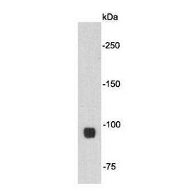
Anti-Transferrin antibody性能
形态Liquid
浓度2 mg/mL.
存放说明Store at +4℃ after thawing. Aliquot store at -20℃. Avoid repeated freeze / thaw cycles.
存储缓冲液1*PBS (pH7.4), 0.2% BSA, 50% Glycerol. Preservative: 0.05% Sodium Azide.
亚型IgG1
纯化方式Protein G purified.
亚细胞定位Secreted.
其它名称
moreApotransferrin antibody
Beta 1 metal binding globulin antibody
Beta-1 metal-binding globulin antibody
Anti-Transferrin antibody应用
IHC-P:1:100
FC:1:50-1:100
Fig1: Immunohistochemical analysis of paraffin-embedded human liver tissue using anti-Transferrin antibody. The section was pre-treated using heat mediated antigen retrieval with sodium citrate buffer (pH 6.0) for 20 minutes. The tissues were blocked in 5% BSA for 30 minutes at room temperature, washed with ddH2O and PBS, and then probed with the antibody at 1/50 dilution, for 30 minutes at room temperature and detected using an HRP conjugated compact polymer system. DAB was used as the chrogen. Counter stained with hematoxylin and mounted with DPX.

Fig2: Immunohistochemical analysis of paraffin-embedded human kidney tissue using anti-Transferrin antibody. The section was pre-treated using heat mediated antigen retrieval with sodium citrate buffer (pH 6.0) for 20 minutes. The tissues were blocked in 5% BSA for 30 minutes at room temperature, washed with ddH2O and PBS, and then probed with the antibodat 1/50 dilution, for 30 minutes at room temperature and detected using an HRP conjugated compact polymer system. DAB was used as the chrogen. Counter stained with hematoxylin and mounted with DPX.

Fig3: Flow cytometric analysis of Transferrin was done on HepG2 cells. The cells were fixed, permeabilized and stained with Transferrin antibody at 1/100 dilution (red) compared with an unlabelled control (cells without incubation with primary antibody; black). After incubation of the primary antibody on room temperature for an hour, the cells was stained with a Alexa Fluor™ 488-conjugated goat anti-mouse IgG Secondary antibody at 1/500 dilution for 30 minutes.
特别提示:本公司的所有产品仅可用于科研实验,严禁用于临床医疗及其他非科研用途!