| 供货周期: | 现货 |
| 品牌: | 康朗生物 |
| 规格: | 100ul/50ul/25ul |
| 货号: | KL600149 |
| CAS号: |


Anti-β-Actin 抗体
Product Properties
Cat CodeProduct NameClonalityCross reactivityApplicationMW(kDa)
600149Anti-β-Actin Mouse mAbMouse mAbHuman,Mouse,Rat.IHC-P, FC, WB, IF42kDa
Sensitivity
Anti-β-Actin 抗体Immunogen This ACTB Monoclonal antibody is generated from mouse immunized with ACTB recombinant protein.
Host Mouse
Clone Number 3B12-B7-B7
Antibody type Monoclonal antibody
Anti-β-Actin 抗体Purified method Affinity purified
Isotype IgG1,κ
Formulation Purified monoclonal antibody supplied in PBS with 0.09% (W/V) sodium azide. This antibody is purified through a protein G column, eluted with high and low pH buffers and neutralized immediately, followed by dialysis against PBS.
Dilution
WB~~1:1000
ICC~~1:10~50
IHC~~1:25
Anti-β-Actin 抗体FCM~~1:25
Application Images-expand |

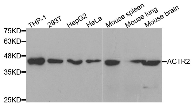
Western blot detection of β-Actin in A549,MCF7,3T3,C6 and Hela cell lysates using β-Actin mouse mAb (1:10000 diluted).Predicted band size:42KDa.Observed band size:42KDa.

Confocal immunofluorescent analysis of ACTB Antibody (Cat#AM1829b) with Hela cell followed by Alexa Fluor? 488-conjugated goat anti-mouse lgG (green). DAPI was used to stain the cell nuclear (blue).

AM1829b staining ACTB in human heart tissue sections by Immunohistochemistry (IHC-P - paraformaldehyde-fixed, paraffin-embedded sections). Tissue was fixed with formaldehyde and blocked with 3% BSA for 0. 5 hour at room temperature; antigen retrieval wa

Overlay histogram showing A431 cells stained with AM1829b(green line). The cells were fixed with 2% paraformaldehyde (10 min) and then permeabilized with 90% methanol for 10 min. The cells were then icubated in 2% bovine serum albumin to block non-speci

All lanes : Anti-ACTB Antibody at 1:1000 dilution Lane 1: A431 whole cell lysate Lane 2: C2C12 whole cell lysate Lane 3: C6 whole cell lysate Lane 4: Hela whole cell lysate Lane 5: MCF-7 whole cell lysate Lysates/proteins at 20 μg per lane. Secondary G

Immunohistochemical analysis of paraffin-embedded H.spleen section using Beta-Actin Antibody(Cat#AM1829b). AM1829b was diluted at 1:25 dilution. A peroxidase-conjugated goat anti-rabbit IgG at 1:400 dilution was used as the secondary antibody, followed b

Immunohistochemical analysis of paraffin-embedded H.spleen section using Beta-Actin Antibody(Cat#AM1829b). AM1829b was diluted at 1:25 dilution. A peroxidase-conjugated goat anti-mouse IgG at 1:400 dilution was used as the secondary antibody, followed by
相关产品

关注

拨打电话

留言咨询