| 供货周期: | 现货 |
| 品牌: | 泽叶生物 |
| 规格: | 兔多抗 |
| 货号: | ZY6901-65R |
| CAS号: |


Anti-GAPDH antibody
种属反应性Human, Mouse, Rat
验证应用WB, IHC-P, ICC, FC
抗体类型兔多抗
免疫原Recombinant protein within Human GAPDH aa 15-171.
偶联Non-conjugated
形态Liquid
浓度1 mg/mL.
存放说明Store at +4℃ after thawing. Aliquot store at -20℃. Avoid repeated freeze / thaw cycles.
存储缓冲液1*PBS (pH7.4), 0.2% BSA, 50% Glycerol. Preservative: 0.05% Sodium Azide.
亚型IgG
纯化方式Protein A affinity purified.
亚细胞定位Cytoplasm, Nucleus, Membrane.
其它名称
38 kDa BFA-dependent ADP-ribosylation substrate antibody
aging associated gene 9 protein antibody
Aging-associated gene 9 protein antibody
WB:1:500-1:2,000
ICC:1:50-1:200
IHC-P:1:50-1:200
FC:1:50-1:100

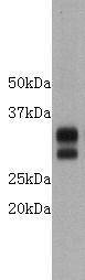
Fig1: Western blot analysis of GAPDH on PC-12 cell lysate. Proteins were transferred to a PVDF membrane and blocked with 5% BSA in PBS for 1 hour at room temperature. The primary antibody was used at a 1:2,000 dilution in 5% BSA at room temperature for 2 hours. Goat Anti-Rabbit IgG - HRP Secondary Antibody (HA1001) at 1:5,000 dilution was used for 1 hour at room temperature.

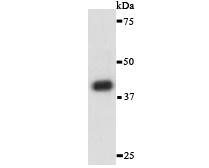
Fig2: Western blot analysis of GAPDH on different lysates. Proteins were transferred to a PVDF membrane and blocked with 5% BSA in PBS for 1 hour at room temperature. The primary antibody was used in 5% BSA at room temperature for 2 hours. Goat Anti-Rabbit IgG - HRP Secondary Antibody (HA1001) at 1:5,000 dilution was used for 1 hour at room temperature.
Positive control:
Lane 1: PC-12 cell lysate
Lane 2: Hela cell lysate
Lane 3: Mouse ovary tissue lysate
Lane 4: Human placenta tissue lysate
Lane 5: Rat brain tissue lysate
Lane 6: NCCIT cell lysate

Fig3: ICC staining of GAPDH in A549 cells (green). Formalin fixed cells were permeabilized with 0.1% Triton X-100 in TBS for 10 minutes at room temperature and blocked with 1% Blocker BSA for 15 minutes at room temperature. Cells were probed with the antibody at a dilution of 1:50 for 1 hour at room temperature, washed with PBS. Alexa Fluor®488 Goat anti-Rabbit IgG was used as the secondary antibody at 1/100 dilution. The nuclear counter stain is DAPI (blue).

Fig4: ICC staining of GAPDH in LoVo cells (green). Formalin fixed cells were permeabilized with 0.1% Triton X-100 in TBS for 10 minutes at room temperature and blocked with 1% Blocker BSA for 15 minutes at room temperature. Cells were probed with the antibodat a dilution of 1:50 for 1 hour at room temperature, washed with PBS. Alexa Fluor®488 Goat anti-Rabbit IgG was used as the secondary antibody at 1/100 dilution. The nuclear counter stain is DAPI (blue).

Fig5: ICC staining of GAPDH in MCF-7 cells (green). Formalin fixed cells were permeabilized with 0.1% Triton X-100 in TBS for 10 minutes at room temperature and blocked with 1% Blocker BSA for 15 minutes at room temperature. Cells were probed with the antibody at a dilution of 1:100 for 1 hour at room temperature, washed with PBS. Alexa Fluor®488 Goat anti-Rabbit IgG was used as the secondary antibody at 1/100 dilution. The nuclear counter stain is DAPI (blue).

Fig6: Immunohistochemical analysis of paraffin-embedded rat kidney tissue using anti-GAPDH antibody. The section was pre-treated using heat mediated antigen retrieval with sodium citrate buffer (pH 6.0) for 20 minutes. The tissues were blocked in 5% BSA for 30 minutes at room temperature, washed with ddH2O and PBS, and then probed with the antibody at 1/50 dilution, for 30 minutes at room temperature and detected using an HRP conjugated compact polymer system. DAB was used as the chromogen. Counter stained with hematoxylin and mounted with DPX.

Fig7: Immunohistochemical analysis of paraffin-embedded human colon cancer tissue using anti-GAPDH antibody. The section was pre-treated using heat mediated antigen retrieval with sodium citrate buffer (pH 6.0) for 20 minutes. The tissues were blocked in 5% BSA for 30 minutes at room temperature, washed with ddH2O and PBS, and then probed with the antibody at 1/50 dilution, for 30 minutes at room temperature and detected using an HRP conjugated compact polymer system. DAB was used as the chromogen. Counter stained with hematoxylin and mounted with DPX.

Fig8: Immunohistochemical analysis of paraffin-embedded human spleen tissue using anti-GAPDH antibody. The section was pre-treated using heat mediated antigen retrieval with sodium citrate buffer (pH 6.0) for 20 minutes. The tissues were blocked in 5% BSA for 30 minutes at room temperature, washed with ddH2O and PBS, and then probed with the antibody at 1/50 dilution, for 30 minutes at room temperature and detected using an HRP conjugated compact polymer system. DAB was used as the chromogen. Counter stained with hematoxylin and mounted with DPX.

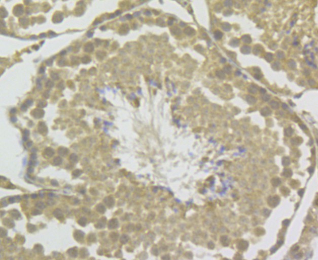
Fig9: Immunohistochemical analysis of paraffin-embedded mouse testis tissue using anti-GAPDH antibody. The section was pre-treated using heat mediated antigen retrieval with sodium citrate buffer (pH 6.0) for 20 minutes. The tissues were blocked in 5% BSA for 30 minutes at room temperature, washed with ddH2O and PBS, and then probed with the antibody at 1/50 dilution, for 30 minutes at room temperature and detected using an HRP conjugated compact polymer system. DAB was used as the chromogen. Counter stained with hematoxylin and mounted with DPX.

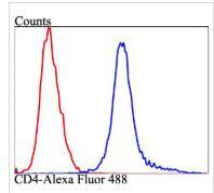
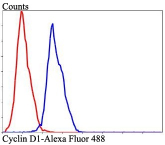
Fig10: Flow cytometric analysis of GAPDH was done on MCF-7 cells. The cells were fixed, permeabilized and stained with GAPDH antibody at 1/50 dilution (red) compared with an unlabelled control (cells without incubation with primary antibody; black). After incubation of the primary antibody on room temperature for an hour, the cells was stained with a Alexa Fluor 488-conjugated goat anti-rabbit IgG Secondary antibody at 1/500 dilution for 30 minutes.
特别提示:本公司的所有产品仅可用于科研实验,严禁用于临床医疗及其他非科研用途!
相关产品
彩色预染蛋白Marker(10-180 kDa,三色)
Super GelBlueTM 核酸染料, 10,000× in water
彩色预染蛋白Marker(10-180 kDa,三色)
彩色预染蛋白Marker(10-250kDa, 双色)
琼脂糖 A2015
Super ECL Plus(超敏化学发光检测试剂盒)
PAGE 彩色快速凝胶制备试剂盒(10%)
Minerva Super Fusion Cloning Kit(无缝克隆试剂盒)
Cell Counting Kit-8(CCK-8)细胞增殖检测试剂盒
FITC-Annexin V/PI 细胞凋亡试剂盒
呋喃西林快速检测卡
呋喃妥因快速检测卡
呋喃它酮快速检测试剂盒
呋喃妥因快速检测试剂盒
呋喃西林快速检测试剂盒

关注

拨打电话

留言咨询