[font=宋体][font=宋体]在生物学和医学研究中,细胞增殖是一个关键过程,对于理解生命活动的基本规律以及疾病的发病机理具有重要意义。随着科技的发展,流式细胞仪作为一种高效、灵敏的分析工具,广泛应用于细胞增殖的检测。流式细胞仪通过快速分析单个细胞,可以对细胞周期、细胞增殖活性、细胞凋亡等多个方面进行研究。本文将探讨流式细胞仪在检测细胞增殖方面的主要方法,包括但不限于溴脱氧尿苷([/font][font=Calibri]BrdU[/font][font=宋体])掺入法、细胞周期蛋白检测法以及细胞大小分析法等,以期为读者提供全面的技术应用概览。流式细胞仪检测细胞增殖方法:[/font][/font][b][font=宋体][font=Calibri]1[/font][font=宋体]、[/font][font=Calibri]3H[/font][font=宋体](氚离子)掺入法[/font][/font][/b][font=宋体][font=宋体]原理:是在细胞[/font][font=Calibri]DNA[/font][font=宋体]合成时,用[/font][font=Calibri]3H[/font][font=宋体]脱氧胸腺嘧啶核苷代替普通的脱氧胸腺嘧啶核苷掺入新合成的[/font][font=Calibri]DNA[/font][font=宋体]中,增殖的细胞因为掺入[/font][font=Calibri]3H[/font][font=宋体]而具有放射性,通过定量检测样品细胞的放射性大小而反映细胞的增值活性[/font][/font][font=宋体][font=宋体]缺点:[/font][font=Calibri]1[/font][font=宋体])使用的是具有放射性的同位素,操作较为复杂,同时需要采取放射性保护措施 [/font][font=Calibri]2[/font][font=宋体])低比例高活跃增殖和高比例低活跃增殖可能得到的是相同的结果,用此方法无法进行鉴别 [/font][font=Calibri]3[/font][font=宋体])此方法无法进一步得到具有活性的增值细胞用于下一步的研究 [/font][font=Calibri]4[/font][font=宋体]) 此方法时间较短,无法检测加入前细胞的增殖情况,而且检测到放射性只能说明细胞[/font][font=Calibri]DNA[/font][font=宋体]合成,而不能提供合成[/font][font=Calibri]DNA[/font][font=宋体]的细胞是否进入增殖阶段的信息[/font][/font][b][font=宋体][font=Calibri]2[/font][font=宋体]、相对计数法[/font][/font][/b][font=宋体]原理:将对照组和各实验组控制在相同条件下直接计数然后比较计数结果得到增殖结论[/font][font=宋体]注意点:[/font][font=宋体][font=宋体]对照组与实验组每种细胞所加浓度必须相同,每组至少设置[/font][font=Calibri]3[/font][font=宋体]个复孔,这样每个孔可以得到[/font][font=Calibri]1[/font][font=宋体]个细胞数,将[/font][font=Calibri]3[/font][font=宋体]个复孔取平均值后就是这个组的结果。如果同时需要得到每孔目标细胞增殖后的绝对参数,在每孔细胞中加入[/font][font=Calibri]1*105PE[/font][font=宋体]标记的人工微球作为内参[/font][/font][font=宋体] [/font][font=宋体][font=宋体]收集各组的细胞于[/font][font=Calibri]EP[/font][font=宋体]管中,注意必须尽量将各组的所有细胞都收集起来。标记需要计数细胞的标志表型的荧光素偶联抗体,[/font][font=Calibri]4[/font][font=宋体]℃静置[/font][font=Calibri]30min[/font][/font][font=宋体] [/font][font=宋体][font=Calibri]PBS[/font][font=宋体]洗涤一次,洗去游离的抗体[/font][/font][b][font=宋体][font=Calibri]3[/font][font=宋体]、示踪染料标记法[/font][/font][/b][font=宋体][font=宋体]示踪染料与细胞结合的方式:[/font][font=Calibri]1[/font][font=宋体])能够与细胞内的蛋白质上的氨基发生非特异性的共价结合 [/font][font=Calibri]2[/font][font=宋体])能够非特异性地嵌入细胞膜的脂质双分子层中与细胞发生非共价性结合[/font][/font][font=宋体] [/font][font=宋体][font=宋体]原理:示踪染料的荧光信号都很强,当细胞分裂时,母细胞内的染料会被平均分配到子细胞中,细胞荧光信号会被减弱一半,所以通过检测减弱的、发射示踪染料荧光信号的细胞比例就可以判断细胞增殖的强弱。当荧光强度减弱到标记时的[/font][font=Calibri]1/2[/font][font=宋体]以及以下的细胞都是增殖后的细胞,这些细胞所占比例越高则代表细胞增殖越活跃[/font][/font][font=宋体] [/font][font=宋体]标记方法:[/font][font=宋体][font=宋体]①纯化增殖反应的目标细胞,将细胞的浓度调整为[/font][font=Calibri]1*106/ml[/font][font=宋体],加入[/font][font=Calibri]CFSE[/font][font=宋体],其标记浓度为[/font][font=Calibri]5[/font][font=宋体]微摩尔[/font][font=Calibri]/[/font][font=宋体]升。置于[/font][font=Calibri]37[/font][font=宋体]℃水浴中标记[/font][font=Calibri]15min[/font][font=宋体],在标记过程中每隔一段时间混匀细胞一次[/font][/font][font=宋体] [/font][font=宋体][font=宋体]②加入预冷、含有血清的培养基终止标记,在[/font][font=Calibri]4[/font][font=宋体]℃冰箱中静置[/font][font=Calibri]5min[/font][font=宋体],离心沉淀[/font][/font][font=宋体] [/font][font=宋体][font=宋体]③用培养基再洗涤一次,尽量洗净未结合的游离的[/font][font=Calibri]CFSE[/font][font=宋体],然后将目标细胞静置在增殖体系中[/font][/font][font=宋体] [/font][b][font=宋体][font=Calibri]4[/font][font=宋体]、[/font][font=Calibri]BrdU[/font][font=宋体]和[/font][font=Calibri]EdU[/font][font=宋体]掺入法[/font][/font][/b][font=宋体][font=Calibri]BrdU[/font][font=宋体]:[/font][font=Calibri]5-[/font][font=宋体]溴脱氧尿嘧啶核苷是胸腺嘧啶核苷的类似物,其特点是胸腺嘧啶环上[/font][font=Calibri]5[/font][font=宋体]位[/font][font=Calibri]C[/font][font=宋体]连接的甲基被溴取代,在细胞增殖[/font][font=Calibri]DNA[/font][font=宋体]合成时可以与内源性的胸腺嘧啶核苷竞争掺入到新合成的[/font][font=Calibri]DNA[/font][font=宋体]中,而[/font][font=Calibri]BrdU[/font][font=宋体]抗体可以特异性的识别[/font][font=Calibri]BrdU[/font][font=宋体],不与胸腺嘧啶核苷结合,所以可以用于检测细胞增殖[/font][/font][font=宋体][font=宋体]适用范围:适用于体内检测目标细胞的增殖,一般将[/font][font=Calibri]BrdU[/font][font=宋体]掺入小鼠的应用水中或经腹腔注射,经过一段时间后,取出目标细胞制成单细胞悬液然后用多聚甲醛固定细胞,后用打孔剂皂苷在细胞膜上打孔,最后标记荧光素偶联抗[/font][font=Calibri]BrdU[/font][font=宋体]抗体,目标细胞的[/font][font=Calibri]BrdU[/font][font=宋体]阳性细胞就是增殖的细胞,阳性比例越高,增殖越活跃。[/font][/font][font=宋体] [/font][b][font=宋体][font=Calibri]5[/font][font=宋体]、其他方法[/font][/font][/b][font=宋体][font=宋体]细胞周期法检测细胞增殖:流式细胞术能够检测细胞内[/font][font=Calibri]DNA[/font][font=宋体]的含量,所以可以检测细胞周期。处于[/font][font=Calibri]S[/font][font=宋体]期的细胞,[/font][font=Calibri]DNA[/font][font=宋体]的量处于二倍体和四倍体之间[/font][font=Calibri] [/font][font=宋体]处于[/font][font=Calibri]G2/M[/font][font=宋体]期时,[/font][font=Calibri]DNA[/font][font=宋体]量为四倍体。处于[/font][font=Calibri]S[/font][font=宋体]期和[/font][font=Calibri]G2/M[/font][font=宋体]期的细胞比例越高说明细胞增殖越活跃[/font][/font][font=宋体] [/font][font=宋体][font=Calibri]PCNA[/font][font=宋体]检测细胞增殖:[/font][font=Calibri]PCNA[/font][font=宋体](增殖细胞核抗原),在细胞核合成且只存在于细胞核内,是[/font][font=Calibri]DNA[/font][font=宋体]聚合酶的辅助蛋白,所以与细胞[/font][font=Calibri]DNA[/font][font=宋体]的合成关系密切,是反映细胞增殖状态的良好指标[/font][/font][font=宋体] [/font][font=宋体][font=Calibri]Ki-67[/font][font=宋体]检测细胞增殖:是一种与细胞增殖特异相关的核抗原[/font][/font][font=宋体] [/font][font=宋体][font=Calibri]CD71[/font][font=宋体]检测细胞增殖:是转铁蛋白受体,表达于细胞的表面,该受体广泛表达于各种恶性肿瘤细胞表面,正常细胞表达较少,与肿瘤细胞的增殖密切相关[/font][/font][font=宋体] [/font][font=宋体][font=宋体]义翘神州提供[url=https://cn.sinobiological.com/services/flow-cytometry-service][b]流式细胞检测技术服务[/b][/url],更多关于流式细胞仪检测细胞增殖详情欢迎咨询,详情可以关注:[/font][font=Calibri]https://cn.sinobiological.com/services/flow-cytometry-service[/font][/font][b][font=宋体] [/font][font=宋体][font=宋体]义翘神州:蛋白与抗体的专业引领者,欢迎通过百度搜索[/font][font=宋体]“义翘神州”与我们取得联系。[/font][/font][/b]
WST-8是MTT的一种升级替代产品,和MTT、WST-1或其它MTT类似产品如XTT、MTS等相比有明显的优点,成为细胞增殖与活性测定新选择。国际知名生化试剂供应商Cayman Chemical提供的WST-8 Cell Proliferation Assay Kit(WST-8细胞增殖分析试剂盒)及WST-8,因其超高性价比,受到国内外科研工作者的追捧! WST-8是一种水溶性四唑盐,常用于评估细胞的代谢活性。在中性pH值及中间电子受体存在的情况下,被细胞线粒体中的脱氢酶还原为具有高度水溶性的蓝/紫色甲臜产物(formazan)。生成的甲臜物的数量与活细胞的数量成正比。用酶联免疫检测仪在450nm波长处测定其光吸收值,可间接反映活细胞数量。该方法已被广泛用于一些生物活性因子的活性检测、大规模的抗肿瘤药物筛选、细胞增殖试验、细胞毒性试验以及药敏试验等。http://www.bio-review.com/wp-content/uploads/2016/04/WST.jpg图1:WST-8作用原理以前,客户习惯用MTT、WST-1法测定细胞增殖,WST-8是MTT的一种升级替代产品,和MTT或其它MTT类似产品如XTT、MTS等相比有明显的优点:WST-8溶液对细胞的毒性非常低,细胞在WST-8法检测后仍然可以正常生长MTT被线粒体内的一些脱氢酶还原生成的formazan不是水溶性的,需要有特定的溶解液来溶解;而WST-8和XTT、MTS产生的formazan都是水溶性的,可以省去后续的溶解步骤。WST-8产生的formazan比XTT和MTS产生的formazan更易溶解。WST-8比XTT和MTS更加稳定,使实验结果更加稳定。另外,WST-8和MTT、XTT等相比线性范围更宽,灵敏度更高。WST-8和WST-1相比,检测灵敏度更高,更易溶解,并且更加稳定。WST-8 Cell Proliferation Assay Kit(WST-8细胞增殖分析试剂盒,货号:10010199)及WST-8(货号:18721)。http://www.bio-review.com/wp-content/uploads/2010/06/ads1-earthox.gif产品名称及描述品牌货号产品说明WST-8 Cell Proliferation Assay KitCayman10010199 WST-8 Cell Proliferation Assay Kit特点:比色法、无放射性测定细胞活性及增殖可定量的细胞密度高达5×10⁶cells/ml,每孔可定量2,000-500,000的细胞快速分析,2-4h得结果操作简便、灵敏度高、数据可靠、重现性好这么无敌的试剂盒,价格还便宜!该试剂盒含两种Cayman Chemical专利组分:WST-8 Developer Reagent及Electron Mediator Solution。简便的试剂盒操作步骤如下:将WST-8 Developer Reagent和Electron Mediator Solution等比混合,制成混合物将混合物加入细胞(如果需要测细胞毒性,此过程中加待测药物),共孵育450nm,读取吸光值。http://www.bio-review.com/wp-content/uploads/2016/04/result-1.png图2:白血病细胞活性测定结果建议参考文献:Li, Li, et al. "Protective effects of decursin and decursinol angelate against amyloid β-protein-induced oxidative stress in the PC12 cell line: the role of Nrf2 and antioxidant enzymes." Bioscience, biotechnology, and biochemistry 75.3 (2011): 434-442.Lin, Tzu-yin, et al. "Targeting canine bladder transitional cell carcinoma with a human bladder cancer-specific ligand." Molecular cancer 10.1 (2011): 1.Li, Li, et al. "Decursin Isolated from Angelica gigas Nakai Rescues PC12 Cells from Amyloid-Protein-Induced Neurotoxicity through Nrf2-Mediated Upregulation of Heme Oxygenase-1: Potential Roles of MAPK." Evidence-Based Complementary and Alternative Medicine 2013 (2013).Martinesi, M., et al. "Biocompatibility studies of low temperature nitrided and collagen-I coated AISI 316L austenitic stainless steel." Journal of Materials Science: Materials in Medicine 24.6 (2013): 1501-1513.l向全球科学工作者提供多研究领域的生化、免疫试剂和分析试剂盒,其产品被广泛应用于肿瘤、氧化氮、神经学、凋亡、氧化性损伤、内分泌学等不同研究领域。Cayman Chemical还提供各种有机化合物和生物化合物的定制合成服务,被视为全球最复杂和最不稳定化合物合成的唯一供应商。作为Cayman Chemical在中国的区域总代理,将为中国客户提供最全面的Cayman Chemical产品及客户订制化服务。如果您对以上产品及Cayman Chemical产品感兴趣,
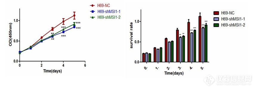
[align=center][size=18px]敲低[/size][size=18px] MSI1 对小细胞肺癌细胞生长增殖的影响[/size][/align][size=16px]检测 MSI1 对人小细胞肺癌细胞生长增殖的影响[/size][size=16px]收集[/size][size=16px] H69、H82、H526、SW1271 的对照组和实验组[/size][size=16px]细胞细胞[/size][size=16px]离心,并用完全培养基调整细胞浓度,H69-NC、H69-shMSI1-1、H69-shMSI1-2、H82-NC、H82-shMSI1-1、[/size][size=16px] [/size][size=16px]H82-shMSI1-2、H526-NC、H526-shMSI1-1、H526-shMSI1-2 以每孔 1×10⁴ [/size][size=16px]个[/size][size=16px]细胞平铺于 96 孔板中,SW1271-NC、SW1271-shMSI1-1、SW1271-shMSI1-2,以每孔 1.3×[/size][size=16px]104 [/size][size=16px]个[/size][size=16px]细胞平铺于 96 孔板中,37℃ 恒温培养箱中培养。铺板后,分别于 24 h、48 h、72 h、96 h、120h 在每孔加入 10 [/size][size=16px]μL[/size][size=16px] CCK-8 溶液,37℃ 恒温培养箱中孵育 4h。并用酶标仪测定波长 450 nm 处 OD 值,利用 [/size][size=16px]Graphpad[/size][size=16px] prism5 计算增殖情况。[/size][size=16px]检测 MSI1 对人小细胞肺癌细胞药物敏感性的影响[/size][size=16px]收集[/size][size=16px] H69 、H82 、H526 、SW1271 的对照组和实验组细胞, 其中 H69-NC 、H69-shMSI1-1、H69-shMSI1-2、H82-NC、H82-shMSI1-1、H82-shMSI1-2、H526-NC、[/size][size=16px]H526-shMSI1-1、H526-shMSI1-2 细胞系以 1×10⁴ [/size][size=16px]个[/size][size=16px]细胞/孔的细胞密度接种于 96 孔板中,SW1271-NC、SW1271-shMSI1-1、SW1271-shMSI1-2 以 1.3×10⁴ [/size][size=16px]个[/size][size=16px]细胞/孔的细[/size][size=16px]胞[/size][size=16px]密度接种于[/size][size=16px] 96 孔板中。待细胞融合率约 80%,加入不同浓度顺[/size][size=16px]铂[/size][size=16px]。每组均设置对照组及空白组(仅有同体积培养基)。H69、H82、H526 的对照组和实验组[/size][size=16px]加药浓度梯度为 0、1、2、4、8、16、32、64 nmol/mL,SW1271 对照组和实验组细胞加药浓度梯度为 0、2、4、8、16、32、64、128、256 nmol/mL,(加药浓度梯度根据细胞类型、前期预实验结果及细胞对药物的敏感程度而定)。每种浓度设 6 [/size][size=16px]个[/size][size=16px]复孔,每孔总体积为 100 [/size][size=16px]μL[/size][size=16px],培养 24、48、72、96、120 h 后[/size][size=16px]分别检测细胞活力。每孔加入[/size][size=16px] 10 [/size][size=16px]μL[/size][size=16px] 的 CCK-8[/size][size=16px](避光),培养箱中孵育[/size][size=16px] 4 h 后取出,使用酶标仪测定波长为 450 nm 的吸光度(OD 值)。利用公式:抑制率=(加药组-空白组)/(对照组-空白组)计算增殖抑制率。实验重复 3 次,取平均值。以药物浓度为横坐标,细胞增殖抑制率为纵坐标,利用[/size][size=16px]Graphpad[/size][size=16px] prism5 绘图。[/size] [size=16px]敲低[/size][size=16px] MSI1 对人小细胞肺癌细胞增殖能力的影响[/size][size=16px]CCK-8 是一种基于 WST-8 而广泛应用于细胞增殖和细胞毒性的快速、高灵敏度、无放射性的比色检测试剂盒。WST-8 在电子耦合试剂存在的情况下,可以被线粒体内的一些脱氢酶还原生成橙黄色的甲[/size][size=16px]瓒[/size][size=16px],生成的甲[/size][size=16px]瓒[/size][size=16px]物的数量与活细胞的数量呈正比,因此可以直接进行细胞增殖和毒性分析。[/size][size=16px]CCK-8 法 测 生 长 曲 线 实 验 结 果 如 图[/size] [size=16px]3-1 显 示 , 实 验 组 H69-shMSI1-1 、[/size][size=16px]H69-shMSI1-2 、 H82-shMSI1-1 、 H82-shMSI1-2 、 H526-shMSI1-1 、 H526-shMSI1-2 、[/size][size=16px]SW1271-shMSI1-1、SW1271-shMSI1-2 细胞的 OD [/size][size=16px]值明显[/size][size=16px]低于对照组。[/size][size=16px]表明敲低[/size][size=16px] MSI1[/size][size=16px]抑制了[/size][size=16px] SCLC 细胞的生长增殖。[/size][img]https://ng1.17img.cn/bbsfiles/images/2022/11/202211302321138249_2126_5887180_3.png[/img][size=16px] [/size][size=16px]图[/size][size=16px] [/size] [size=16px]MSI1 低表达对 H69、H82、H526、SW1271 对照组和实验组细胞增殖的抑制情况。应用 [/size][size=16px]Graphpad[/size][size=16px] prism5 作图所示(*P0.05,**P0.01,***P0.001,表示与对照组相比,[/size][size=16px]敲低组[/size][size=16px] OD 值减小[/size][size=16px]具有统计学意义)。[/size] [size=16px]测敲低[/size][size=16px] MSI1 对人小细胞肺癌细胞药物敏感性的影响[/size][size=16px]药敏实验结果如图[/size][size=16px] 3-2 所示,与对照组相比,实验组 H69-shMSI1-1、H69-shMSI1-2、H82-shMSI1-1、H82-shMSI1-2、H526-shMSI1-1、H526-shMSI1-2、SW1271-shMSI1-1、[/size][size=16px]SW1271-shMSI1-2 经不同浓度[/size][size=16px]顺铂处理[/size][size=16px] 24、48、72、96、120 h 后细胞的药物敏感性无明显变化。[/size][size=16px] [/size][img]https://ng1.17img.cn/bbsfiles/images/2022/11/202211302321124223_9282_5887180_3.png[/img]
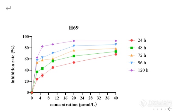
[font=黑体]西达本胺抑制[/font]SCLC[font=黑体]细胞增殖和凋亡[/font]CCK-8[font=宋体]药敏实验结果表明,[/font]SCLC[font=宋体]细胞系[/font]H69[font=宋体]、[/font]H446[font=宋体]、[/font]H526[font=宋体]、[/font]DMS114[font=宋体]经不同浓度西达本胺处理[/font] 24[font=宋体]、[/font]48[font=宋体]、[/font]72[font=宋体]、[/font]96[font=宋体]、[/font]120 h[font=宋体]后,细胞产生明显的增殖抑制现象,且随着药物浓度增加及作用时间延长,抑制作用逐渐增强,呈现出时间[/font]-[font=宋体]浓度依赖性,如图[/font]2[font=宋体]所示。同时得到西达本胺对四种细胞系作用[/font]72h[font=宋体]后的[/font]IC10[font=宋体]、[/font]IC20[font=宋体]、[/font]IC50[font=宋体]见表[/font]1-8[font=宋体]。结果显示四种细胞系对西达本胺均较为敏感,其中,与[/font]H69[font=宋体]相比,[/font]DMS114[font=宋体]对西达本胺相对不敏感。[/font][align=center][img]file:///C:/Users/Wang/AppData/Local/Temp/msohtmlclip1/01/clip_image002.png[/img] [img=,579,366]https://ng1.17img.cn/bbsfiles/images/2022/12/202212020948338518_7449_3237657_3.png!w579x366.jpg[/img][/align][align=center][font=宋体]不同浓度西达本胺作用不同时间后对[/font]H69[font=宋体]的增殖抑制情况[/font][/align][align=center] [/align][align=center][font=宋体]表[/font]1-8 [font=宋体]西达本胺作用[/font]72 h[font=宋体]后达到不同抑制效果的药物浓度([/font]μmol/L[font=宋体])[/font][/align] [table=95%][tr][td] [font=宋体]细胞名称[/font] [/td][td] [align=center] IC10[/align] [/td][td] [align=center] IC20[/align] [/td][td] [align=center] IC50[/align] [/td][/tr][tr][td] [align=center]H69[/align] [/td][td] [align=center][font='Times New Roman',serif]0.423[/font][/align] [/td][td] [align=center][font='Times New Roman',serif]0.632[/font][/align] [/td][td] [align=center][font='Times New Roman',serif]2.916[/font][/align] [/td][/tr][tr][td] [align=center]H446[/align] [/td][td] [align=center][font='Times New Roman',serif]0.404[/font][/align] [/td][td] [align=center][font='Times New Roman',serif]0.571[/font][/align] [/td][td] [align=center][font='Times New Roman',serif]1.033[/font][/align] [/td][/tr][tr][td] [align=center]H526[/align] [/td][td] [align=center][font='Times New Roman',serif]0.118[/font][/align] [/td][td] [align=center][font='Times New Roman',serif]0.261[/font][/align] [/td][td] [align=center][font='Times New Roman',serif]1.015[/font][/align] [/td][/tr][tr][td] [align=center]DMS114[/align] [/td][td] [align=center][font='Times New Roman',serif]1.272[/font][/align] [/td][td] [align=center][font='Times New Roman',serif]2.815[/font][/align] [/td][td] [align=center][font='Times New Roman',serif] 10.943[/font][/align] [/td][/tr][/table][font=黑体]西达本胺改变[/font]SCLC[font=黑体]细胞形态[/font][font=宋体]不同浓度([/font]0[font=宋体]、[/font]IC20[font=宋体]、[/font]IC50[font=宋体])西达本胺作用于[/font]H69[font=宋体]、[/font]H446[font=宋体]、[/font]H526[font=宋体]、[/font]DMS114[font=宋体]细胞[/font]48[font=宋体]及[/font]72 h[font=宋体]后在显微镜下观察细胞形态改变如图[/font]1-3[font=宋体]所示。随着药物浓度及作用时间的增加,[/font]SCLC[font=宋体]细胞系形态发生了变化,细胞增殖率减低。[/font]H69[font=宋体]团状细胞减少,单个凋亡细胞增多;[/font]H446[font=宋体]贴壁细胞减少,凋亡细胞增多,触角伸长,形状变得不规则;[/font]H526[font=宋体]细胞体积缩小,由片状变为球形团块,周围散在大量凋亡细胞;[/font]DMS114[font=宋体]由椭圆形变为长梭形,细胞内颗粒物增多,可见空泡,出现凋亡小体。由此可见,低剂量西达本胺即可影响[/font]SCLC[font=宋体]细胞形态,促进细胞凋亡,四种细胞系对西达本胺均较为敏感。[/font][img]file:///C:/Users/Wang/AppData/Local/Temp/msohtmlclip1/01/clip_image004.png[/img][align=center][img=,690,437]https://ng1.17img.cn/bbsfiles/images/2022/12/202212020948563335_146_3237657_3.png!w690x437.jpg[/img][/align][align=center] [/align][font=黑体]西达本胺诱导[/font]SCLC[font=黑体]细胞凋亡[/font][font=宋体]流式结果显示,用不同浓度([/font]0[font=宋体]、[/font]IC20[font=宋体]、[/font]IC50[font=宋体])西达本胺处理[/font]SCLC[font=宋体]细胞系[/font]48 h[font=宋体]后,四种亚型细胞系凋亡率均上升,且与加药浓度成正比,如图[/font]1-4 A[font=宋体]所示。[/font]48 h[font=宋体]检测在[/font]IC20[font=宋体]、[/font]IC50[font=宋体]浓度下[/font]H69[font=宋体]细胞凋亡率为[/font]8.45%[font=宋体]和[/font]14.46%[font=宋体],[/font]H446[font=宋体]细胞凋亡率为[/font]8.88%[font=宋体]和[/font]41.6%[font=宋体],[/font]H526[font=宋体]细胞凋亡率为[/font]11.48%[font=宋体]和[/font]20.77%[font=宋体],[/font]DMS114[font=宋体]细胞凋亡率为[/font]11.83%[font=宋体]和[/font]16.07%[font=宋体],与对照组相比,差异具有统计学意义([/font]P0.05[font=宋体])(图[/font]1-4 B[font=宋体])。为了进一步检测西达本胺在[/font]H69[font=宋体]、[/font]H446[font=宋体]、[/font]H526[font=宋体]、[/font]DMS114[font=宋体]四种细胞系中的作用差异,[/font][font=宋体]我们用[/font]1 μmol/L[font=宋体]的西达本胺分别处理[/font]H69[font=宋体]、[/font]H446[font=宋体]、[/font]H526[font=宋体]、[/font]DMS114[font=宋体]细胞[/font]48h[font=宋体]后进行流式细胞仪检测,结果如图[/font]1-4 C[font=宋体]所示,与[/font]DMS114[font=宋体]比较,[/font]H69[font=宋体]、[/font]H446[font=宋体]、[/font]H526[font=宋体]对西达本胺更敏感。[/font][img]file:///C:/Users/Wang/AppData/Local/Temp/msohtmlclip1/01/clip_image006.png[/img][align=center][img=,690,711]https://ng1.17img.cn/bbsfiles/images/2022/12/202212020949136557_2690_3237657_3.png!w690x711.jpg[/img][/align][align=center][font=黑体]图[/font][font=宋体]不同浓度([/font]0[font=宋体]、[/font]IC20[font=宋体]、[/font]IC50[font=宋体])西达本胺作用[/font]48 h[font=宋体]后[/font]H69[font=宋体]、[/font]H446[font=宋体]、[/font]H526[font=宋体]、[/font]DMS114[font=宋体]细胞凋亡率柱状图[/font][/align][align=center]. 1 μmol /L[font=宋体]西达本胺作用[/font]48 h[font=宋体]后对[/font]H69[font=宋体]、[/font]H446[font=宋体]、[/font]H526[font=宋体]、[/font]DMS114[font=宋体]细胞凋亡的影响[/font][/align][align=center] [/align] [font=黑体]西达本胺抑制[/font]SCLC[font=黑体]细胞克隆[/font][font=宋体]用不同浓度([/font]0[font=宋体]、[/font]IC20[font=宋体]、[/font]IC50[font=宋体])西达本胺处理[/font]SCLC[font=宋体]细胞系[/font]48 h[font=宋体]后,[/font][font=宋体]在细胞克隆第[/font]14[font=宋体]天,镜下观察细胞克隆情况并拍照(图[/font]1-5 A[font=宋体]),细胞单克隆数量随加药浓度增加而减少。各组细胞克隆形成率绘制成柱状图,如图[/font]1-5 B[font=宋体]所示,随着加药浓度增加细胞克隆形成率逐渐减小,与对照组相比,差异具有统计学意义([/font]P0.05[font=宋体])。[/font]H446[font=宋体]、[/font]DMS114[font=宋体]细胞[/font][font=宋体]进行了平板克隆实验,随着加药浓度的增加,克隆数明显减少,结果[/font][font=宋体]如图所示。[/font][b][img]file:///C:/Users/Wang/AppData/Local/Temp/msohtmlclip1/01/clip_image008.png[/img][/b][align=center][b][img=,690,425]https://ng1.17img.cn/bbsfiles/images/2022/12/202212020949347589_4119_3237657_3.png!w690x425.jpg[/img][/b][/align]
CCK-8检测试剂盒的优势有很多,看下面的详细介绍如下表,CCK-8检测试剂盒对细胞增殖/毒性检测优势比较,结果不言而喻! 检测方法MTT法XTT法WST-1法CCK-8法甲臜产物的水溶性差(需加有机溶剂溶解后再检测)好好好产品性状粉末2瓶溶液溶液1瓶溶液使用方法配成溶液后使用现配现用即开即用即开即用检测灵敏度高很高很高高检测时间较长较短较短最短检测波长560-600nm420-480nm420-480nm430-490nm细胞毒性高,细胞形态完全消失很低,细胞形态不变很低,细胞形态不变很低,细胞形态不变试剂稳定性一般较差一般很好批量样品检测可以非常适合非常适合非常适合便捷程度一般便捷便捷非常便捷* 细胞毒性非常低,因此加入WST-8显色后,可以在不同时间反复用酶标仪读数从而找到最佳测定时间* 酚红和血清对CCK法的检测不会造成干扰(扣除空白孔即可)如何选择合适的CCK-8检测试剂盒?目前市面上能提供的【CCK-8】细胞增殖/毒性检测试剂盒有国产分装或原产的,国产分装的CCK-8试剂盒在质量稳定性上有不足。而原产于国内实验室的CCK-8试剂盒有些具有较好的检测结果,在批次之间的稳定性上较难控制。至于原装进口供应CCK-8试剂盒的进口品牌并不多,就连Abcam、Thermo,Sigma等等几个品牌都不能够提供CCK-8试剂盒,而其它品牌在性价比上远逊于Abbkine产品。CCK-8检测试剂盒综合对比品牌(规格)AbbkineSigmaDojindo100次200 元703.17 元350 元500次640 元2779.92 元890 元2000次2050 元13899.6 元(3000次)——10000次7180 元——8380 元
【序号】:4【作者】:王栋何静吴方【题名】:羧甲基壳聚糖水凝胶的合成及其对人皮肤成纤维细胞增殖的影响【期刊】:中国科技论文. 【年、卷、期、起止页码】:2017,12(06)【全文链接】:https://kns.cnki.net/kcms2/article/abstract?v=3uoqIhG8C44YLTlOAiTRKibYlV5Vjs7iAEhECQAQ9aTiC5BjCgn0RtO1XN7r3ud8G6vNHx7e9SFQ2wwOcBY8uF-I-dwKOw9x&uniplatform=NZKPT
[b][size=15px][color=#595959]多囊卵巢综合征(PCOS)[/color][/size][/b][size=15px][color=#595959]患者和动物模型表现出闭锁卵泡增加、[b]颗粒细胞(GCs)[/b]紊乱、细胞层变薄和黄体数量减少。一些研究表明,卵泡发育不良与GCs存在相关性。GCs为正常卵泡发育提供必要的激素和营养支持。目前关于PCOS和卵泡发育的大量实验研究都是为了了解GCs的功能。[/color][/size] [b][size=15px][color=#595959]滋肾清热利湿化瘀方(ZQLHR)[/color][/size][/b][size=15px][color=#595959]由知母、山萸肉、丹参、桃仁、薏苡仁、白芥子、黄柏、玄参、甘草组成,是在中医理论和临床实践的基础上创制的中药方剂,主要用于PCOS。前期研究发现,ZQLHR能有效提高PCOS患者自主排卵率及有排卵月经的发生率,并能改善痤疮及降低血清睾酮水平。然而,ZQLHR治疗PCOS的潜在机制尚未清楚阐明。[/color][/size] [size=15px][color=#595959]验证ZQLHR是否通过Krüppel样因子4[b](KLF4)[/b]-CCATT增强子结合蛋白β [b](C/EBPβ)[/b]通路调控GCs[b]增殖和凋亡[/b],为ZQLHR促进卵泡发育和治疗PCOS患者的作用提供体外分子机制支持。 [/color][/size] [size=15px][color=#595959]在前期实验的基础上,首先用不同浓度的ZQLHR处理KGN细胞(一种类固醇性人颗粒样[b]肿瘤细胞[/b]系)48 h,观察各组细胞的凋亡情况。其次,采用实时荧光定量[url=https://insevent.instrument.com.cn/t/jp][color=#3333ff]PCR[/color][/url](q-[url=https://insevent.instrument.com.cn/t/jp][color=#3333ff]PCR[/color][/url])和Western blot检测ZQLHR给药后KGN细胞中KLF4和C/EBPβ mRNA和蛋白的表达水平。第三,利用siRNA敲除KLF4和C/EBPβ后,检测KGN细胞中KLF4和C/EBPβ的关系。[/color][/size] [size=15px][color=#595959]进一步通过细胞计数试剂盒-8、集落形成实验和流式细胞术验证ZQLHR是否通过[b]KLF4-C/EBPβ通路[/b]促进KGN细胞增殖和凋亡。最后,采用q-[url=https://insevent.instrument.com.cn/t/jp][color=#3333ff]PCR[/color][/url]和Western blot检测ZQLHR是否通过KLF4-C/EBPβ通路介导b细胞[b]淋巴瘤[/b]-2 (Bcl-2)、Bcl-2相关X (BAX)、增殖细胞核抗原(PCNA)和裂解caspase-3等增殖和凋亡相关因子影响KGN细胞的增殖和凋亡。[/color][/size] [b][size=15px][color=#595959][/color][/size][size=15px][color=#595959][/color][/size][size=15px][color=#595959][/color][/size][size=15px][color=#595959][/color][/size][size=15px][color=#595959][/color][/size][size=15px][color=#595959][/color][/size][/b][font=&][size=16px][color=#232323][/color][/size][/font][b][size=15px][color=#595959][/color][/size][size=15px][color=#595959][/color][/size][size=15px][color=#595959][/color][/size][size=15px][color=#595959][/color][/size][size=15px][color=#595959][/color][/size][size=15px][color=#595959][/color][/size][size=15px][color=#595959][/color][/size][/b][font=&][size=16px][color=#232323][/color][/size][/font][size=15px][color=#595959][font=&][/font][font=&][/font][/color][/size][b][size=15px][color=#595959][/color][/size][size=15px][color=#595959][/color][/size][size=15px][color=#595959][/color][/size][/b][font=&][size=16px][color=#232323][/color][/size][/font][b][size=15px][color=#595959][/color][/size][size=15px][color=#595959][/color][/size][size=15px][color=#595959][/color][/size][/b][size=15px][color=#595959][font=&][/font][/color][/size][size=15px][color=#595959]体积0.2%的ZQLHR对KGN细胞的凋亡率最低。ZQLHR处理后,KGN细胞中KLF4和C/EBPβ的表达水平升高。此外,ZQLHR通过KLF4-C/EBPβ通路调节增殖和凋亡相关因子,促进KGN细胞增殖和抑制凋亡。此外,[b]证实了KLF4和C/EBPβ在KGN细胞中相互调节[/b]。这些结果表明,[b]ZQLHR可增强GCs的增殖并抑制其凋亡,这可能是治疗PCOS患者的一种治疗机制[/b]。[/color][/size][size=15px][color=#595959][font=&][/font][/color][/size][size=15px][color=#595959][/color][/size][size=15px][color=#595959][/color][/size][size=15px][color=#595959][/color][/size][font=&][size=16px][color=#232323][/color][/size][/font][size=15px][color=#595959][/color][/size][size=15px][color=#595959][/color][/size]
【序号】:2【作者】:孔丽欣李静徐丹阳【题名】:凝胶样支架材料对三维培养的人牙髓干细胞增殖的影响【期刊】:口腔医学. 【年、卷、期、起止页码】:2018,38(07)【全文链接】:https://kns.cnki.net/kcms2/article/abstract?v=3uoqIhG8C44YLTlOAiTRKibYlV5Vjs7i0-kJR0HYBJ80QN9L51zrP1ziWAsvXz63X4iwE86ahO77cv-GmOIa_NaCUi6fN7f2&uniplatform=NZKPT
【序号】:3【作者】:朱琳邹德庆范作卿【题名】:蚕蛹羧甲基壳聚糖对小鼠成纤维细胞增殖与生长的影响【期刊】:蚕业科学. 【年、卷、期、起止页码】:2017,43(03)【全文链接】:https://kns.cnki.net/kcms2/article/abstract?v=3uoqIhG8C44YLTlOAiTRKibYlV5Vjs7iAEhECQAQ9aTiC5BjCgn0RurLuDBf62EN5lCHC3tB7EA_NCYlyZxEZRugsuO02raR&uniplatform=NZKPT
自去年我公司推出全自动核酸分析系统的免费试用后,今天我们又再次为各位老师奉上强大的细胞分析平台:一款可以解决您MTT实验烦恼、无需标记、全自动化,带给您高信息量、高灵敏度和高准确性的iCELLigence全自动细胞分析仪将会出现在您的面前。iCELLigence全自动细胞分析仪通过嵌在E-plate板上孔底的微电子感应器阻抗变化去感受细胞的有无以及贴壁、黏附和生长程度的改变,无需标记即可实时、直观的反应细胞增殖、存活、凋亡、形态变化等细胞生物学变化。iCELLigence全自动细胞分析仪的传感器阻抗技术在细胞分析中具有其独特的优势:它为整个的实验分析过程包括细胞黏附、细胞增殖和细胞融合提供了全程无损伤的监控,实时、连续显示的数据让您可以更加自信更加清楚的进行细胞操作和细胞增殖等分析,而不是假定细胞处于合适的处理阶段。一连串实时获取和显示的数据让您处理每一步结果都可以通过机理来预测,同时也可以结合iCELLigence全自动细胞分析仪连续的读数来决定传统终点细胞分析的最佳时间点。只需几个简单的操作步骤您就可以获得高信息量的、直观的、准确的结果,就可以让您的细胞增殖等分析实验变得更加省时与高效。莫再犹豫,快来参加体验吧,经历过你就会发现有时候细胞增殖等实验会是这么的easy。赶快报名,免费的试用在等着您哦···活动期间,凡是参与试用的用户均可获得昊诺斯8GU盘或瑞士军刀背包一个(奖品以实物为准)。http://ng1.17img.cn/bbsfiles/images/2017/01/201701191656_647437_1622715_3.jpg真心英雄活动第二关试用报名网址:http://www.instrument.com.cn/custom/SH100700/20130522/free.shtml另外,您也可以致电北京昊诺斯科技有限公司市场部产品负责人孙健13710746995sunjian@herosbio.com(因为区域划分,活动仅限北方区域,具体问题欢迎来电垂询)。
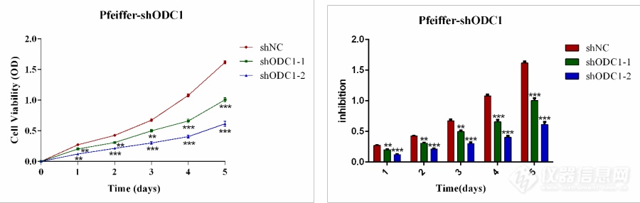
[font='times new roman'][size=18px]敲低[/size][/font][font='times new roman'][size=18px]ODC1[/size][/font][font='times new roman'][size=18px]对[/size][/font][font='times new roman'][size=18px]DLBCL[/size][/font][font='times new roman'][size=18px]细胞增殖、周期、凋亡的影响[/size][/font][font='times new roman'][size=18px]敲低[/size][/font][font='times new roman'][size=18px]ODC1[/size][/font][font='times new roman'][size=18px]对[/size][/font][font='times new roman'][size=18px]DLBCL[/size][/font][font='times new roman'][size=18px]细胞系增殖的影响[/size][/font][font='times new roman'][size=16px][color=#000000]CCK-8[/color][/size][/font][font='times new roman'][size=16px][color=#000000]是一种基于[/color][/size][/font][font='times new roman'][size=16px][color=#000000]WST-8[/color][/size][/font][font='times new roman'][size=16px][color=#000000]而广泛应用于细胞增殖和细胞毒性的快速、高灵敏度、无放射性的比色检测试剂盒。[/color][/size][/font][font='times new roman'][size=16px][color=#000000]WST-8[/color][/size][/font][font='times new roman'][size=16px][color=#000000]在电子耦合试剂存在的情况下,可以被线粒体内的一些脱氢酶还原生成橙黄色的甲[/color][/size][/font][font='times new roman'][size=16px][color=#000000]瓒[/color][/size][/font][font='times new roman'][size=16px][color=#000000],生成的甲[/color][/size][/font][font='times new roman'][size=16px][color=#000000]瓒[/color][/size][/font][font='times new roman'][size=16px][color=#000000]物的数量与活细胞的数量呈正比,因此可以直接进行细胞增殖和毒性分析。[/color][/size][/font][img]https://ng1.17img.cn/bbsfiles/images/2023/10/202310082309292993_3451_4239500_3.png[/img][font='times new roman'][size=16px][color=#000000]CCK-8[/color][/size][/font][font='times new roman'][size=16px][color=#000000]法测生长曲线实验结果如图[/color][/size][/font][font='times new roman'][size=16px][color=#000000]3-1[/color][/size][/font][font='times new roman'][size=16px][color=#000000]显示,实验组[/color][/size][/font][font='times new roman'][size=16px][color=#000000]Pfeiffer-shODC1[/color][/size][/font][font='times new roman'][size=16px][color=#000000]细胞的[/color][/size][/font][font='times new roman'][size=16px][color=#000000]OD[/color][/size][/font][font='times new roman'][size=16px][color=#000000]值明显[/color][/size][/font][font='times new roman'][size=16px][color=#000000]低于对照组。[/color][/size][/font][font='times new roman'][size=16px][color=#000000]表明敲低[/color][/size][/font][font='times new roman'][size=16px][color=#000000]ODC1[/color][/size][/font][font='times new roman'][size=16px][color=#000000]抑制了[/color][/size][/font][font='times new roman'][size=16px][color=#000000]DLBCL[/color][/size][/font][font='times new roman'][size=16px][color=#000000]细胞的生长增殖。[/color][/size][/font][align=center][font='times new roman'][color=#000000]图[/color][/font][font='times new roman'][color=#000000]ODC1[/color][/font][font='times new roman'][color=#000000]低表达对[/color][/font][font='times new roman'][color=#000000]Pfeiffer[/color][/font][font='times new roman'][color=#000000]对照组和实验组细胞增殖的抑制情况。应用[/color][/font][font='times new roman'][color=#000000] [/color][/font][font='times new roman'][color=#000000]Graphpad[/color][/font][font='times new roman'][color=#000000] prism5 [/color][/font][font='times new roman'][color=#000000]作图所示([/color][/font][font='times new roman'][color=#000000]*[/color][/font][font='times new roman'][color=#000000]P[/color][/font][font='times new roman'][color=#000000] 0.05,**[/color][/font][font='times new roman'][color=#000000]P [/color][/font][font='times new roman'][color=#000000]0.01,***[/color][/font][font='times new roman'][color=#000000]P[/color][/font][font='times new roman'][color=#000000] 0.001,[/color][/font][font='times new roman'][color=#000000]表示与对照组相比,[/color][/font][font='times new roman'][color=#000000]敲低组[/color][/font][font='times new roman'][color=#000000]OD[/color][/font][font='times new roman'][color=#000000]值减小具有统计学意义)。[/color][/font][/align][font='times new roman'][size=18px]敲低[/size][/font][font='times new roman'][size=18px]ODC1[/size][/font][font='times new roman'][size=18px]对[/size][/font][font='times new roman'][size=18px]DLBCL[/size][/font][font='times new roman'][size=18px]细胞系凋亡的影响[/size][/font][font='times new roman'][size=16px][color=#000000]流式结果如图[/color][/size][/font][font='times new roman'][size=16px][color=#000000]所示,与对照组相比,[/color][/size][/font][font='times new roman'][size=16px][color=#000000]实验组[/color][/size][/font][font='times new roman'][size=16px][color=#000000]Pfeiffer-shODC1[/color][/size][/font][font='times new roman'][size=16px][color=#000000]的[/color][/size][/font][font='times new roman'][size=16px][color=#000000]早期[/color][/size][/font][font='times new roman'][size=16px][color=#000000]凋亡[/color][/size][/font][font='times new roman'][size=16px][color=#000000]和晚期凋亡均[/color][/size][/font][font='times new roman'][size=16px][color=#000000]显著增加。[/color][/size][/font][font='times new roman'][size=16px][color=#000000]表明敲低[/color][/size][/font][font='times new roman'][size=16px][color=#000000]ODC1[/color][/size][/font][font='times new roman'][size=16px][color=#000000]促进了[/color][/size][/font][font='times new roman'][size=16px][color=#000000]DLBCL[/color][/size][/font][font='times new roman'][size=16px][color=#000000]细胞的凋[/color][/size][/font][font='times new roman'][size=16px][color=#000000]亡。[/color][/size][/font][font='times new roman'][size=16px][color=#000000]a[/color][/size][/font][align=center][/align][align=center][/align][align=center][/align][align=center][/align][align=center][/align][align=center][/align][font='times new roman'][size=16px][color=#000000]b[/color][/size][/font][align=center][img]" style="max-width: 100% max-height: 100% [/img][/align][align=center][font='times new roman'][color=#000000]图[/color][/font][font='times new roman'][color=#000000] a [/color][/font][font='times new roman'][color=#000000]流式细胞术结果图[/color][/font][font='times new roman'][color=#000000] b [/color][/font][font='times new roman'][color=#000000]早期和晚期凋亡柱状图[/color][/font][/align][font='times new roman'][size=18px] [/size][/font][font='times new roman'][size=18px]敲低[/size][/font][font='times new roman'][size=18px]ODC1[/size][/font][font='times new roman'][size=18px]对[/size][/font][font='times new roman'][size=18px]DLBCL[/size][/font][font='times new roman'][size=18px]细胞系线粒体凋亡途径关键蛋白的影响[/size][/font][font='times new roman'][size=16px][color=#000000]提取对数生长期的[/color][/size][/font][font='times new roman'][size=16px][color=#000000]Pfeiffer[/color][/size][/font][font='times new roman'][size=16px][color=#000000]对照组及实验组细胞总蛋白,利用[/color][/size][/font][font='times new roman'][size=16px][color=#000000]Western blot [/color][/size][/font][font='times new roman'][size=16px][color=#000000]检测敲低[/color][/size][/font][font='times new roman'][size=16px][color=#000000]ODC1[/color][/size][/font][font='times new roman'][size=16px][color=#000000]对[/color][/size][/font][font='times new roman'][size=16px][color=#000000]Pfeiffer[/color][/size][/font][font='times new roman'][size=16px][color=#000000]细胞凋亡的影响,因此我们检测了[/color][/size][/font][font='times new roman'][size=16px][color=#000000] Caspase [/color][/size][/font][font='times new roman'][size=16px][color=#000000]信号通路相关蛋白[/color][/size][/font][font='times new roman'][size=16px][color=#000000] BCL-2[/color][/size][/font][font='times new roman'][size=16px][color=#000000],[/color][/size][/font][font='times new roman'][size=16px][color=#000000]BAX[/color][/size][/font][font='times new roman'][size=16px][color=#000000],[/color][/size][/font][font='times new roman'][size=16px][color=#000000]Cytochrome c[/color][/size][/font][font='times new roman'][size=16px][color=#000000],[/color][/size][/font][font='times new roman'][size=16px][color=#000000]Caspase 3[/color][/size][/font][font='times new roman'][size=16px][color=#000000],[/color][/size][/font][font='times new roman'][size=16px][color=#000000]cleaved Caspase 3[/color][/size][/font][font='times new roman'][size=16px][color=#000000],[/color][/size][/font][font='times new roman'][size=16px][color=#000000]PARP[/color][/size][/font][font='times new roman'][size=16px][color=#000000],[/color][/size][/font][font='times new roman'][size=16px][color=#000000]cleaved PARP[/color][/size][/font][font='times new roman'][size=16px][color=#000000]。结果如图[/color][/size][/font][font='times new roman'][size=16px][color=#000000]3-3[/color][/size][/font][font='times new roman'][size=16px][color=#000000]所示,与对照组相比,实验组的抗凋亡蛋白[/color][/size][/font][font='times new roman'][size=16px][color=#000000] BCL-2 [/color][/size][/font][font='times new roman'][size=16px][color=#000000]表达下调,促凋亡蛋白[/color][/size][/font][font='times new roman'][size=16px][color=#000000] BAX [/color][/size][/font][font='times new roman'][size=16px][color=#000000]显著上调,[/color][/size][/font][font='times new roman'][size=16px][color=#000000] Cytochrome c[/color][/size][/font][font='times new roman'][size=16px][color=#000000],[/color][/size][/font][font='times new roman'][size=16px][color=#000000]cleaved Caspase 3[/color][/size][/font][font='times new roman'][size=16px][color=#000000],[/color][/size][/font][font='times new roman'][size=16px][color=#000000]cleaved PARP [/color][/size][/font][font='times new roman'][size=16px][color=#000000]显著上调,[/color][/size][/font][font='times new roman'][size=16px][color=#000000]Caspase 3 [/color][/size][/font][font='times new roman'][size=16px][color=#000000]的表达下调,[/color][/size][/font][font='times new roman'][size=16px][color=#000000]PARP [/color][/size][/font][font='times new roman'][size=16px][color=#000000]无明显变化。这些结果表明,在[/color][/size][/font][font='times new roman'][size=16px][color=#000000]DLBCL[/color][/size][/font][font='times new roman'][size=16px][color=#000000]细胞中,[/color][/size][/font][font='times new roman'][size=16px][color=#000000]敲低[/color][/size][/font][font='times new roman'][size=16px][color=#000000] ODC1 [/color][/size][/font][font='times new roman'][size=16px][color=#000000]诱导[/color][/size][/font][font='times new roman'][size=16px][color=#000000]Pfeiffer[/color][/size][/font][font='times new roman'][size=16px][color=#000000]细胞凋亡,且诱导凋亡的[/color][/size][/font][font='times new roman'][size=16px][color=#000000]的[/color][/size][/font][font='times new roman'][size=16px][color=#000000]机制可能是激活了[/color][/size][/font][font='times new roman'][size=16px][color=#000000] Caspase [/color][/size][/font][font='times new roman'][size=16px][color=#000000]凋亡途径。[/color][/size][/font][font='times new roman'][size=18px]敲低[/size][/font][font='times new roman'][size=18px]ODC1[/size][/font][font='times new roman'][size=18px]对[/size][/font][font='times new roman'][size=18px]DLBCL[/size][/font][font='times new roman'][size=18px]细胞系周期的影响[/size][/font][font='times new roman'][size=16px][color=#000000]提取对数生长期的[/color][/size][/font][font='times new roman'][size=16px][color=#000000]Pfeiffer [/color][/size][/font][font='times new roman'][size=16px][color=#000000]对照组及实验组总蛋白,利用[/color][/size][/font][font='times new roman'][size=16px][color=#000000]Western blot [/color][/size][/font][font='times new roman'][size=16px][color=#000000]检测敲低[/color][/size][/font][font='times new roman'][size=16px][color=#000000]ODC1[/color][/size][/font][font='times new roman'][size=16px][color=#000000]对[/color][/size][/font][font='times new roman'][size=16px][color=#000000]Pfeiffer[/color][/size][/font][font='times new roman'][size=16px][color=#000000]细胞周期的影响。结果如图显示,在细胞系[/color][/size][/font][font='times new roman'][size=16px][color=#000000]Pfeiffer[/color][/size][/font][font='times new roman'][size=16px][color=#000000]中,与对照组相比,实验组[/color][/size][/font][font='times new roman'][size=16px][color=#000000] Cyclin B1 [/color][/size][/font][font='times new roman'][size=16px][color=#000000]表达上调,[/color][/size][/font][font='times new roman'][size=16px][color=#000000]Cyclin A2[/color][/size][/font][font='times new roman'][size=16px][color=#000000]的表达下调,[/color][/size][/font][font='times new roman'][size=16px][color=#000000]说明敲低[/color][/size][/font][font='times new roman'][size=16px][color=#000000]ODC1[/color][/size][/font][font='times new roman'][size=16px][color=#000000]后,细胞阻滞于[/color][/size][/font][font='times new roman'][size=16px][color=#000000]G2 /M[/color][/size][/font][font='times new roman'][size=16px][color=#000000]期[/color][/size][/font][font='times new roman'][size=16px][color=#000000]。[/color][/size][/font][align=center][img]https://ng1.17img.cn/bbsfiles/images/2023/10/202310082309294706_2597_4239500_3.png[/img][/align][align=center][font='times new roman'][color=#000000]图与对照组相比,实验组[/color][/font][font='times new roman'][color=#000000] Cyclin B1 [/color][/font][font='times new roman'][color=#000000]表达上调,[/color][/font][font='times new roman'][color=#000000]Cyclin A2[/color][/font][font='times new roman'][color=#000000]的表达下调,[/color][/font][font='times new roman'][color=#000000]说明敲低[/color][/font][font='times new roman'][color=#000000] ODC1 [/color][/font][font='times new roman'][color=#000000]后,细胞阻滞于[/color][/font][font='times new roman'][color=#000000] G2 /M [/color][/font][font='times new roman'][color=#000000]期。[/color][/font][/align]
Cellscreen系统第一次实现了可重复对细胞培养进行观察。无需染色、无需制样,通过光学图像分析将细胞培养的生长曲线保存;与其它现有测试方法相比,Cellscreen系统对细胞培养无损伤性,独立性。第一次实现了对同一细胞培养区域进行多次测量。Cellscreen技术证明是一种精确的、可靠的、自定义实验条件、操作方便、节省成本的方法。Cellscreen能优化和加速新产品和测试程序的开发。 Cellscreen应用领域 Cellscreen模块化设计能适用于更广范的领域,例如: 制药研究:Cellscreen系统能缩短常规科研究时间,能拍摄细胞生长因子的各种因素,如毒性测试及生物适应性的测试。 生物技术研究:Cellscreen系统适用于增殖研究、过程(培养基)最优化、质量控制。另外,用于拍摄克隆细胞实验的新性质,如应用在新的治疗蛋白和抗体的研究。 Cellscreen系统的优势: l缩短制药研发的时间周期 l对细胞培养无损伤—细胞可再用于其它研究 l可扩展的详细的结果描述,对细胞培养过程的文档和图像存档 l与现有的方法相比,更精确——可靠、重复性、自定义 l很容易融入到日常实验 l很容易操作Multiwellplate l特别低的操作成本-对所有的测量只需要一个培养皿,不需试剂、不需对细胞染色。 l节省时间—不需样品制备 l技术成熟—innovatesAG图像识别技术 l模块化设计—系统可扩展其它分析模块 Cellscreen系统模块化设计: 为满足广泛的分析需求,Cellscreen系统是按模块化设计,能运行不同的软件系统。硬件由一个双处理器电脑及控制单元组成,控制单元通过高精度电机台自动聚焦、自动调光来控制显微镜;不同的软件模块对数码相机获取的图像进行分析,分析所得的图像和数据存储在终端数据库。 软件模块包括: l悬浮细胞的增殖研究模块(PS模块) l贴壁细胞的增殖研究模块(PA) l克隆细胞实验模块(CL) 细胞增殖研究模块(PS和PA)能重复观测细胞培养的生长因子,CL模块观察克隆细胞,并能追随到起源的单克隆细胞。 克隆细胞实验模块(CL) 在整个培养过程中,CL模块自动监控单个克隆细胞到群体的生长过程;为了证明细胞群体的单克隆细胞起源,需要监控整个生长过程。 Cellscreen系统用40倍放大系数抓取容器低部的16幅图像,它能代表整个容器的状态。 所有获得的图像和数据存档到数据库,因此可以跟踪任何生物群体的成长过程,证明生物群体起源的单克隆细胞。 很好的保护—Cellscreen的培养器 Cellscreen的培养器精确的安装在显微镜上,在测量过程中,对您贵重的细胞培养,它保持一个稳定的环境。对温度和CO2的浓度能精确的控制。培养器可以长时间或频繁的监控微量滴定盘,而不需移动它。 贴壁细胞的增殖研究模块(PA) PA模块通过测量细胞覆盖区域,用户可以观察到贴壁细胞的增殖。PA以40放大倍数获得图像。系统可以自由选择培养皿的区域。这系统适用于所有通用的微量滴定盘规格(6-96眼)。结果以图像和曲线的形式表示出来。PA模块将所有的图像和结果存档,输出格式CSV(兼容Excel格式)。 PA模块精确的测量至少80%细胞的生长因子,用于科研、发展、研制新产品。 悬浮细胞的增殖研究模块(PS) PS模块通常应用在对细胞生长因子影响的研究。为了获得生长曲线,悬浮液里培育的细胞数量被重复的量化——时间、原料、操作的消耗成本。对同一培养皿里的细胞生长,PS模块能重复计数、消除对每次测量都需要更换盘子的影响。 另外,Cellscreen系统对每个细胞进行计数,相对现存的细胞计数方法,它有很高的精确性。PS模块用的是100放大倍数,它获得的图像有很好的分辨率,能提供出细胞直径和细胞形状的一些信息。 Cellscreen系统概述 很容易综合到您的日常工作中——易学易用 实验和测量的标准化 在准备阶段,用户按要求设定实验配置。对实验条件有详细选择和描述,例如:微量滴定盘上的哪个培养皿,培养皿里哪个区域需要检测;另外一些参数需要选定,如:体积、细胞类型、培养方法、细胞直径。因为无需吸液管,其它方法中隐含的误差就很容易避免。 图像清晰、分析准确 现在的测量方法里,用CCD相机拍摄图像,对每一幅图片用相同的技术指标聚焦。 Cellscreen精确的控制技术保证,在每一个测量过程中,准确的拍摄培养皿的同一区域;因此对每一选择的区域,用户可以跟踪细胞的生长过程;PS软件对选定区域的细胞进行计数;CL和PA软件用来测定克隆细胞和悬浮细胞的表面区域。 广泛和详细的结果陈述 用户可以选择结果的描述方式:如照片、生长曲线、细胞浓度曲线图或幻灯片,用来证明细胞生长发育的全过程。所有的信息,如实验设置、图像的获得、处理结果以及一些简单的实验文档都自动保存在终端数据库。
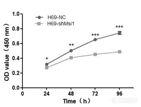
[font='times new roman'][size=21px][color=#000000]MSI1[/color][/size][/font][font='times new roman'][size=21px][color=#000000]对小细胞肺癌细胞生长增殖的影响及相关机制研究[/color][/size][/font][font='times new roman'][size=20px][color=#000000]实验方法[/color][/size][/font][font='times new roman'][size=18px][color=#000000]检测[/color][/size][/font][font='times new roman'][size=18px][color=#000000]MSI1[/color][/size][/font][font='times new roman'][size=18px][color=#000000]对人小细胞肺癌细胞的生长增殖的影响[/color][/size][/font][font='times new roman'][size=16px][color=#000000]CCK-8[/color][/size][/font][font='times new roman'][size=16px][color=#000000]实验检测小细胞肺癌细胞系生长增殖情况[/color][/size][/font][font='times new roman'][size=16px][color=#000000]分别将对数生长期的[/color][/size][/font][font='times new roman'][size=16px][color=#000000]H69[/color][/size][/font][font='times new roman'][size=16px][color=#000000]-NC[/color][/size][/font][font='times new roman'][size=16px][color=#000000]、[/color][/size][/font][font='times new roman'][size=16px][color=#000000]H69[/color][/size][/font][font='times new roman'][size=16px][color=#000000]-sh[/color][/size][/font][font='times new roman'][size=16px][color=#000000]MSI1[/color][/size][/font][font='times new roman'][size=16px][color=#000000]细胞离心,并用[/color][/size][/font][font='times new roman'][size=16px][color=#000000]完全培养基调整细胞浓度,以每孔[/color][/size][/font][font='times new roman'][size=16px][color=#000000]1.5[/color][/size][/font][font='times new roman'][size=16px][color=#000000]×[/color][/size][/font][font='times new roman'][size=16px][color=#000000]10[/color][/size][/font][font='times new roman'][sup][size=16px][color=#000000]4[/color][/size][/sup][/font][font='times new roman'][sup][size=16px][color=#000000] [/color][/size][/sup][/font][font='times new roman'][size=16px][color=#000000]个细胞平铺于[/color][/size][/font][font='times new roman'][size=16px][color=#000000]96[/color][/size][/font][font='times new roman'][size=16px][color=#000000]孔板中,[/color][/size][/font][font='times new roman'][size=16px][color=#000000]37[/color][/size][/font][font='times new roman'][size=16px][color=#000000]℃恒温培养箱中培养。铺板后,分别于[/color][/size][/font][font='times new roman'][size=16px][color=#000000]24 h[/color][/size][/font][font='times new roman'][size=16px][color=#000000]、[/color][/size][/font][font='times new roman'][size=16px][color=#000000]48 h[/color][/size][/font][font='times new roman'][size=16px][color=#000000]、[/color][/size][/font][font='times new roman'][size=16px][color=#000000]72 h[/color][/size][/font][font='times new roman'][size=16px][color=#000000]、[/color][/size][/font][font='times new roman'][size=16px][color=#000000]96 h[/color][/size][/font][font='times new roman'][size=16px][color=#000000]在每孔加入[/color][/size][/font][font='times new roman'][size=16px][color=#000000]10 [/color][/size][/font][font='times new roman'][size=16px][color=#000000]μL[/color][/size][/font][font='times new roman'][size=16px][color=#000000] CCK-8[/color][/size][/font][font='times new roman'][size=16px][color=#000000]溶液,[/color][/size][/font][font='times new roman'][size=16px][color=#000000]37[/color][/size][/font][font='times new roman'][size=16px][color=#000000]℃、[/color][/size][/font][font='times new roman'][size=16px][color=#000000]5%CO[/color][/size][/font][font='times new roman'][sub][size=16px][color=#000000]2[/color][/size][/sub][/font][font='times new roman'][size=16px][color=#000000]培养箱中孵育[/color][/size][/font][font='times new roman'][size=16px][color=#000000]4h[/color][/size][/font][font='times new roman'][size=16px][color=#000000]。并用酶标仪测定波长[/color][/size][/font][font='times new roman'][size=16px][color=#000000]450 nm[/color][/size][/font][font='times new roman'][size=16px][color=#000000]处[/color][/size][/font][font='times new roman'][size=16px][color=#000000]OD[/color][/size][/font][font='times new roman'][size=16px][color=#000000]值,结果应用[/color][/size][/font][font='times new roman'][size=16px][color=#000000]t[/color][/size][/font][font='times new roman'][size=16px][color=#000000]检验,利用[/color][/size][/font][font='times new roman'][size=16px][color=#000000]Graphpad prism5[/color][/size][/font][font='times new roman'][size=16px][color=#000000]计算增殖情况。[/color][/size][/font][font='times new roman'][size=16px][color=#000000]软琼脂克隆形成实验检测单细胞克隆形成能力[/color][/size][/font][font='times new roman'][size=16px][color=#000000]用琼脂糖及[/color][/size][/font][font='times new roman'][size=16px][color=#000000]ddH[/color][/size][/font][font='times new roman'][sub][size=16px][color=#000000]2[/color][/size][/sub][/font][font='times new roman'][size=16px][color=#000000]O[/color][/size][/font][font='times new roman'][size=16px][color=#000000]分别配置[/color][/size][/font][font='times new roman'][size=16px][color=#000000]0.3%[/color][/size][/font][font='times new roman'][size=16px][color=#000000]与[/color][/size][/font][font='times new roman'][size=16px][color=#000000]0.7%[/color][/size][/font][font='times new roman'][size=16px][color=#000000]的软琼脂凝胶,高压灭菌后维持[/color][/size][/font][font='times new roman'][size=16px][color=#000000]40[/color][/size][/font][font='times new roman'][size=16px][color=#000000]℃不会凝固,按[/color][/size][/font][font='times new roman'][size=16px][color=#000000]1:1[/color][/size][/font][font='times new roman'][size=16px][color=#000000]比例将[/color][/size][/font][font='times new roman'][size=16px][color=#000000]0.7%[/color][/size][/font][font='times new roman'][size=16px][color=#000000]的软琼脂与[/color][/size][/font][font='times new roman'][size=16px][color=#000000]2[/color][/size][/font][font='times new roman'][size=16px][color=#000000]×[/color][/size][/font][font='times new roman'][size=16px][color=#000000]1640[/color][/size][/font][font='times new roman'][size=16px][color=#000000]培养基混合,将混合液置入[/color][/size][/font][font='times new roman'][size=16px][color=#000000]6[/color][/size][/font][font='times new roman'][size=16px][color=#000000]孔板中冷却凝固半小时,作为底层琼脂置于培养箱中备用。分别将对数生长期的[/color][/size][/font][font='times new roman'][size=16px][color=#000000]H69[/color][/size][/font][font='times new roman'][size=16px][color=#000000]-NC[/color][/size][/font][font='times new roman'][size=16px][color=#000000]、[/color][/size][/font][font='times new roman'][size=16px][color=#000000]H69[/color][/size][/font][font='times new roman'][size=16px][color=#000000]-sh[/color][/size][/font][font='times new roman'][size=16px][color=#000000]MSI1[/color][/size][/font][font='times new roman'][size=16px][color=#000000]细胞计数,按[/color][/size][/font][font='times new roman'][size=16px][color=#000000]1:1[/color][/size][/font][font='times new roman'][size=16px][color=#000000]比例将[/color][/size][/font][font='times new roman'][size=16px][color=#000000]0.3%[/color][/size][/font][font='times new roman'][size=16px][color=#000000]软琼脂凝胶与细胞悬液混合铺入上层,每孔细胞数为[/color][/size][/font][font='times new roman'][size=16px][color=#000000]1000[/color][/size][/font][font='times new roman'][size=16px][color=#000000]个,注意不要产生气泡。每[/color][/size][/font][font='times new roman'][size=16px][color=#000000]2-3[/color][/size][/font][font='times new roman'][size=16px][color=#000000]天滴加少量培养基,防止干燥。待上层凝固后放入恒温培养箱中培养[/color][/size][/font][font='times new roman'][size=16px][color=#000000]2-3[/color][/size][/font][font='times new roman'][size=16px][color=#000000]周后拍照计数克隆形成数及单个细胞团细胞数目,比较两种细胞克隆形成能力的差异,结果应用[/color][/size][/font][font='times new roman'][size=16px][color=#000000]t[/color][/size][/font][font='times new roman'][size=16px][color=#000000]检验,利用[/color][/size][/font][font='times new roman'][size=16px][color=#000000]Graphpad prism5[/color][/size][/font][font='times new roman'][size=16px][color=#000000]作图计算两种细胞克隆形成能力的差异。[/color][/size][/font][font='times new roman'][size=18px][color=#000000]MSI1[/color][/size][/font][font='times new roman'][size=18px][color=#000000]影响小细胞肺癌细胞的相关机制研究[/color][/size][/font][font='times new roman'][size=16px][color=#000000] Western blot[/color][/size][/font][font='times new roman'][size=16px][color=#000000]检测相关蛋白的表达[/color][/size][/font][font='times new roman'][size=16px][color=#000000]提取适量[/color][/size][/font][font='times new roman'][size=16px][color=#000000]H69[/color][/size][/font][font='times new roman'][size=16px][color=#000000]-NC[/color][/size][/font][font='times new roman'][size=16px][color=#000000]、[/color][/size][/font][font='times new roman'][size=16px][color=#000000]H69[/color][/size][/font][font='times new roman'][size=16px][color=#000000]-sh[/color][/size][/font][font='times new roman'][size=16px][color=#000000]MSI1[/color][/size][/font][font='times new roman'][size=16px][color=#000000]细胞总蛋白,[/color][/size][/font][font='times new roman'][size=16px][color=#000000]20[/color][/size][/font][font='times new roman'][size=16px][color=#000000]:[/color][/size][/font][font='times new roman'][size=16px][color=#000000]1[/color][/size][/font][font='times new roman'][size=16px][color=#000000]稀释后,与[/color][/size][/font][font='times new roman'][size=16px][color=#000000]200[/color][/size][/font][font='times new roman'][size=16px][color=#000000] [/color][/size][/font][font='times new roman'][size=16px][color=#000000]μ[/color][/size][/font][font='times new roman'][size=16px][color=#000000]L BCA[/color][/size][/font][font='times new roman'][size=16px][color=#000000]工作液混合均匀。[/color][/size][/font][font='times new roman'][size=16px][color=#000000]37[/color][/size][/font][font='times new roman'][size=16px][color=#000000]℃孵育[/color][/size][/font][font='times new roman'][size=16px][color=#000000]30 min[/color][/size][/font][font='times new roman'][size=16px][color=#000000],用酶标仪测定波长[/color][/size][/font][font='times new roman'][size=16px][color=#000000]562 nm[/color][/size][/font][font='times new roman'][size=16px][color=#000000]处[/color][/size][/font][font='times new roman'][size=16px][color=#000000]OD[/color][/size][/font][font='times new roman'][size=16px][color=#000000]值,根据标准曲线计算出样品中的蛋白浓度。在蛋白原液中加入相应体积[/color][/size][/font][font='times new roman'][size=16px][color=#000000]4[/color][/size][/font][font='times new roman'][size=16px][color=#000000]×[/color][/size][/font][font='times new roman'][size=16px][color=#000000]SDS Loading Buffer[/color][/size][/font][font='times new roman'][size=16px][color=#000000],沸水浴煮[/color][/size][/font][font='times new roman'][size=16px][color=#000000]5 min[/color][/size][/font][font='times new roman'][size=16px][color=#000000],分别取[/color][/size][/font][font='times new roman'][size=16px][color=#000000]20[/color][/size][/font][font='times new roman'][size=16px][color=#000000] μ[/color][/size][/font][font='times new roman'][size=16px][color=#000000]g[/color][/size][/font][font='times new roman'][size=16px][color=#000000]细胞总蛋白,在[/color][/size][/font][font='times new roman'][size=16px][color=#000000]10% SDS-PAGE[/color][/size][/font][font='times new roman'][size=16px][color=#000000]分离胶电泳。电泳结束后,将蛋白转至[/color][/size][/font][font='times new roman'][size=16px][color=#000000]PVDF[/color][/size][/font][font='times new roman'][size=16px][color=#000000]膜上。用含[/color][/size][/font][font='times new roman'][size=16px][color=#000000]5%[/color][/size][/font][font='times new roman'][size=16px][color=#000000]脱脂奶粉的封闭液[/color][/size][/font][font='times new roman'][size=16px][color=#000000]37[/color][/size][/font][font='times new roman'][size=16px][color=#000000]℃封闭[/color][/size][/font][font='times new roman'][size=16px][color=#000000]1.5 h[/color][/size][/font][font='times new roman'][size=16px][color=#000000]。弃去封闭液,用[/color][/size][/font][font='times new roman'][size=16px][color=#000000]TBST[/color][/size][/font][font='times new roman'][size=16px][color=#000000]洗[/color][/size][/font][font='times new roman'][size=16px][color=#000000]3[/color][/size][/font][font='times new roman'][size=16px][color=#000000]次,加入相应抗体,[/color][/size][/font][font='times new roman'][size=16px][color=#000000]4[/color][/size][/font][font='times new roman'][size=16px][color=#000000]℃孵育过夜,[/color][/size][/font][font='times new roman'][size=16px][color=#000000]TBST[/color][/size][/font][font='times new roman'][size=16px][color=#000000] [/color][/size][/font][font='times new roman'][size=16px][color=#000000]洗膜[/color][/size][/font][font='times new roman'][size=16px][color=#000000]3[/color][/size][/font][font='times new roman'][size=16px][color=#000000]次,每次[/color][/size][/font][font='times new roman'][size=16px][color=#000000]10 min[/color][/size][/font][font='times new roman'][size=16px][color=#000000]。加入相应二抗([/color][/size][/font][font='times new roman'][size=16px][color=#000000]1:5000[/color][/size][/font][font='times new roman'][size=16px][color=#000000]),[/color][/size][/font][font='times new roman'][size=16px][color=#000000]37[/color][/size][/font][font='times new roman'][size=16px][color=#000000]℃敷育[/color][/size][/font][font='times new roman'][size=16px][color=#000000]1 h[/color][/size][/font][font='times new roman'][size=16px][color=#000000],[/color][/size][/font][font='times new roman'][size=16px][color=#000000]TBST[/color][/size][/font][font='times new roman'][size=16px][color=#000000]洗膜[/color][/size][/font][font='times new roman'][size=16px][color=#000000]3[/color][/size][/font][font='times new roman'][size=16px][color=#000000]次,每次[/color][/size][/font][font='times new roman'][size=16px][color=#000000]10 min[/color][/size][/font][font='times new roman'][size=16px][color=#000000]。用增敏化学发光底物试剂检测,暗室曝光显影。重复多次实验,应用[/color][/size][/font][font='times new roman'][size=16px][color=#000000]ImageJ[/color][/size][/font][font='times new roman'][size=16px][color=#000000]计算出各个蛋白条带的灰度对比,并应用[/color][/size][/font][font='times new roman'][size=16px][color=#000000]Graphpad prism5[/color][/size][/font][font='times new roman'][size=16px][color=#000000]作出柱状图。[/color][/size][/font][font='times new roman'][size=18px][color=#000000] MSI1[/color][/size][/font][font='times new roman'][size=18px][color=#000000]对人小细胞肺癌细胞生长增殖能力的影响[/color][/size][/font][font='times new roman'][size=16px][color=#000000]实验检测[/color][/size][/font][font='times new roman'][size=16px][color=#000000]MSI1[/color][/size][/font][font='times new roman'][size=16px][color=#000000]对人小细胞肺癌细胞增殖能力的影响[/color][/size][/font][font='times new roman'][size=16px][color=#000000]CCK-8[/color][/size][/font][font='times new roman'][size=16px][color=#000000]试剂可用于检测细胞生长增殖能力,过程简便精确。其基本原理为:试剂中含有的[/color][/size][/font][font='times new roman'][size=16px][color=#000000]WST-8[/color][/size][/font][font='times new roman'][size=16px][color=#000000]([/color][/size][/font][font='times new roman'][size=16px][color=#000000]2-(2-[/color][/size][/font][font='times new roman'][size=16px][color=#000000]甲氧基[/color][/size][/font][font='times new roman'][size=16px][color=#000000]-4-[/color][/size][/font][font='times new roman'][size=16px][color=#000000]硝基苯基[/color][/size][/font][font='times new roman'][size=16px][color=#000000])-3-(4-[/color][/size][/font][font='times new roman'][size=16px][color=#000000]硝基苯基[/color][/size][/font][font='times new roman'][size=16px][color=#000000])-5-(2,4-[/color][/size][/font][font='times new roman'][size=16px][color=#000000]二磺酸苯[/color][/size][/font][font='times new roman'][size=16px][color=#000000])-2H-[/color][/size][/font][font='times new roman'][size=16px][color=#000000]四唑单钠盐),在电子载体的作用下被细胞中的脱氢酶还原为黄色甲瓒产物。生成的甲瓒物的数量与活细胞的数量成正比。[/color][/size][/font][font='times new roman'][size=16px][color=#000000]用[/color][/size][/font][font='times new roman'][size=16px][color=#000000]酶标仪测定[/color][/size][/font][font='times new roman'][size=16px][color=#000000]450 nm[/color][/size][/font][font='times new roman'][size=16px][color=#000000]处[/color][/size][/font][font='times new roman'][size=16px][color=#000000]OD[/color][/size][/font][font='times new roman'][size=16px][color=#000000]值,[/color][/size][/font][font='times new roman'][size=16px][color=#000000]可间接反映活细胞数量。[/color][/size][/font][font='times new roman'][size=16px][color=#000000]利用[/color][/size][/font][font='times new roman'][size=16px][color=#000000]CCK-8[/color][/size][/font][font='times new roman'][size=16px][color=#000000]实验检测[/color][/size][/font][font='times new roman'][size=16px][color=#000000]H69-NC[/color][/size][/font][font='times new roman'][size=16px][color=#000000]、[/color][/size][/font][font='times new roman'][size=16px][color=#000000]H69-shMSI1[/color][/size][/font][font='times new roman'][size=16px][color=#000000]细胞增殖能力,[/color][/size][/font][font='times new roman'][size=16px][color=#000000]结果如图[/color][/size][/font][font='times new roman'][size=16px][color=#000000]3-1[/color][/size][/font][font='times new roman'][size=16px][color=#000000]显示,[/color][/size][/font][font='times new roman'][size=16px][color=#000000]H69-shMSI1[/color][/size][/font][font='times new roman'][size=16px][color=#000000]组[/color][/size][/font][font='times new roman'][size=16px][color=#000000]OD[/color][/size][/font][font='times new roman'][size=16px][color=#000000]值明显低于对照组,并且增长变化不明显。表明,[/color][/size][/font][font='times new roman'][size=16px][color=#000000]MSI1[/color][/size][/font][font='times new roman'][size=16px][color=#000000]低表达抑制小细胞肺癌细胞的生长增殖。[/color][/size][/font][align=center][img]https://ng1.17img.cn/bbsfiles/images/2022/10/202210102203024978_3594_5389809_3.png[/img][/align][font='times new roman'][color=#000000] H69-NC[/color][/font][font='times new roman'][color=#000000]、[/color][/font][font='times new roman'][color=#000000]H69-MSI1[/color][/font][font='times new roman'][color=#000000]细胞生长曲线。应用[/color][/font][font='times new roman'][color=#000000]Graphpad prism5[/color][/font][font='times new roman'][color=#000000]作图所示([/color][/font][font='times new roman'][color=#000000]*[/color][/font][font='times new roman'][color=#000000]代表[/color][/font][font='times new roman'][color=#000000]P[/color][/font][font='times new roman'][color=#000000]0.05,**[/color][/font][font='times new roman'][color=#000000]代表[/color][/font][font='times new roman'][color=#000000]P[/color][/font][font='times new roman'][color=#000000]0.01,***[/color][/font][font='times new roman'][color=#000000]代表[/color][/font][font='times new roman'][color=#000000]P[/color][/font][font='times new roman'][color=#000000]0.001,[/color][/font][font='times new roman'][color=#000000]表示与对照组相比,[/color][/font][font='times new roman'][color=#000000]H69-shMSI1[/color][/font][font='times new roman'][color=#000000]组[/color][/font][font='times new roman'][color=#000000]OD[/color][/font][font='times new roman'][color=#000000]值减小具有统计学意义)。[/color][/font][align=center][/align][font='times new roman'][size=16px][color=#000000]软琼脂克隆形成实验检测[/color][/size][/font][font='times new roman'][size=16px][color=#000000]MSI1[/color][/size][/font][font='times new roman'][size=16px][color=#000000]对人小细胞肺癌细胞单细胞克隆能力的影响[/color][/size][/font][font='times new roman'][size=16px][color=#000000]软琼脂克隆形成实验能够检测悬浮细胞单个细胞克隆形成的能力,培养[/color][/size][/font][font='times new roman'][size=16px][color=#000000]2[/color][/size][/font][font='times new roman'][size=16px][color=#000000]周后肉眼可见克隆形成,拍照并分别计数两种细胞克隆形成数量及单个克隆细胞团的细胞数目。[/color][/size][/font][font='times new roman'][size=16px][color=#000000]结果如图[/color][/size][/font][font='times new roman'][size=16px][color=#000000]3-2[/color][/size][/font][font='times new roman'][size=16px][color=#000000]所示,[/color][/size][/font][font='times new roman'][size=16px][color=#000000]与对照组相比,[/color][/size][/font][font='times new roman'][size=16px][color=#000000]H69[/color][/size][/font][font='times new roman'][size=16px][color=#000000]-sh[/color][/size][/font][font='times new roman'][size=16px][color=#000000]MSI1[/color][/size][/font][font='times new roman'][size=16px][color=#000000]组[/color][/size][/font][font='times new roman'][size=16px][color=#000000]形成的[/color][/size][/font][font='times new roman'][size=16px][color=#000000]克隆数目[/color][/size][/font][font='times new roman'][size=16px][color=#000000]明显[/color][/size][/font][font='times new roman'][size=16px][color=#000000]减少[/color][/size][/font][font='times new roman'][size=16px][color=#000000]([/color][/size][/font][font='times new roman'][size=16px][color=#000000]P[/color][/size][/font][font='times new roman'][size=16px][color=#000000]<[/color][/size][/font][font='times new roman'][size=16px][color=#000000]0.01[/color][/size][/font][font='times new roman'][size=16px][color=#000000])[/color][/size][/font][font='times new roman'][size=16px][color=#000000],克隆细胞团体积明显较小,[/color][/size][/font][font='times new roman'][size=16px][color=#000000]并且通过计数单个克隆的细胞数发现,[/color][/size][/font][font='times new roman'][size=16px][color=#000000]H69[/color][/size][/font][font='times new roman'][size=16px][color=#000000]-sh[/color][/size][/font][font='times new roman'][size=16px][color=#000000]MSI1[/color][/size][/font][font='times new roman'][size=16px][color=#000000]组单个克隆的细胞数明显低于对照组([/color][/size][/font][font='times new roman'][size=16px][color=#000000]P[/color][/size][/font][font='times new roman'][size=16px][color=#000000]<[/color][/size][/font][font='times new roman'][size=16px][color=#000000]0.01[/color][/size][/font][font='times new roman'][size=16px][color=#000000])。该实验同样表明[/color][/size][/font][font='times new roman'][size=16px][color=#000000]MSI1[/color][/size][/font][font='times new roman'][size=16px][color=#000000]低表达对小细胞肺癌细胞的增殖具有抑制作用。[/color][/size][/font][font='times new roman'][color=#000000] [/color][/font][font='times new roman'][color=#000000] [/color][/font][table][tr][td][align=center][img]https://ng1.17img.cn/bbsfiles/images/2022/10/202210102203030673_3184_5389809_3.png[/img][/align][/td][/tr][tr][td][align=center][img]https://ng1.17img.cn/bbsfiles/images/2022/10/202210102203032295_6922_5389809_3.png[/img][/align][/td][/tr][/table][font='times new roman'][color=#000000]琼脂克隆形成实验。([/color][/font][font='times new roman'][color=#000000]a[/color][/font][font='times new roman'][color=#000000])软琼脂克隆形成实验镜下观察;([/color][/font][font='times new roman'][color=#000000]b[/color][/font][font='times new roman'][color=#000000])克隆形成率;([/color][/font][font='times new roman'][color=#000000]c[/color][/font][font='times new roman'][color=#000000])单个克隆形成的细胞数([/color][/font][font='times new roman'][color=#000000]***[/color][/font][font='times new roman'][color=#000000]表示与对照组相比,[/color][/font][font='times new roman'][color=#000000]H69-shMSI1[/color][/font][font='times new roman'][color=#000000]组克隆形成率及单个克隆的细胞数减小具有统计学意义,[/color][/font][font='times new roman'][color=#000000]P[/color][/font][font='times new roman'][color=#000000]0.001[/color][/font][font='times new roman'][color=#000000])。[/color][/font][font='times new roman'][size=18px][color=#000000]MSI1[/color][/size][/font][font='times new roman'][size=18px][color=#000000]影响人小细胞肺癌细胞的相关机制研究[/color][/size][/font][font='times new roman'][size=16px][color=#000000]MSI1[/color][/size][/font][font='times new roman'][size=16px][color=#000000]低表达后肿瘤干细胞标志物[/color][/size][/font][font='times new roman'][size=16px][color=#000000]CD133[/color][/size][/font][font='times new roman'][size=16px][color=#000000]、[/color][/size][/font][font='times new roman'][size=16px][color=#000000]CD44[/color][/size][/font][font='times new roman'][size=16px][color=#000000]表达量下降[/color][/size][/font][font='times new roman'][size=16px][color=#000000] [/color][/size][/font][font='times new roman'][size=16px][color=#000000] [/color][/size][/font][font='times new roman'][size=16px][color=#000000]CD133[/color][/size][/font][font='times new roman'][size=16px][color=#000000]、[/color][/size][/font][font='times new roman'][size=16px][color=#000000]CD44[/color][/size][/font][font='times new roman'][size=16px][color=#000000]为目前已知研究较为广泛的的肿瘤干细胞标志物,为了研究[/color][/size][/font][font='times new roman'][size=16px][color=#000000]MSI1[/color][/size][/font][font='times new roman'][size=16px][color=#000000]在人小细胞肺癌细胞系中与[/color][/size][/font][font='times new roman'][size=16px][color=#000000]CD133[/color][/size][/font][font='times new roman'][size=16px][color=#000000]、[/color][/size][/font][font='times new roman'][size=16px][color=#000000]CD44[/color][/size][/font][font='times new roman'][size=16px][color=#000000]的相关性,分别提取对数生长期[/color][/size][/font][font='times new roman'][size=16px][color=#000000]H69[/color][/size][/font][font='times new roman'][size=16px][color=#000000]-NC[/color][/size][/font][font='times new roman'][size=16px][color=#000000]、[/color][/size][/font][font='times new roman'][size=16px][color=#000000]H69[/color][/size][/font][font='times new roman'][size=16px][color=#000000]-sh[/color][/size][/font][font='times new roman'][size=16px][color=#000000]MSI1[/color][/size][/font][font='times new roman'][size=16px][color=#000000]细胞总蛋白,应用[/color][/size][/font][font='times new roman'][size=16px][color=#000000]Western blot[/color][/size][/font][font='times new roman'][size=16px][color=#000000]验证[/color][/size][/font][font='times new roman'][size=16px][color=#000000]CD133[/color][/size][/font][font='times new roman'][size=16px][color=#000000]、[/color][/size][/font][font='times new roman'][size=16px][color=#000000]CD44[/color][/size][/font][font='times new roman'][size=16px][color=#000000]在蛋白质水平的表达量。结果如图[/color][/size][/font][font='times new roman'][size=16px][color=#000000]3-3[/color][/size][/font][font='times new roman'][size=16px][color=#000000]所示,与对照组相比,[/color][/size][/font][font='times new roman'][size=16px][color=#000000]H69[/color][/size][/font][font='times new roman'][size=16px][color=#000000]-sh[/color][/size][/font][font='times new roman'][size=16px][color=#000000]MSI1[/color][/size][/font][font='times new roman'][size=16px][color=#000000]组[/color][/size][/font][font='times new roman'][size=16px][color=#000000]CD133[/color][/size][/font][font='times new roman'][size=16px][color=#000000]、[/color][/size][/font][font='times new roman'][size=16px][color=#000000]CD44[/color][/size][/font][font='times new roman'][size=16px][color=#000000]的表达量明显降低。表明[/color][/size][/font][font='times new roman'][size=16px][color=#000000]MSI1[/color][/size][/font][font='times new roman'][size=16px][color=#000000]的敲低降低了小细胞肺癌细胞的干性,并且[/color][/size][/font][font='times new roman'][size=16px][color=#000000]CD133[/color][/size][/font][font='times new roman'][size=16px][color=#000000]、[/color][/size][/font][font='times new roman'][size=16px][color=#000000]CD44[/color][/size][/font][font='times new roman'][size=16px][color=#000000]的表达与[/color][/size][/font][font='times new roman'][size=16px][color=#000000]MSI1[/color][/size][/font][font='times new roman'][size=16px][color=#000000]具有相关性。[/color][/size][/font][table][tr][td][img]https://ng1.17img.cn/bbsfiles/images/2022/10/202210102203030163_8814_5389809_3.png[/img][/td][td][img]https://ng1.17img.cn/bbsfiles/images/2022/10/202210102203031179_6656_5389809_3.png[/img][/td][/tr][/table][font='times new roman'][color=#000000]CD133[/color][/font][font='times new roman'][color=#000000]、[/color][/font][font='times new roman'][color=#000000]CD44[/color][/font][font='times new roman'][color=#000000]蛋白的表达[/color][/font][font='times new roman'][color=#000000]:[/color][/font][font='times new roman'][color=#000000]([/color][/font][font='times new roman'][color=#000000]a[/color][/font][font='times new roman'][color=#000000])[/color][/font][font='times new roman'][color=#000000]CD133[/color][/font][font='times new roman'][color=#000000]、[/color][/font][font='times new roman'][color=#000000]CD44[/color][/font][font='times new roman'][color=#000000]蛋白条带;([/color][/font][font='times new roman'][color=#000000]b[/color][/font][font='times new roman'][color=#000000])[/color][/font][font='times new roman'][color=#000000]CD133[/color][/font][font='times new roman'][color=#000000]、[/color][/font][font='times new roman'][color=#000000]CD44[/color][/font][font='times new roman'][color=#000000]蛋白[/color][/font][font='times new roman'][color=#000000]相对表达量[/color][/font][font='times new roman'][color=#000000]([/color][/font][font='times new roman'][color=#000000]*[/color][/font][font='times new roman'][color=#000000]代表与对照组相比,[/color][/font][font='times new roman'][color=#000000]H69-shMSI1[/color][/font][font='times new roman'][color=#000000]组[/color][/font][font='times new roman'][color=#000000]CD133[/color][/font][font='times new roman'][color=#000000]、[/color][/font][font='times new roman'][color=#000000]CD44[/color][/font][font='times new roman'][color=#000000]蛋白表达下降具有统计学意义,[/color][/font][font='times new roman'][color=#000000]P[/color][/font][font='times new roman'][color=#000000]0.05[/color][/font][font='times new roman'][color=#000000])。[/color][/font][align=center][/align][font='times new roman'][size=16px][color=#000000]MSI1[/color][/size][/font][font='times new roman'][size=16px][color=#000000]对人小细胞肺癌细胞[/color][/size][/font][font='times new roman'][size=16px][color=#000000]EMT[/color][/size][/font][font='times new roman'][size=16px][color=#000000]过程的影响[/color][/size][/font][font='times new roman'][size=16px][color=#000000]为了研究[/color][/size][/font][font='times new roman'][size=16px][color=#000000]MSI1[/color][/size][/font][font='times new roman'][size=16px][color=#000000]是否对小细胞肺癌[/color][/size][/font][font='times new roman'][size=16px][color=#000000]EMT[/color][/size][/font][font='times new roman'][size=16px][color=#000000]过程产生影响,分别提取对数生长期的[/color][/size][/font][font='times new roman'][size=16px][color=#000000]H69[/color][/size][/font][font='times new roman'][size=16px][color=#000000]-NC[/color][/size][/font][font='times new roman'][size=16px][color=#000000]、[/color][/size][/font][font='times new roman'][size=16px][color=#000000]H69[/color][/size][/font][font='times new roman'][size=16px][color=#000000]-sh[/color][/size][/font][font='times new roman'][size=16px][color=#000000]MSI1[/color][/size][/font][font='times new roman'][size=16px][color=#000000]细胞总蛋白,利用[/color][/size][/font][font='times new roman'][size=16px][color=#000000]Western blot[/color][/size][/font][font='times new roman'][size=16px][color=#000000]验证[/color][/size][/font][font='times new roman'][size=16px][color=#000000]E-cadherin[/color][/size][/font][font='times new roman'][size=16px][color=#000000]、[/color][/size][/font][font='times new roman'][size=16px][color=#000000]N-cadherin[/color][/size][/font][font='times new roman'][size=16px][color=#000000]、[/color][/size][/font][font='times new roman'][size=16px][color=#000000]Vimentin[/color][/size][/font][font='times new roman'][size=16px][color=#000000]的表达量,结果如图[/color][/size][/font][font='times new roman'][size=16px][color=#000000]3-4[/color][/size][/font][font='times new roman'][size=16px][color=#000000]显示,与对照组相比,[/color][/size][/font][font='times new roman'][size=16px][color=#000000]H69[/color][/size][/font][font='times new roman'][size=16px][color=#000000]-sh[/color][/size][/font][font='times new roman'][size=16px][color=#000000]MSI1[/color][/size][/font][font='times new roman'][size=16px][color=#000000]组[/color][/size][/font][font='times new roman'][size=16px][color=#000000]N-cadherin[/color][/size][/font][font='times new roman'][size=16px][color=#000000]、[/color][/size][/font][font='times new roman'][size=16px][color=#000000]Vimentin[/color][/size][/font][font='times new roman'][size=16px][color=#000000]表达量明显下降,而[/color][/size][/font][font='times new roman'][size=16px][color=#000000]E-cadherin[/color][/size][/font][font='times new roman'][size=16px][color=#000000]表达量增加,表明[/color][/size][/font][font='times new roman'][size=16px][color=#000000]MSI1[/color][/size][/font][font='times new roman'][size=16px][color=#000000]低表达后,小细胞肺癌细胞的[/color][/size][/font][font='times new roman'][size=16px][color=#000000]EMT[/color][/size][/font][font='times new roman'][size=16px][color=#000000]过程受到明显抑制。[/color][/size][/font][table][tr][td][img]https://ng1.17img.cn/bbsfiles/images/2022/10/202210102203035586_9939_5389809_3.png[/img][/td][td][img]https://ng1.17img.cn/bbsfiles/images/2022/10/202210102203034079_6990_5389809_3.png[/img][/td][/tr][/table][font='times new roman'][color=#000000]EMT[/color][/font][font='times new roman'][color=#000000]相关蛋白表达:([/color][/font][font='times new roman'][color=#000000]a[/color][/font][font='times new roman'][color=#000000])[/color][/font][font='times new roman'][color=#000000]EMT[/color][/font][font='times new roman'][color=#000000]相关蛋白表达条带;([/color][/font][font='times new roman'][color=#000000]b[/color][/font][font='times new roman'][color=#000000])[/color][/font][font='times new roman'][color=#000000]EMT[/color][/font][font='times new roman'][color=#000000]相关蛋白[/color][/font][font='times new roman'][color=#000000]相对表达量([/color][/font][font='times new roman'][color=#000000]*[/color][/font][font='times new roman'][color=#000000]代表与对照组相比,[/color][/font][font='times new roman'][color=#000000]H69-shMSI1[/color][/font][font='times new roman'][color=#000000]组[/color][/font][font='times new roman'][color=#000000]EMT[/color][/font][font='times new roman'][color=#000000]相关蛋白表达下降具有统计学意义,[/color][/font][font='times new roman'][color=#000000]P[/color][/font][font='times new roman'][color=#000000]0.05[/color][/font][font='times new roman'][color=#000000])。[/color][/font][font='times new roman'][size=16px][color=#000000]MSI1[/color][/size][/font][font='times new roman'][size=16px][color=#000000]对人小细胞肺癌细胞凋亡过程的影响[/color][/size][/font][font='times new roman'][size=16px][color=#000000]细胞凋亡是多基因调控的细胞程序性的死亡,为了研究[/color][/size][/font][font='times new roman'][size=16px][color=#000000]MSI1[/color][/size][/font][font='times new roman'][size=16px][color=#000000]在人小细胞肺癌细胞中对细胞凋亡的影响,提取对数生长期的[/color][/size][/font][font='times new roman'][size=16px][color=#000000]H69[/color][/size][/font][font='times new roman'][size=16px][color=#000000]-NC[/color][/size][/font][font='times new roman'][size=16px][color=#000000]、[/color][/size][/font][font='times new roman'][size=16px][color=#000000]H69[/color][/size][/font][font='times new roman'][size=16px][color=#000000]-sh[/color][/size][/font][font='times new roman'][size=16px][color=#000000]MSI1[/color][/size][/font][font='times new roman'][size=16px][color=#000000]细胞总蛋白后,通过[/color][/size][/font][font='times new roman'][size=16px][color=#000000]Western blot[/color][/size][/font][font='times new roman'][size=16px][color=#000000]验证细胞凋亡相关蛋白的表达,结果如图[/color][/size][/font][font='times new roman'][size=16px][color=#000000]3-5[/color][/size][/font][font='times new roman'][size=16px][color=#000000]显示,与对照组相比,[/color][/size][/font][font='times new roman'][size=16px][color=#000000]H69[/color][/size][/font][font='times new roman'][size=16px][color=#000000]-sh[/color][/size][/font][font='times new roman'][size=16px][color=#000000]MSI1[/color][/size][/font][font='times new roman'][size=16px][color=#000000]组凋亡相关蛋白[/color][/size][/font][font='times new roman'][size=16px][color=#000000]cleaved caspase9[/color][/size][/font][font='times new roman'][size=16px][color=#000000]、[/color][/size][/font][font='times new roman'][size=16px][color=#000000]cleaved[/color][/size][/font][font='times new roman'][size=16px][color=#000000] c[/color][/size][/font][font='times new roman'][size=16px][color=#000000]aspase[/color][/size][/font][font='times new roman'][size=16px][color=#000000]3[/color][/size][/font][font='times new roman'][size=16px][color=#000000]、[/color][/size][/font][font='times new roman'][size=16px][color=#000000]cleaved PARP[/color][/size][/font][font='times new roman'][size=16px][color=#000000]表达量明显上调,抗凋亡蛋白[/color][/size][/font][font='times new roman'][size=16px][color=#000000]Bcl-2[/color][/size][/font][font='times new roman'][size=16px][color=#000000]表达量下调,[/color][/size][/font][font='times new roman'][size=16px][color=#000000]PARP[/color][/size][/font][font='times new roman'][size=16px][color=#000000]表达下降,表明[/color][/size][/font][font='times new roman'][size=16px][color=#000000]MSI1[/color][/size][/font][font='times new roman'][size=16px][color=#000000]低表达促进了小细胞肺癌细胞系凋亡过程。[/color][/size][/font][align=center][/align][align=center][/align][font='times new roman'][color=#000000]图[/color][/font][font='times new roman'][color=#000000]3-5 [/color][/font][font='times new roman'][color=#000000]凋亡相关蛋白[/color][/font][font='times new roman'][color=#000000]表达:([/color][/font][font='times new roman'][color=#000000]a[/color][/font][font='times new roman'][color=#000000])凋亡相关蛋白表达条带;([/color][/font][font='times new roman'][color=#000000]b[/color][/font][font='times new roman'][color=#000000])凋亡相关蛋白相对表达量([/color][/font][font='times new roman'][color=#000000]*[/color][/font][font='times new roman'][color=#000000]代表[/color][/font][font='times new roman'][color=#000000]P[/color][/font][font='times new roman'][color=#000000]0.05,**[/color][/font][font='times new roman'][color=#000000]代表[/color][/font][font='times new roman'][color=#000000]P[/color][/font][font='times new roman'][color=#000000]0.01,[/color][/font][font='times new roman'][color=#000000]表示与对照组相比,[/color][/font][font='times new roman'][color=#000000]H69-shMSI1[/color][/font][font='times new roman'][color=#000000]组凋亡相关蛋白表达下降具有统计学意义)。[/color][/font][align=center][/align][font='times new roman'][size=16px][color=#000000]主要研究[/color][/size][/font][font='times new roman'][size=16px][color=#000000]MSI1[/color][/size][/font][font='times new roman'][size=16px][color=#000000]对小细胞肺癌细胞生长增殖的影响及相关机制,首先利用[/color][/size][/font][font='times new roman'][size=16px][color=#000000]CCK-8[/color][/size][/font][font='times new roman'][size=16px][color=#000000]实验及软琼脂克隆形成实验验证[/color][/size][/font][font='times new roman'][size=16px][color=#000000]MSI1[/color][/size][/font][font='times new roman'][size=16px][color=#000000]低表达后小细胞肺癌细胞的生长增殖能力。结果显示,[/color][/size][/font][font='times new roman'][size=16px][color=#000000]H69-shMSI1[/color][/size][/font][font='times new roman'][size=16px][color=#000000]组细胞生长速度明显减慢,单个细胞克隆能力明显降低,表明[/color][/size][/font][font='times new roman'][size=16px][color=#000000]MSI1[/color][/size][/font][font='times new roman'][size=16px][color=#000000]低表达后抑制了小细胞肺癌细胞系的生长增殖。通过[/color][/size][/font][font='times new roman'][size=16px][color=#000000]western blot[/color][/size][/font][font='times new roman'][size=16px][color=#000000]实验检测肿瘤干细胞标志物[/color][/size][/font][font='times new roman'][size=16px][color=#000000]CD133[/color][/size][/font][font='times new roman'][size=16px][color=#000000]、[/color][/size][/font][font='times new roman'][size=16px][color=#000000]CD44[/color][/size][/font][font='times new roman'][size=16px][color=#000000]的表达。结果显示,[/color][/size][/font][font='times new roman'][size=16px][color=#000000]MSI1[/color][/size][/font][font='times new roman'][size=16px][color=#000000]低表达后肿瘤干细胞标志物[/color][/size][/font][font='times new roman'][size=16px][color=#000000]CD133[/color][/size][/font][font='times new roman'][size=16px][color=#000000]、[/color][/size][/font][font='times new roman'][size=16px][color=#000000]CD44[/color][/size][/font][font='times new roman'][size=16px][color=#000000]表达量下降,表明[/color][/size][/font][font='times new roman'][size=16px][color=#000000]MSI1[/color][/size][/font][font='times new roman'][size=16px][color=#000000]的敲低降低了小细胞肺癌细胞的干性,[/color][/size][/font][font='times new roman'][size=16px][color=#000000]CD133[/color][/size][/font][font='times new roman'][size=16px][color=#000000]、[/color][/size][/font][font='times new roman'][size=16px][color=#000000]CD44[/color][/size][/font][font='times new roman'][size=16px][color=#000000]的表达量可能与[/color][/size][/font][font='times new roman'][size=16px][color=#000000]MSI1[/color][/size][/font][font='times new roman'][size=16px][color=#000000]具有相关性,但具体机制尚不清楚,需进一步研究。[/color][/size][/font][font='times new roman'][size=16px][color=#000000]EMT[/color][/size][/font][font='times new roman'][size=16px][color=#000000]过程是肿瘤转移[/color][/size][/font][font='times new roman'][size=16px][color=#000000]过程的关键步骤,[/color][/size][/font][font='times new roman'][size=16px][color=#000000]MSI1[/color][/size][/font][font='times new roman'][size=16px][color=#000000]敲低后间充质标志物[/color][/size][/font][font='times new roman'][size=16px][color=#000000]N-cadherin[/color][/size][/font][font='times new roman'][size=16px][color=#000000]、[/color][/size][/font][font='times new roman'][size=16px][color=#000000]Vimentin[/color][/size][/font][font='times new roman'][size=16px][color=#000000]表达量下降,而上皮标志物[/color][/size][/font][font='times new roman'][size=16px][color=#000000]E-cadherin[/color][/size][/font][font='times new roman'][size=16px][color=#000000]表达量增加,表明[/color][/size][/font][font='times new roman'][size=16px][color=#000000]MSI1[/color][/size][/font][font='times new roman'][size=16px][color=#000000]敲低抑制了[/color][/size][/font][font='times new roman'][size=16px][color=#000000]EMT[/color][/size][/font][font='times new roman'][size=16px][color=#000000]过程。通过验证凋亡相关蛋白发现,凋亡相关蛋白[/color][/size][/font][font='times new roman'][size=16px][color=#000000]cleaved caspase9[/color][/size][/font][font='times new roman'][size=16px][color=#000000]、[/color][/size][/font][font='times new roman'][size=16px][color=#000000]cleaved[/color][/size][/font][font='times new roman'][size=16px][color=#000000] c[/color][/size][/font][font='times new roman'][size=16px][color=#000000]aspase[/color][/size][/font][font='times new roman'][size=16px][color=#000000]3[/color][/size][/font][font='times new roman'][size=16px][color=#000000]、[/color][/size][/font][font='times new roman'][size=16px][color=#000000]cleaved PARP[/color][/size][/font][font='times new roman'][size=16px][color=#000000]表达量明显上调,抗凋亡蛋白[/color][/size][/font][font='times new roman'][size=16px][color=#000000]Bcl-2[/color][/size][/font][font='times new roman'][size=16px][color=#000000]表达量下调,[/color][/size][/font][font='times new roman'][size=16px][color=#000000]PARP[/color][/size][/font][font='times new roman'][size=16px][color=#000000]表达下降,表明敲低[/color][/size][/font][font='times new roman'][size=16px][color=#000000]MSI1[/color][/size][/font][font='times new roman'][size=16px][color=#000000]后促进了凋亡的产生。[/color][/size][/font][font='times new roman'][size=16px][color=#000000]Wnt/[/color][/size][/font][font='times new roman'][size=16px][color=#000000]β[/color][/size][/font][font='times new roman'][size=16px][color=#000000]-Catenin[/color][/size][/font][font='times new roman'][size=16px][color=#000000]通路在细胞生长发育过程中具有重要作用,在多种肿瘤中表达失调,[/color][/size][/font][font='times new roman'][size=16px][color=#000000]β[/color][/size][/font][font='times new roman'][size=16px][color=#000000]-Catenin[/color][/size][/font][font='times new roman'][size=16px][color=#000000]是[/color][/size][/font][font='times new roman'][size=16px][color=#000000]Wnt[/color][/size][/font][font='times new roman'][size=16px][color=#000000]信号通路关键下游靶点,[/color][/size][/font][font='times new roman'][size=16px][color=#000000]MSI1[/color][/size][/font][font='times new roman'][size=16px][color=#000000]低表达使[/color][/size][/font][font='times new roman'][size=16px][color=#000000]β[/color][/size][/font][font='times new roman'][size=16px][color=#000000]-Catenin[/color][/size][/font][font='times new roman'][size=16px][color=#000000]表达量下降,表明[/color][/size][/font][font='times new roman'][size=16px][color=#000000]MSI1[/color][/size][/font][font='times new roman'][size=16px][color=#000000]在人小细胞肺癌细胞中也通过激活[/color][/size][/font][font='times new roman'][size=16px][color=#000000]Wnt[/color][/size][/font][font='times new roman'][size=16px][color=#000000]通路发挥作用。[/color][/size][/font]
[size=18px]实时荧光定量[/size][size=18px][url=https://insevent.instrument.com.cn/t/jp][color=#3333ff]PCR[/color][/url][/size][size=18px]([/size][size=18px]RT-[/size][size=18px][url=https://insevent.instrument.com.cn/t/jp][color=#3333ff]PCR[/color][/url][/size][size=18px])[/size][size=18px]及细胞增殖实验操作[/size][size=16px]选择管家基因[/size][size=16px]GAPDH[/size][size=16px]作为内参基因,以[/size][size=16px]H[/size][size=16px]69[/size][size=16px]作为对照组,检测四种亚型标志物[/size][size=16px][color=#000000]ASCL1[/color][/size][size=16px]、[/size][size=16px]NEUROD1[/size][size=16px]、[/size][size=16px]YAP1[/size][size=16px]、[/size][size=16px][color=#000000]POU2F3[/color][/size][size=16px]在[/size][size=16px]H69[/size][size=16px]、[/size][size=16px]H[/size][size=16px]446[/size][size=16px]、[/size][size=16px]H[/size][size=16px]526[/size][size=16px]、[/size][size=16px]D[/size][size=16px]MS114[/size][size=16px]细胞系中的相对表达量。并根据实验得到的[/size][size=16px]CT[/size][size=16px]值,采用[/size][size=16px]2[/size][font='times new roman'][sup][size=16px]-[/size][/sup][/font][font='times new roman'][sup][size=16px]△△[/size][/sup][/font][font='times new roman'][sup][size=16px]CT[/size][/sup][/font][size=16px]法计算相对表达量。[/size][size=16px]本实验所用引物如下:[/size][align=center]表1 引物序列[/align][align=center][img=,690,474]https://ng1.17img.cn/bbsfiles/images/2022/12/202212011425345641_7273_3237657_3.png!w690x474.jpg[/img][/align][size=16px]本研究应用[/size][size=16px]10 [/size][size=16px]μL[/size][size=16px]的[/size][size=16px][url=https://insevent.instrument.com.cn/t/jp][color=#3333ff]PCR[/color][/url][/size][size=16px]反应体系。[/size][size=16px]([/size][size=16px]1[/size][size=16px])将试剂解冻,上下颠倒混匀(勿剧烈摇动)后置于冰上。[/size][size=16px]([/size][size=16px]2[/size][size=16px])将引物工作液解冻,混合均匀后置于冰上,[/size][size=16px]DEPC[/size][size=16px]水也置于冰上。[/size][size=16px]([/size][size=16px]3[/size][size=16px])冰上配制[/size][size=16px][url=https://insevent.instrument.com.cn/t/jp][color=#3333ff]PCR[/color][/url][/size][size=16px]反应混合液。[/size][size=16px] [/size][align=center][font=宋体]表[/font] RT-qPCR[font=宋体]反应试剂[/font][/align][align=center][img=,690,347]https://ng1.17img.cn/bbsfiles/images/2022/12/202212011426306271_8553_3237657_3.png!w690x347.jpg[/img][/align][size=16px]([/size][size=16px]4[/size][size=16px])将[/size][size=16px][url=https://insevent.instrument.com.cn/t/jp][color=#3333ff]PCR[/color][/url][/size][size=16px]混合液混匀,不要涡旋,吸取[/size][size=16px]9 [/size][size=16px]L[/size][size=16px]至反应管内,随后加入[/size][size=16px]1 [/size][size=16px]μL[/size][size=16px] cDNA[/size][size=16px]模板,阴性对照不加[/size][size=16px]cDNA[/size][size=16px]模板,[/size][size=16px]用[url=https://insevent.instrument.com.cn/t/9p][color=#3333ff]移液枪[/color][/url]小心[/size][size=16px]混匀。[/size][size=16px]([/size][size=16px]5[/size][size=16px])加[/size][size=16px]样结束[/size][size=16px]后,八连管封盖,并将反应管内的气泡甩掉,收集反应液于管底。[/size][size=16px]([/size][size=16px]6[/size][size=16px])设置[/size][size=16px][url=https://insevent.instrument.com.cn/t/jp][color=#3333ff]PCR[/color][/url][/size][size=16px]反应程序。[/size][align=center]定量[url=https://insevent.instrument.com.cn/t/jp][color=#3333ff]PCR[/color][/url]反应程序[/align][align=center][img=,690,336]https://ng1.17img.cn/bbsfiles/images/2022/12/202212011427507339_8055_3237657_3.png!w690x336.jpg[/img][/align][size=16px]([/size][size=16px]7[/size][size=16px])将[/size][size=16px]8[/size][size=16px]连管或[/size][size=16px][url=https://insevent.instrument.com.cn/t/jp][color=#3333ff]PCR[/color][/url] 96[/size][size=16px]孔板放入仪器开始反应。[/size][align=left][size=18px]CCK-8[/size][size=18px]法检测细胞增殖[/size][/align][align=left] [size=16px]取对数生长期的[/size][size=16px]H[/size][size=16px]69[/size][size=16px]、[/size][size=16px]H446[/size][size=16px]、[/size][size=16px]H[/size][size=16px]526[/size][size=16px]、[/size][size=16px]D[/size][size=16px]MS114[/size][size=16px]细胞,[/size][size=16px]其中[/size][size=16px]H[/size][size=16px]446[/size][size=16px]、[/size][size=16px]H[/size][size=16px]69[/size][size=16px]、[/size][size=16px]H[/size][size=16px]526[/size][size=16px]以[/size][size=16px]1[/size][size=16px]×[/size][size=16px]1[/size][size=16px]0[/size][font='times new roman'][sup][size=16px]4[/size][/sup][/font][size=16px]个细胞[/size][size=16px]/[/size][size=16px]孔的细胞密度[/size][size=16px]接种于[/size][size=16px] 96 [/size][size=16px]孔板中,[/size][size=16px]D[/size][size=16px]MS114[/size][size=16px]以[/size][size=16px]4[/size][font='arial'][size=16px]×[/size][/font][size=16px]1[/size][size=16px]0[/size][font='times new roman'][sup][size=16px]3[/size][/sup][/font][size=16px]个细胞[/size][size=16px]/[/size][size=16px]孔的细胞密度[/size][size=16px]接种于[/size][size=16px] 96 [/size][size=16px]孔板中[/size][size=16px]。[/size][size=16px]待[/size][size=16px]细胞融合率约[/size][size=16px]80%[/size][size=16px],[/size][size=16px]加入不同浓度西达本胺。每组均设置[/size][size=16px]D[/size][size=16px]MSO[/size][size=16px]对照组及空白组(仅有同体积培养基)。[/size][size=16px]H[/size][size=16px]446[/size][size=16px]加药浓度梯度为[/size][size=16px] 0[/size][size=16px]、[/size][size=16px]0[/size][size=16px].5[/size][size=16px]、[/size][size=16px]1[/size][size=16px]、[/size][size=16px]2[/size][size=16px]、[/size][size=16px]4[/size][size=16px]、[/size][size=16px]8[/size][size=16px]、[/size][size=16px]16[/size][size=16px]、[/size][size=16px]3[/size][size=16px]2[/size][size=16px] [/size][size=16px]μmol/L[/size][size=16px],[/size][size=16px]H[/size][size=16px]69[/size][size=16px]加药浓度梯度为[/size][size=16px]0[/size][size=16px]、[/size][size=16px]2[/size][size=16px].[/size][size=16px]5[/size][size=16px]、[/size][size=16px]5[/size][size=16px]、[/size][size=16px]10[/size][size=16px]、[/size][size=16px]2[/size][size=16px]0[/size][size=16px]、[/size][size=16px]4[/size][size=16px]0[/size][size=16px] [/size][size=16px]μmol/L[/size][size=16px],[/size][size=16px]H526[/size][size=16px]加药浓度梯度为[/size][size=16px]0[/size][size=16px]、[/size][size=16px]1.25[/size][size=16px]、[/size][size=16px]2[/size][size=16px].[/size][size=16px]5[/size][size=16px]、[/size][size=16px]5[/size][size=16px]、[/size][size=16px]10[/size][size=16px]、[/size][size=16px]2[/size][size=16px]0[/size][size=16px]、[/size][size=16px]4[/size][size=16px]0[/size][size=16px] [/size][size=16px]μmol/L[/size][size=16px],[/size][size=16px]D[/size][size=16px]MS114[/size][size=16px]加药浓度梯度为[/size][size=16px]0[/size][size=16px]、[/size][size=16px]1[/size][size=16px]、[/size][size=16px]2[/size][size=16px]、[/size][size=16px]4[/size][size=16px]、[/size][size=16px]8[/size][size=16px]、[/size][size=16px]16[/size][size=16px]、[/size][size=16px]3[/size][size=16px]2[/size][size=16px]、[/size][size=16px]6[/size][size=16px]4[/size][size=16px] [/size][size=16px]μmol/L[/size][size=16px](加药浓度梯度根据细胞类型、前期预实验结果及细胞对药物的敏感程度而定)。[/size][size=16px]每[/size][size=16px]种[/size][size=16px]浓度设[/size][size=16px]6[/size][size=16px]个复孔,每孔总体积为[/size][size=16px]100[/size] [size=16px]μL[/size][size=16px],培养[/size][size=16px]24[/size][size=16px]、[/size][size=16px]48[/size][size=16px]、[/size][size=16px]72[/size][size=16px]、[/size][size=16px]9[/size][size=16px]6[/size][size=16px]、[/size][size=16px]1[/size][size=16px]20[/size][size=16px] [/size][size=16px]h[/size][size=16px]后[/size][size=16px]检测细胞[/size][size=16px]活力[/size][size=16px]。每孔加入[/size][size=16px]10[/size] [size=16px]μL[/size][size=16px]的[/size][size=16px]CCK-8[/size][size=16px](避光),培养箱中孵育[/size][size=16px]2-[/size][size=16px]4[/size][size=16px] [/size][size=16px]h[/size][size=16px]后取出,使[/size][size=16px]用酶标仪测定波长为[/size][size=16px]450 nm[/size][size=16px]的吸光度[/size][size=16px]([/size][size=16px]O[/size][size=16px]D[/size][size=16px]值[/size][size=16px])[/size][size=16px]。[/size][size=16px]利用公式:抑制率[/size][size=16px]=[/size][size=16px](加药组[/size][font='宋体'][size=16px]-[/size][/font][size=16px]空白组)[/size][size=16px]/[/size][size=16px](对照组[/size][font='宋体'][size=16px]-[/size][/font][size=16px]空白组)计算增殖抑制率。[/size][size=16px]实验重复[/size][size=16px]3[/size][size=16px]次,[/size][size=16px]取平均值。[/size][size=16px]以药物[/size][size=16px]浓度[/size][size=16px]为横坐标,细胞增殖抑制率为纵坐标[/size][size=16px]绘图[/size],[size=16px]使用[/size][size=16px]S[/size][size=16px]PSS23.0[/size][size=16px]计算[/size][size=16px]IC10[/size][size=16px]、[/size][size=16px]I[/size][size=16px]C20[/size][size=16px]、[/size][size=16px]I[/size][size=16px]C50[/size][size=16px],取平均值作为后续实验药物浓度。[/size][/align]
iCELLigence全自动细胞分析仪让您远离MTT实验不断重复还无法得到统一结果的烦恼,让您不再因只看到其中的一个点而损失了其它的细胞生物学信息而无计可施,因为它可以清楚的记录下细胞完整的一生! 一:全自动细胞分析仪仪器原理 iCELLigence实时无标记全自动细胞分析仪是一款新型的细胞分析平台,具有实时监测、高信息量、无需标记、全自动化、高灵敏度和高准确性等独特优点。该细胞分析仪通过嵌在E-plate板上孔底的微电子感应器阻抗变化去感受细胞的有无以及贴壁、黏附和生长程度的改变。在细胞毒性检测中,可实时、直观的反应细胞增殖、存活、凋亡、形态变化等细胞生物学变化。 二:全自动细胞分析仪仪器优势 iCELLigence全自动细胞分析仪的传感器阻抗技术在细胞分析中具有其独特的优势:它为整个的细胞毒性检测分析过程中提供了全程无损伤的监控,实时、连续显示的数据让您可以更加自信更加清楚的进行细胞毒性检测操作和其它的细胞分析,而不是假定细胞处于合适的处理阶段。一连串实时获取和显示的数据让您处理每一步结果都可以通过机理来预测,同时也可以结合全自动细胞分析仪实时的读数来决定传统终点细胞毒性检测分析的最佳时间点。只需几个简单的操作步骤您就可以获得高信息量的、直观的、准确的结果,就可以让您的细胞实验变得更加省时高效。 三:全自动细胞分析仪的应用领域基于iCELLigence全自动细胞分析仪的技术优势,该系统在基础生命科学领域具有广泛的应用,如细胞质量控制、细胞毒性检测、细胞粘附和细胞伸展等。
[font='times new roman'][size=14px]MSI1 调节小细胞肺癌增殖凋亡[/size][/font][font='times new roman'][size=14px]简介[/size][/font][font='times new roman'][size=14px]肺癌是最常见的恶性肿瘤之一,也是全球癌症死亡的主要原因[/size][/font][font='times new roman'][size=14px]。小细胞肺癌[/size][/font][font='times new roman'][size=14px]([/size][/font][font='times new roman'][size=14px]small cell lung cancer, SCLC[/size][/font][font='times new roman'][size=14px])[/size][/font][font='times new roman'][size=14px]发生率占肺癌的[/size][/font][font='times new roman'][size=14px] [/size][/font][font='times new roman'][size=14px]10%-15%[/size][/font][font='times new roman'][size=14px],[/size][/font][font='times new roman'][size=14px]5 [/size][/font][font='times new roman'][size=14px]年生存率仅为[/size][/font][font='times new roman'][size=14px] [/size][/font][font='times new roman'][size=14px]7%[/size][/font][font='times new roman'][size=14px]。[/size][/font][font='times new roman'][size=14px]SCLC [/size][/font][font='times new roman'][size=14px]具[/size][/font][font='times new roman'][size=14px]有生长迅速、容易耐药、早期转移等特点,恶性程度在所有肺癌类型中最高,总体预后[/size][/font][font='times new roman'][size=14px]差,未经治疗的[/size][/font][font='times new roman'][size=14px] [/size][/font][font='times new roman'][size=14px]SCLC [/size][/font][font='times new roman'][size=14px]患者生存期仅[/size][/font][font='times new roman'][size=14px] [/size][/font][font='times new roman'][size=14px]2-4[/size][/font][font='times new roman'][size=14px] [/size][/font][font='times new roman'][size=14px]个[/size][/font][font='times new roman'][size=14px]月[/size][/font][font='times new roman'][size=14px]。大多数[/size][/font][font='times new roman'][size=14px] [/size][/font][font='times new roman'][size=14px]SCLC[/size][/font][font='times new roman'][size=14px] [/size][/font][font='times new roman'][size=14px]患者在诊断时已处于广泛期,其预后极差,因此[/size][/font][font='times new roman'][size=14px][color=#000007]对于[/color][/size][/font][font='times new roman'][size=14px][color=#000007] [/color][/size][/font][font='times new roman'][size=14px][color=#000007]SCLC [/color][/size][/font][font='times new roman'][size=14px][color=#000007]转移侵袭相关的分子机制的研究,有助于识别[/color][/size][/font][font='times new roman'][size=14px][color=#000007]SCLC[/color][/size][/font][font='times new roman'][size=14px][color=#000007] [/color][/size][/font][font='times new roman'][size=14px][color=#000007]新的生物标记物和治疗靶点。[/color][/size][/font][font='times new roman'][size=14px]SCLC [/size][/font][font='times new roman'][size=14px]是一种高度恶性的肺神经内分泌肿瘤。有学者通过对[/size][/font][font='times new roman'][size=14px] [/size][/font][font='times new roman'][size=14px]SCLC[/size][/font][font='times new roman'][size=14px] [/size][/font][font='times new roman'][size=14px]表观遗传学和[/size][/font][font='times new roman'][size=14px]基因表达的研究发现,[/size][/font][font='times new roman'][size=14px]ASCL1 [/size][/font][font='times new roman'][size=14px]和[/size][/font][font='times new roman'][size=14px] [/size][/font][font='times new roman'][size=14px]NEUROD1 [/size][/font][font='times new roman'][size=14px]揭示了[/size][/font][font='times new roman'][size=14px] [/size][/font][font='times new roman'][size=14px]SCLC [/size][/font][font='times new roman'][size=14px]的异质性[/size][/font][font='times new roman'][size=14px][5][/size][/font][font='times new roman'][size=14px],而最新研究基[/size][/font][font='times new roman'][size=14px]于[/size][/font][font='times new roman'][size=14px] [/size][/font][font='times new roman'][size=14px]SCLC [/size][/font][font='times new roman'][size=14px]中四个关键转录因子[/size][/font][font='times new roman'][size=14px] [/size][/font][font='times new roman'][size=14px]ASCL1[/size][/font][font='times new roman'][size=14px]、[/size][/font][font='times new roman'][size=14px]NEUROD1[/size][/font][font='times new roman'][size=14px]、[/size][/font][font='times new roman'][size=14px]POU2F3[/size][/font][font='times new roman'][size=14px] [/size][/font][font='times new roman'][size=14px]和[/size][/font][font='times new roman'][size=14px] [/size][/font][font='times new roman'][size=14px]YAP1[/size][/font][font='times new roman'][size=14px] [/size][/font][font='times new roman'][size=14px]的差异表达,[/size][/font][font='times new roman'][size=14px] [/size][/font][font='times new roman'][size=14px]将其定义为[/size][/font][font='times new roman'][size=14px] [/size][/font][font='times new roman'][size=14px]SCLC-A[/size][/font][font='times new roman'][size=14px]、[/size][/font][font='times new roman'][size=14px]SCLC-N[/size][/font][font='times new roman'][size=14px]、[/size][/font][font='times new roman'][size=14px]SCLC-P [/size][/font][font='times new roman'][size=14px]和[/size][/font][font='times new roman'][size=14px] [/size][/font][font='times new roman'][size=14px]SCLC-Y [/size][/font][font='times new roman'][size=14px]四种亚型[/size][/font][font='times new roman'][size=14px]。尽管在早期发现和[/size][/font][font='times new roman'][size=14px]治疗方面取得了进展,但[/size][/font][font='times new roman'][size=14px] [/size][/font][font='times new roman'][size=14px]SCLC [/size][/font][font='times new roman'][size=14px]的死亡率仍在上升。因此,了解肿瘤发生发展的分子机制对于更好的诊断和预后是必需的。[/size][/font][font='times new roman'][size=14px]Musashi1(MSI1) [/size][/font][font='times new roman'][size=14px]是一种保守的[/size][/font][font='times new roman'][size=14px] [/size][/font][font='times new roman'][size=14px]RNA [/size][/font][font='times new roman'][size=14px]结合蛋白[/size][/font][font='times new roman'][size=14px], [/size][/font][font='times new roman'][size=14px]最初在果蝇中被发现,位于染色体[/size][/font][font='times new roman'][size=14px]12q24 [/size][/font][font='times new roman'][size=14px]上,编码一个[/size][/font][font='times new roman'][size=14px] [/size][/font][font='times new roman'][size=14px]362-[/size][/font][font='times new roman'][size=14px]氨基酸多肽;[/size][/font][font='times new roman'][size=14px]MSI1 [/size][/font][font='times new roman'][size=14px]与真核转录起始因子[/size][/font][font='times new roman'][size=14px] [/size][/font][font='times new roman'][size=14px]4G[/size][/font][font='times new roman'][size=14px]([/size][/font][font='times new roman'][size=14px]eIF4G[/size][/font][font='times new roman'][size=14px])[/size][/font][font='times new roman'][size=14px] [/size][/font][font='times new roman'][size=14px]竞争[/size][/font][font='times new roman'][size=14px]多聚[/size][/font][font='times new roman'][size=14px] [/size][/font][font='times new roman'][size=14px]A [/size][/font][font='times new roman'][size=14px]结合蛋白[/size][/font][font='times new roman'][size=14px]([/size][/font][font='times new roman'][size=14px]PABP[/size][/font][font='times new roman'][size=14px])[/size][/font][font='times new roman'][size=14px],以[/size][/font][font='times new roman'][size=14px]结合其靶[/size][/font][font='times new roman'][size=14px] [/size][/font][font='times new roman'][size=14px]mRNA [/size][/font][font='times new roman'][size=14px]的[/size][/font][font='times new roman'][size=14px] [/size][/font][font='times new roman'][size=14px]3[/size][/font][font='times new roman'][size=14px]′[/size][/font][font='times new roman'][size=14px]-[/size][/font][font='times new roman'][size=14px]非翻译区[/size][/font][font='times new roman'][size=14px]([/size][/font][font='times new roman'][size=14px]UTR[/size][/font][font='times new roman'][size=14px])[/size][/font][font='times new roman'][size=14px] [/size][/font][font='times new roman'][size=14px]中的特定序列,从而抑制靶基因的翻译[/size][/font][font='times new roman'][size=14px]。作为一种[/size][/font][font='times new roman'][size=14px] [/size][/font][font='times new roman'][size=14px]RNA [/size][/font][font='times new roman'][size=14px]结合蛋白,[/size][/font][font='times new roman'][size=14px]MSI1[/size][/font][font='times new roman'][size=14px] [/size][/font][font='times new roman'][size=14px]已被证明参与许多与癌症有关的信号通路,其表达与肿瘤的发生发展密切相关[/size][/font][font='times new roman'][size=14px][9-11][/size][/font][font='times new roman'][size=14px]。[/size][/font][font='times new roman'][size=14px]Akt [/size][/font][font='times new roman'][size=14px]信号通路在肿瘤[/size][/font][font='times new roman'][size=14px]的发生发展中发挥重要作用,参与调节肿瘤细胞增殖、周期、凋亡以及肿瘤细胞与细胞外基质之间的相互作用[/size][/font][font='times new roman'][size=14px];研究发现[/size][/font][font='times new roman'][size=14px] [/size][/font][font='times new roman'][size=14px]MSI1 [/size][/font][font='times new roman'][size=14px]通过调节[/size][/font][font='times new roman'][size=14px] [/size][/font][font='times new roman'][size=14px]Akt [/size][/font][font='times new roman'][size=14px]信号通路的活性来[/size][/font][font='times new roman'][size=14px]调节非[/size][/font][font='times new roman'][size=14px]小细胞肺癌的恶性和化疗耐药性[/size][/font][font='times new roman'][size=14px][14][/size][/font][font='times new roman'][size=14px],[/size][/font][font='times new roman'][size=14px] [/size][/font][font='times new roman'][size=14px]MSI1 [/size][/font][font='times new roman'][size=14px]也可以通过激活[/size][/font][font='times new roman'][size=14px] [/size][/font][font='times new roman'][size=14px]Wnt [/size][/font][font='times new roman'][size=14px]信号通路促进宫颈癌细胞的[/size][/font][font='times new roman'][size=14px] [/size][/font][font='times new roman'][size=14px]EMT[/size][/font][font='times new roman'][size=14px]、侵袭和转移[/size][/font][font='times new roman'][size=14px]。另有研究发现[/size][/font][font='times new roman'][size=14px] [/size][/font][font='times new roman'][size=14px]MSI1 [/size][/font][font='times new roman'][size=14px]表达增强会导致肿瘤的扩散和复发,[/size][/font][font='times new roman'][size=14px]MSI1 [/size][/font][font='times new roman'][size=14px]高表达[/size][/font][font='times new roman'][size=14px]被认为是生存率低和预后差的指标。[/size][/font][font='times new roman'][size=14px]Moreira[/size][/font][font='times new roman'][size=14px]等人发现[/size][/font][font='times new roman'][size=14px] [/size][/font][font='times new roman'][size=14px]MSI1 [/size][/font][font='times new roman'][size=14px]在[/size][/font][font='times new roman'][size=14px]SCLC[/size][/font][font='times new roman'][size=14px] [/size][/font][font='times new roman'][size=14px]组织中普遍表达,因此本研究对[/size][/font][font='times new roman'][size=14px] [/size][/font][font='times new roman'][size=14px]MSI1 [/size][/font][font='times new roman'][size=14px]在[/size][/font][font='times new roman'][size=14px] [/size][/font][font='times new roman'][size=14px]SCLC[/size][/font][font='times new roman'][size=14px] [/size][/font][font='times new roman'][size=14px]中的生物学功能和分子机制研[/size][/font][font='times new roman'][size=14px]究进行了初步探索,旨在为[/size][/font][font='times new roman'][size=14px] [/size][/font][font='times new roman'][size=14px]SCLC[/size][/font][font='times new roman'][size=14px] [/size][/font][font='times new roman'][size=14px]诊治提供新思路,为[/size][/font][font='times new roman'][size=14px] [/size][/font][font='times new roman'][size=14px]SCLC[/size][/font][font='times new roman'][size=14px] [/size][/font][font='times new roman'][size=14px]的治疗以及改善患者预后提供有力的临床前研究依据。[/size][/font][font='times new roman'][size=14px]上皮间充质转化([/size][/font][font='times new roman'][size=14px]epithelial-mesenchymal transition[/size][/font][font='times new roman'][size=14px],[/size][/font][font='times new roman'][size=14px]EMT[/size][/font][font='times new roman'][size=14px])是肿瘤转移侵袭的主要[/size][/font][font='times new roman'][size=14px]机制之一,具有促进肿瘤细胞恶性增殖、减少细胞凋亡和衰老、促进免疫抑制等作用[/size][/font][font='times new roman'][size=14px][19][/size][/font][font='times new roman'][size=14px]。[/size][/font][font='times new roman'][size=14px]E-[/size][/font][font='times new roman'][size=14px]粘连蛋白[/size][/font][font='times new roman'][size=14px]([/size][/font][font='times new roman'][size=14px]E-cadherin[/size][/font][font='times new roman'][size=14px])[/size][/font][font='times new roman'][size=14px]表达下调或缺失、细胞极性的缺失都是[/size][/font][font='times new roman'][size=14px] [/size][/font][font='times new roman'][size=14px]EMT [/size][/font][font='times new roman'][size=14px]的重要标志[/size][/font][font='times new roman'][size=14px][20][/size][/font][font='times new roman'][size=14px]。[/size][/font][font='times new roman'][size=14px]N-[/size][/font][font='times new roman'][size=14px]钙粘蛋白[/size][/font][font='times new roman'][size=14px]([/size][/font][font='times new roman'][size=14px]N-cadherin[/size][/font][font='times new roman'][size=14px])[/size][/font][font='times new roman'][size=14px]、波形蛋白[/size][/font][font='times new roman'][size=14px]([/size][/font][font='times new roman'][size=14px]Vimentin[/size][/font][font='times new roman'][size=14px])[/size][/font][font='times new roman'][size=14px]属于间质细胞表型标记物,在[/size][/font][font='times new roman'][size=14px] [/size][/font][font='times new roman'][size=14px]EMT [/size][/font][font='times new roman'][size=14px]过程中,[/size][/font][font='times new roman'][size=14px]E-cadherin[/size][/font][font='times new roman'][size=14px] [/size][/font][font='times new roman'][size=14px]的下调会伴随着[/size][/font][font='times new roman'][size=14px] [/size][/font][font='times new roman'][size=14px]N-cadherin[/size][/font][font='times new roman'][size=14px] [/size][/font][font='times new roman'][size=14px]及[/size][/font][font='times new roman'][size=14px] [/size][/font][font='times new roman'][size=14px]Vimentin [/size][/font][font='times new roman'][size=14px]表达的上调,从而导致肿[/size][/font][font='times new roman'][size=14px]瘤细胞骨架重排,促进肿瘤的转移[/size][/font][font='times new roman'][size=14px][21[/size][/font][font='times new roman'][size=14px],[/size][/font][font='times new roman'][size=14px]22][/size][/font][font='times new roman'][size=14px]。大量研究表明,在乳腺癌、结直肠癌、肺癌等[/size][/font][font='times new roman'][size=14px]多种恶性肿瘤中,具有[/size][/font][font='times new roman'][size=14px] [/size][/font][font='times new roman'][size=14px]EMT[/size][/font][font='times new roman'][size=14px] [/size][/font][font='times new roman'][size=14px]表型的肿瘤细胞可表现出高度的侵袭和转移能力,导致患者的预后极差[/size][/font][font='times new roman'][size=14px]。大量研究证明,[/size][/font][font='times new roman'][size=14px]SCLC [/size][/font][font='times new roman'][size=14px]的[/size][/font][font='times new roman'][size=14px] [/size][/font][font='times new roman'][size=14px]EMT [/size][/font][font='times new roman'][size=14px]过程受到多种通路的调控,进而促进[/size][/font][font='times new roman'][size=14px] [/size][/font][font='times new roman'][size=14px]SCLC[/size][/font][font='times new roman'][size=14px] [/size][/font][font='times new roman'][size=14px]发生发展以及化疗的耐药性。[/size][/font][font='times new roman'][size=14px]Wnt[/size][/font][font='times new roman'][size=14px] [/size][/font][font='times new roman'][size=14px]信号通路调节生物体发育、体内平衡、愈合再生和维持干细胞等各种生物过程[/size][/font][font='times new roman'][size=14px],[/size][/font][font='times new roman'][size=14px]Wnt [/size][/font][font='times new roman'][size=14px]通路传导缺失会导致发育缺陷、骨骼疾病,甚至参与调控乳腺癌、结直肠等恶性肿瘤的发生发展[/size][/font][font='times new roman'][size=14px]。尽管其主要通路成分已被确定,但[/size][/font][font='times new roman'][size=14px] [/size][/font][font='times new roman'][size=14px]Wnt [/size][/font][font='times new roman'][size=14px]信号在肿瘤中的功能十分复杂,目前也只是部分了解。[/size][/font][font='times new roman'][size=14px]Wnt [/size][/font][font='times new roman'][size=14px]信号激活的转录反应是由细胞核内[/size][/font][font='times new roman'][size=14px] [/size][/font][font='times new roman'][size=14px]β-catenin [/size][/font][font='times new roman'][size=14px]结合因子的复杂网络所[/size][/font][font='times new roman'][size=14px]介[/size][/font][font='times new roman'][size=14px]导的[/size][/font][font='times new roman'][size=14px]。大肠腺瘤性息肉病[/size][/font][font='times new roman'][size=14px]([/size][/font][font='times new roman'][size=14px]adenomatous polyposis coli [/size][/font][font='times new roman'][size=14px],[/size][/font][font='times new roman'][size=14px]APC[/size][/font][font='times new roman'][size=14px])[/size][/font][font='times new roman'][size=14px] [/size][/font][font='times new roman'][size=14px]基因的缺失是大肠癌中[/size][/font][font='times new roman'][size=14px] [/size][/font][font='times new roman'][size=14px]Wnt [/size][/font][font='times new roman'][size=14px]信号传导的主要驱动因素,另外[/size][/font][font='times new roman'][size=14px] [/size][/font][font='times new roman'][size=14px]R-spondin/Lgr5/RNF43 [/size][/font][font='times new roman'][size=14px]模块的突变也被认为是依赖[/size][/font][font='times new roman'][size=14px] [/size][/font][font='times new roman'][size=14px]Wnt [/size][/font][font='times new roman'][size=14px]通路的肿瘤生长的驱动因素[/size][/font][font='times new roman'][size=14px][35[/size][/font][font='times new roman'][size=14px],[/size][/font][font='times new roman'][size=14px]36][/size][/font][font='times new roman'][size=14px]。另有研究发现,在[/size][/font][font='times new roman'][size=14px]自体非小细胞肺癌模型中,[/size][/font][font='times new roman'][size=14px]Wnt [/size][/font][font='times new roman'][size=14px]配体抑制剂和[/size][/font][font='times new roman'][size=14px] [/size][/font][font='times new roman'][size=14px]aPD1 [/size][/font][font='times new roman'][size=14px]联合治疗能减弱双重免疫检查点阻断反应[/size][/font][font='times new roman'][size=14px]。[/size][/font][font='times new roman'][size=14px]W[/size][/font][font='times new roman'][size=14px]n[/size][/font][font='times new roman'][size=14px]t[/size][/font][font='times new roman'][size=14px]/β-[/size][/font][font='times new roman'][size=14px]cate[/size][/font][font='times new roman'][size=14px]n[/size][/font][font='times new roman'][size=14px]i[/size][/font][font='times new roman'][size=14px]n[/size][/font][font='times new roman'][size=14px] [/size][/font][font='times new roman'][size=14px]信号通路也是结肠癌的一个公认的驱动因素[/size][/font][font='times new roman'][size=14px],因此对[/size][/font][font='times new roman'][size=14px] [/size][/font][font='times new roman'][size=14px]Wnt[/size][/font][font='times new roman'][size=14px]信号通路的研究,可以为肿瘤的诊断治疗提供了新的方法和思路。[/size][/font]
http://img3.17img.cn/bbs/upfile/images/20100518/201005181701392921.gifImageXpress高内涵成像分析系统在干细胞研究中的应用讲座时间:2014年11月06日 14:00 主讲人:郭海利美谷分子仪器(上海)有限公司http://img3.17img.cn/bbs/upfile/images/20100518/201005181701392921.gif【简介】 干细胞是一类具有自我更新、高度增殖和多向分化潜能的细胞。随着其在临床治疗中的潜力越来越明显,围绕干细胞的科学研究热度不断升高干细胞的研究与其他细胞生物学的研究虽有相似之处,但更强调对分化过程的研究。同时又由于干细胞数量少,难以纯化和大批量培养,同时与周边环境的相互关系密切,使得干细胞相关实验比传统的单线性/单参数的实验需要更多的检测指标,对动态、长时程的观察提出了新的要求。 ImageXpress高内涵成像分析系统具有图像采集方式灵活,成像质量高。同时具有丰富的分析参数,智能化,可拓展的分析软件。满足了干细胞研究的需求。而高内涵成像系统的自动化特点,又为海量信息的采集和分析,提供了细胞信息学研究的坚实基础。-------------------------------------------------------------------------------1、报名条件:只要您是仪器网注册用户均可报名参加。2、报名并参会用户有机会获得100元手机充值卡一张哦~3、报名截止时间:2014年11月06日 13:30 4、报名参会:http://www.instrument.com.cn/webinar/meeting/meetingInsidePage/12195、报名及参会咨询:QQ群—231246773
【线上讲座233期】实时细胞分析技术在肿瘤研究和病毒抗体疫苗检测中的应用 主讲人:周尧 活动时间:2013年10月9日-10月19日 热烈欢迎 周尧 老师光临生命科学仪器版面进行讲座!http://ng1.17img.cn/bbsfiles/images/2017/01/201701191656_647975_2685866_3.gif引言实时无标记细胞分析技术(RTCA, Real Time cell Analysis)是艾森生物全球独有的专利核心技术,该技术采用特殊工艺,将微电极列阵整合在细胞培养板的每个细胞生长孔底部,用以构建实时、动态、定量跟踪细胞形态和增殖分化改变的细胞阻抗检测传感系统。该技术可广泛应用于生物活性因子测定、细胞增殖检测、大规模抗肿瘤药物筛选、细胞毒性检测等研究。http://ng1.17img.cn/bbsfiles/images/2017/01/201701191656_647975_2685866_3.gif提要一、 实时细胞分析技术原理 1.传统终点检测与实时无标记动态检测 2. 实时细胞分析技术原理 3. 实时细胞分析技术优势二、 实时细胞分析技术平台产品简介三、 实时细胞分析技术在肿瘤、药物细胞毒性检测领域的应用 1.RTCA实时动态细胞毒性检测 2.肿瘤与微环境之间的相互作用RTCA实时动态检测 四、 实时细胞分析技术在病毒、细胞毒素、中和抗体及疫苗检测与评估领域的应用 1.RTCA实时动态检测病毒Cytopathic Eff ect效应 2.RTCA实时定量检测病毒侵染效力及评估中和抗体效价http://ng1.17img.cn/bbsfiles/images/2017/01/201701191656_647975_2685866_3.gif提问时间:2013年10月09日--10月19日答疑时间: 2013年10月09日--10月19日特邀佳宾:生命科学仪器版面版主、专家以及同行们参与人员:仪器论坛全体注册用户活动细则:1、请大家就ATR技术知识的相关问题进行提问,直接回复本帖子即可,自即日起提问截至日期2013年10月19日2、凡积极参与且有自己的观点或言论的都有积分奖励(1-50分不等),提问的也有奖励在活动期间我们将评选出20名积极参与奖和5名精彩问答奖。3、提问格式:为了规范大家的提问格式,请按下面的规则来提问 :周尧老师您好!我有以下问题想请教,http://ng1.17img.cn/bbsfiles/images/2017/01/201701191656_647975_2685866_3.gif说明:本讲座内容仅用于个人学习,请勿用于商业用途,由此引发的法律纠纷本人概不负责。虽然讲座的内容主要是对知识与经验的讲解、整理和总结,但是也凝聚着笔者大量心血,版权归tianzhen老师和仪器信息网所有。本讲座是根据笔者对资料的理解写的,理解片面、错误之处肯定是有,欢迎大家指正。http://ng1.17img.cn/bbsfiles/images/2017/01/201701191656_647975_2685866_3.gif
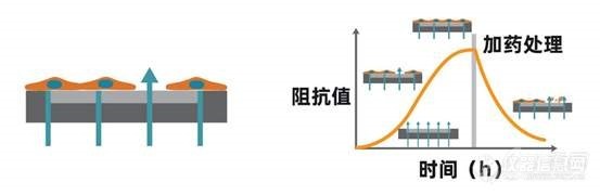
[align=center][font='Segoe UI', sans-serif] -[/font]基于阻抗方法实时、无标记、长期监测细胞表型[/align][align=center]([color=#333333]可用于[/color][b][font=宋体][color=#F76464]细胞增殖、肿瘤免疫、细胞毒性及活力检测、药物筛选、信号通路[/color][/font][font='Segoe UI',sans-serif][color=#F76464](GPCR/CFTR)[/color][/font][font=宋体][color=#F76464]、细胞间相互作用[/color][/font][font='Segoe UI',sans-serif][color=#F76464] ([/color][/font][font=宋体][color=#F76464]屏障功能[/color][/font][font='Segoe UI',sans-serif][color=#F76464])[/color][/font][font=宋体][color=#F76464]、病毒学研究及细胞迁移[/color][/font][/b][color=#333333]等细胞表型研究。[/color])[/align][font=等线][size=16px]细胞表型是涉及基因和蛋白表达的多个细胞过程的集合体,这些过程导致细胞特定的形态和功能。细胞表型检测主要类型有:[b]细胞的增殖、凋亡、迁移、侵袭、活力、信号通路及屏障功能[/b]等。[/size][/font][font=等线][/font][align=left][b][font=宋体][color=#333333]基本原理:[/color][/font][/b][font='Segoe UI',sans-serif][color=black] [/color][/font][font=宋体][color=black]将细胞样本置于[/color][/font][font='Segoe UI',sans-serif][color=black]CytoView-Z[/color][/font][font=宋体][color=black]阻抗板中(底部埋入电极的[/color][/font][font='Segoe UI',sans-serif][color=black]96[/color][/font][font=宋体][color=black]孔培养板)进行培养,当细胞贴附于电极并伸展开后,将微小的电信号施加于电极上,细胞间形成的联接将阻挡这些电信号的通过,导致阻抗值的读数增加,而细胞结构形态上的细微改变(比如源于受体介导的信号传递或细胞形态学变化)也会影响阻抗值。也就是说,细胞的贴壁、黏附、增殖及形变等过程都会引起阻抗的变化,细胞的增殖数量与阻抗呈现一个正相关的关系。[/color][/font][/align][align=left][font=宋体][color=black][img=,553,180]https://ng1.17img.cn/bbsfiles/images/2021/12/202112291044022777_5012_4146479_3.jpg!w553x180.jpg[/img][/color][/font][/align][align=left][font=宋体][color=black]阻抗检测会计算有多少电信号(上图中青色箭头所示)被电极-细胞的界面所阻挡。当电极未被覆盖时,电信号能轻松穿过,这时阻抗值比较低。当细胞盖住电极时,能够通过的电信号就变少了,相应的阻抗值就会增大。当细胞死亡或者脱离电极时,阻抗值就会恢复到基线水平。[/color][/font][/align][font=等线][/font][align=left][font=宋体][color=black]阻抗方法相比于传统的标记方法,具有[/color][/font][/align][align=left][b][font='Segoe UI',sans-serif][color=#F76464]1.[/color][/font][font=宋体][color=#F76464]灵敏度高[/color][/font][/b][/align][align=left][font=宋体][color=black]能够检测出成像技术难以捕捉的、微小的细胞形态、构象变化;[/color][/font][/align][align=left][b][font='Segoe UI',sans-serif][color=#F76464]2.[/color][/font][font=宋体][color=#F76464]长时间持续监测[/color][/font][/b][/align][align=left][font=宋体][color=black]不会错过药物反应时间框,在给药前可通过增殖曲线判断细胞状态;[/color][/font][/align][align=left][b][font='Segoe UI',sans-serif][color=#F76464]3.[/color][/font][font=宋体][color=#F76464]无标记、原位[/color][/font][/b][/align][align=left][font=宋体][color=black]测量过程完全不会影响细胞生物学特性,无需优化抗体用量、染料浓度;[/color][/font][/align][align=left][b][font='Segoe UI',sans-serif][color=#F76464]4.[/color][/font][font=宋体][color=#F76464]孵育时间等参数[/color][/font][/b][/align][align=left][font=宋体][color=black]自动采集数据,中间无需手动操作。[/color][/font][/align][align=left][font=宋体][color=black]目前阻抗平台可用于[/color][/font][b][font=宋体][color=#F76464]细胞增殖、肿瘤免疫、细胞毒性及活力检测、药物筛选、信号通路[/color][/font][font='Segoe UI',sans-serif][color=#F76464](GPCR/CFTR)[/color][/font][font=宋体][color=#F76464]、细胞间相互作用[/color][/font][font='Segoe UI',sans-serif][color=#F76464] ([/color][/font][font=宋体][color=#F76464]屏障功能[/color][/font][font='Segoe UI',sans-serif][color=#F76464])[/color][/font][font=宋体][color=#F76464]、病毒学研究及细胞迁移[/color][/font][/b][font=宋体][color=black]等细胞表型研究。[/color][/font][/align]
【背景】CIK是“Cytokine-Induced Killer Cells”的缩写,中文全称为“细胞因子诱导的杀伤细胞”。 CIK是单个核细胞在CD3单抗和多种细胞因子(包括IFN-g, IL-2等)的作用下培养获得的一群以CD3+CD56+细胞为主要效应细胞的异质细胞群, 其既具有T淋巴细胞强大的抗肿瘤活性,又具有NK细胞(自然杀伤细胞)的非MHC(主要组织相容性抗原)限制性肿瘤杀伤能力。CIK细胞具有杀瘤活性高、杀瘤谱广,对正常组织毒性低,体外可高度扩增等特点,是目前临床上广泛使用的过继性免疫治疗细胞。【培养原理】CIK培养用细胞因子和抗体:nCD3激发型单抗:T细胞活化的第一信号来自于T细胞表面的受体,即T细胞抗原受体(T cell antigen receptor, TCR)与APC提呈的抗原的特异性结合,也就是T细胞对抗原的特异性识别。TCR是由2条不同肽链构成的异二聚体,在T细胞表面,其与CD3分子通过非共价键结合,形成TCR/CD3复合体。TCR识别特异性抗原后会引起CD3和T细胞表面的辅助受体CD4或CD8分子的胞浆尾部聚集,进而激活与胞浆尾部相连的酪氨酸激酶(Lck, Fyn和ZAP-70等),促使CD3分子胞浆区的免疫受体酪氨酸活化基序(immunoreceptor tyrosine-based activation motif, ITAM)中的酪氨酸(Y)磷酸化。磷酸化的酪氨酸(pY)进一步磷酸化下游含酪氨酸的蛋白,从而引起激酶活化的级联反应(磷脂酰肌醇途径或MAP激酶途径等),最终通过激活转录因子,使其进入细胞核内,结合于调控T细胞增殖和活化的靶基因(如IL-2和IFN-g等),引起基因的表达和转录,T细胞因而由静止状态转为增殖和活化状态。由上可见,CD3分子在T细胞活化信号的转导中起着极其关键的作用。CD3激发型单抗与T细胞表面CD3分子特异性结合后,可引起CD3分子胞浆区ITAM基序中酪氨酸的磷酸化,进而导致T细胞增殖和活化的下游信号的激活,从而使T细胞增殖和活化。也就是说,CD3激发型单抗能够模拟抗原与TCR/CD3复合物的识别和激活过程,从而引起T细胞的增殖与活化,因此是CIK细胞培养中不可或缺的刺激因素。此外,CD3激发型单抗在选用时一定要注意克隆号。研究表明,仅克隆号为OKT-3的CD3激发型单抗可以刺激所有人的T细胞的增殖,而其它克隆号的CD3激发型单抗仅能刺激一部分人的T细胞。因此,在进行CIK培养时,最好选用OKT-3克隆,以保证每个患者的T细胞均能被激活。nIL-2 (白细胞介素-2)IL-2最初发现时被称为T细胞生长因子(T cell growth factor, TCGF),是引起T细胞增殖最重要的细胞因子。IL-2既是自分泌细胞因子,也是旁分泌细胞因子,其通过与T细胞表面的IL-2受体(IL-2R)的特异性结合而促使T细胞活化,并进入细胞分裂状态。此外,IL-2还可刺激NK细胞的生长并增强其杀伤能力。因此CIK细胞培养中须添加IL-2,以促进T细胞的增殖与活化。nIFN-g (干扰素-g)IFN-g 具有上调外周血淋巴细胞表面IL-2R表达的作用,因此会增强T细胞对IL-2促增殖反应的敏感度和强度。在诱导CIK细胞形成的过程中加入IFN- g ,可降低IL-2的用量。研究发现,IFN-g加入的顺序与CIK的细胞毒活性密切相关。先加入IFN- g,培养24后再加入IL-2,可明显提高CIK的细胞毒活性。nIL-1a(白细胞介素-1a)IL-1a也可以介导外周血淋巴细胞表面上调表达IL-2R。当IL-1a与IFN-g和激发型CD3单抗合用时,可以明显提高CIK 的细胞毒作用。【细胞制备】1.外周血单个核细胞的采集1.1用血细胞分离机采集患者自身的外周血单个核细胞50-100mL;1.2淋巴细胞分离液密度梯度离心法进一步纯化单个核细胞(PBMC);1.3无血清培养液洗涤2次,获得纯度在90%以上的PBMC。2.CIK细胞的培养及鉴定2.1将PBMC按1-2 x 10⁶/ml的浓度悬浮于无血清培养液中,加入1,000 U/ml 的重组人IFN-g,37oC,5%CO2培养箱中培养;2.224h 后加入50ng/ml 的CD3 单克隆抗体和300 U/ml 的重组人IL-2,刺激CIK 细胞的生长和增殖;注:此时也可同时加入100 U/ml的重组人IL-1a。2.3每3天半量换液或扩瓶一次,并补加重组人IL-2 300 U/ml;2.4在培养的第14d,收获CIK细胞。2.5CIK细胞质控:2.9.1台盼蓝染色检测:活细胞应在80%以上;2.9.2流式细胞仪检测细胞表面CD3、CD8、CD56等分子的表达:CD3+CD56+细胞的比例应在20%以上。2.9.3细胞杀伤实验:以CIK细胞为效应细胞,以肿瘤细胞(可为原代肿瘤细胞或肿瘤细胞株)为靶细胞,将效应细胞与靶细胞按10 : 1(数目比) 的比例加入96 孔U 型板中,每孔含靶细胞1 x 10⁴个,终体积为200 ml,设3个复孔。培养4h,然后取培养上清,用乳酸脱氢酶(LDH) 试剂盒检测效应细胞对靶细胞的杀伤率。2.9.4收获细胞前,取少量培养物进行细菌、真菌培养,并检测支原体、衣原体,及内毒素(标准:病原学检测阴性,内毒素5 Eu)。【步骤简图】http://img.dxycdn.com/trademd/upload/userfiles/image/2013/04/B1366873006_small.jpg 【推荐试剂】http://img.dxycdn.com/trademd/upload/userfiles/image/2013/04/B1366873008_small.jpg 注:Animal Free意为无动物成分。无动物成分的重组细胞因子在生产过程中不会有任何动物源性物质,尤其是牛蛋白的混入,使得最终获得的重组人蛋白中不含任何动物成分。这样可避免动物病原体(如疯牛病,克雅氏病等)的污染及外源蛋白引起的机体异种排斥和过敏反应,因此细胞治疗的体外细胞培养过程中最好使用无动物成分的重组细胞因子。【其它相关试剂】 http://img.dxycdn.com/trademd/upload/userfiles/image/2013/04/B1366873009_small.jpg【参考文献】 Li R, Wang C, et al. Autologous cytokine-induced killer cell immunotherapy in lung cancer: a phase II clinical study. Cancer Immunol Immunother. 2012; 61:2125-2133
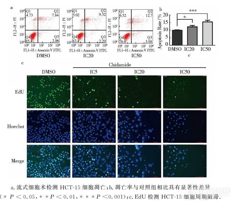
荧光显微镜及流式表征西达本胺诱导细胞凋亡并阻滞细胞周期流式细胞术检测到明显的细胞凋亡,随着加药浓度的升高,细胞凋亡数量增多,早期凋亡细胞、晚期凋亡细胞和坏死细胞 的 数 量 都 随 之 上 升 (图 a).测 得 实 验 组 凋 亡 率 分 别 为 12.32% ±0.84% (P 0.05),15.63%±0.91%(P0.001),与对照组相比,有统计学意义(图b).与此同时通过 EdU 实验检测(图c)其细胞周期的变化,随着加药浓度的增高,Hoechst蓝色荧光染色细胞数目减少,即活细胞数减少,药物对细胞杀伤作用显著 EdU 绿色荧光染色细胞数减少,即进入 DNA 复制期的细胞数量减少.表明西达本胺可以明显促进 HCT-15细胞凋亡、抑制其增殖且阻滞细胞周期.[img]https://ng1.17img.cn/bbsfiles/images/2023/06/202306302205203559_379_5389809_3.png[/img]
来自著名的美国密苏里州斯托瓦斯医学研究所(Stowers Institute for Medical Research),中科院生物物理研究所传染病与免疫学中心,堪萨斯大学医学院,中西大学(Midwestern University)的研究人员揭示了干细胞衰老的奥秘,这一发表在昨天刚刚出版的《Cell Stem Cell》杂志上的文章由中科院海外评审专家解廷(斯托瓦斯医学研究所)领导完成,第一作者是斯托瓦斯医学研究所与中科院生物物理研究所联合培养的博士生潘磊(Lei Pan,音译)。目前普遍认为人类组织衰老与干细胞活性下降和数目减少有关,这些变化在许多譬如皮肤皱纹和器官功能下降等的衰老表现中起着重要的作用。至今为止对于干细胞衰老调控的理解还比较少,但是解廷实验室已经证明了干细胞功能中年龄依赖性得下降有关的特殊因素,以及这些因素的微环境:niche。潘表示,“在这项研究中,我们利用果蝇卵巢生殖干细胞(germline stem cells,GSCs)作为研究模型,证明干细胞功能中年龄依赖性的下降和其niche在干细胞整个衰老过程中扮演着十分重要的角色”,“我们检测了干细胞衰老调控的三个因素,发现并证明衰老过程是受到外在和内在因素调控的”。研究小组首先聚焦在一个称为骨形态发生蛋白(bone morphogenic protein, BMP)的蛋白家族——其在许多组织的发育过程中扮演着重要的角色,他们发现当niche微环境的BMP信号活性随着年龄下降的时候,干细胞增值的能力也会随之降低,干细胞数量也减少了。相反当BMP信号增加,干细胞的寿命以及增值能力也都有所提升。其次研究人员也发现干细胞与niche之间的关联也起到一定作用:强的关联可以延长干细胞的寿命,而降低关联则会增加干细胞衰老。这篇研究报告最后强调了GSCs或者niche中的一个酶(减少自由氧)的过量表达如何延长干细胞的寿命,以及增加干细胞增值的能力。解廷认为,“对成人组织中由于干细胞功能下降导致细胞损耗的长期无效替换也许是人类衰老的一个主要原因”,“如果我们能了解如何通过操纵干细胞和/或niche的功能,来减缓干细胞衰老,我们也许就能够减缓人类衰老,治疗年龄相关性的推行性疾病”。
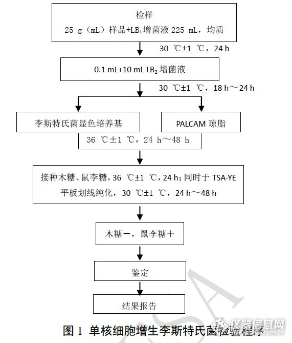
2014年食源性致病菌单核细胞增生李斯特菌风险监测分析单核细胞增生李斯特菌被看作是人畜共患和经食物传播的病原菌只有20多年的历史,由于它能在低温条件下生长,且患者死亡率高达20%-30%,因而受到国际卫生和食品组织以及各国政府的高度重视,多年来它一直是研究胞内致病机制的较好模型。单核细胞增生李斯特菌是通过受污染的食品,如奶酪、牛奶、午餐肉和法兰克福香肠而感染人类。尽管采取了许多预防措施,但近年来欧美等国家还是暴发了多起因单核细胞增生李斯特菌食源性污染导致的人群感染事件。为防范由单核细胞增生李斯特菌引起的食物中毒事件在这里发生,我们2014年共监测了5 类食品180份样品,现将监测结果报道如下。1材料与方法1.1试剂和仪器 使用的培养基有李氏增菌肉汤LB(LB1,LB2)、含0.6%酵母浸膏的胰酪胨大豆琼脂(TSA-YE)、1%盐酸吖啶黄(acriflavine HCl)溶液、1%萘啶酮酸钠盐(naladixic acid)溶液、PALCAM琼脂购买于北京路桥公司。主要仪器包括均质器,电子天平:感量0.1g,VITEK-2 Compact 全自动微生物鉴定系统。1.2依据和方法 根据国家食品安全风险评估中心制定的“食源性致病菌监测工作手册”要求进行增菌、分离、鉴定、菌种保存及送上级实验室复核。试验程序见图1。 http://ng1.17img.cn/bbsfiles/images/2014/12/201412062118_526074_2433088_3.jpg
概念:细胞克隆形成率即细胞接种存活率,表示接种细胞后贴壁的细胞成活并形成克隆的数量。贴壁后的细胞不一定每个都能增殖和形成克隆,而形成克隆的细胞必为贴壁和有增殖活力的细胞。克隆形成率反映细胞群体依赖性和增殖能力两个重要性状。基本步骤:1、取对数生长期的各组细胞,分别用0.25%胰蛋白酶消化并吹打成单个细胞,并把细胞悬浮在10%胎牛血清的DMEM培养液中备用。2、将细胞悬液作梯度倍数稀释,每组细胞分别以每皿50、100、200个细胞的梯度密度分别接种含10mL 37℃预温培养液的皿中,并轻轻转动,使细胞分散均匀。置37℃ 5% CO2及饱和湿度的细胞培养箱中培养2~3周。3、经常观察,当培养皿中出现肉眼可见的克隆时,终止培养。弃去上清液,用PBS小心浸洗2次。加4%多聚甲醛固定细胞5mL固定15分钟。然后去固定液,加适量GIMSA应用染色液染10~30分钟,然后用流水缓慢洗去染色液,空气干燥。4、将平皿倒置并叠加一张带网格的透明胶片,用肉眼直接计数克隆,或在显微镜(低倍镜)计数大于10个细胞的克隆数。最后计算克隆形成率。克隆形成率 =(克隆数/接种细胞数)×100%平板克隆形成试验方法简单,适用于贴壁生长的细胞。适宜底物为玻璃的、塑料瓶皿。试验成功的关键是细胞悬液的制备和接种密度。细胞一定要分散得好,不能有细胞团,接种密度不能过大。
[align=center][font='times new roman'][size=16px]超速离心法[/size][/font][font='times new roman'][size=16px]富集[/size][/font][font='times new roman'][size=16px]细胞分泌的外[/size][/font][font='times new roman'][size=16px]泌[/size][/font][font='times new roman'][size=16px]体[/size][/font][/align][align=left][font='times new roman'][size=16px]引言[/size][/font][/align]本章利用超速离心法富集细胞分泌的外泌体。采用透射电子显微镜,Western Blot实验,纳米颗粒示踪分析等表征了外泌体的形态结构及分布特征。最后,用增殖实验和划痕实验测试了富集得到的外泌体的生物完整性[font='宋体']。[/font][align=left][font='times new roman'][size=16px]细胞分泌的[/size][/font][font='times new roman'][size=16px]外[/size][/font][font='times new roman'][size=16px]泌[/size][/font][font='times new roman'][size=16px]体表征[/size][/font][/align][img]" style="max-width: 100% max-height: 100% [/img]利用透射电子显微镜对超速离心法得到的外泌体进行了表征。如图(a)所示,洗脱后的外泌体具有典型的杯状结构。纳米颗粒示踪分析技术显示富集的外泌体粒径分布较窄,平均粒径为115.3 nm(图 b)。Western blot方法验证了富集方法的有效性。如图(c)所示,经超速离心法富集后,典型的低丰度外泌体标记物HSC70、TSG101、CD63和CD9可得到有效检测。以上结果证明,基于超速离心法对外泌体的富集可行性。[align=center][font='黑体'][size=14px]图[/size][/font][font='黑体'][size=14px] (a)重悬液中外[/size][/font][font='黑体'][size=14px]泌[/size][/font][font='黑体'][size=14px]体的透射电子显微镜表征,(b)[/size][/font][font='黑体'][size=14px]纳米颗粒示踪分析技术[/size][/font][font='黑体'][size=14px]表征外[/size][/font][font='黑体'][size=14px]泌[/size][/font][font='黑体'][size=14px]体的尺寸分布,(c)[/size][/font][font='黑体'][size=14px]Western blot方法[/size][/font][font='黑体'][size=14px]表征[/size][/font][font='黑体'][size=14px]外[/size][/font][font='黑体'][size=14px]泌[/size][/font][font='黑体'][size=14px]体标记物HSC70、TSG101、CD63和CD9[/size][/font][font='黑体'][size=14px]蛋白条带[/size][/font][/align][align=left][font='times new roman'][size=16px][color=#000000] [/color][/size][/font][font='times new roman'][size=16px][color=#000000] [/color][/size][/font][font='times new roman'][size=16px]完整性验证[/size][/font][/align][img]" style="max-width: 100% max-height: 100% [/img]首先用细胞增殖用于评估超速离心法分离的外泌体的生物完整性。不同浓度(10[font='times new roman'][sup][size=16px]6[/size][/sup][/font]、10[font='times new roman'][sup][size=16px]8[/size][/sup][/font]、10[font='times new roman'][sup][size=16px]10[/size][/sup][/font]颗粒/孔)的H1299细胞外泌体分别与H1299细胞共培养24、48和72小时。如图(a)所示,H1299细胞来源的外泌体以剂量依赖性方式显著促进细胞增殖。此外,伤口愈合实验显示,H1299细胞来源的外泌体加速了伤口愈合速度,并以剂量和时间依赖的方式提高了伤口愈合质量(图 b)。上述结果表明,通过超速离心法富集的外泌体具有完整性,可以促进细胞增殖、分化和迁移。[align=center][font='黑体'][size=14px]图[/size][/font][font='黑体'][size=14px](a)H[/size][/font][font='黑体'][size=14px]1299[/size][/font][font='黑体'][size=14px]细胞来源的外[/size][/font][font='黑体'][size=14px]泌[/size][/font][font='黑体'][size=14px]体对细胞增殖的影响结果,(b)[/size][/font][font='黑体'][size=14px]H1299细胞来源的外[/size][/font][font='黑体'][size=14px]泌[/size][/font][font='黑体'][size=14px]体对细胞[/size][/font][font='黑体'][size=14px]划后伤口愈合[/size][/font][font='黑体'][size=14px]的影响结果[/size][/font][/align][align=left][font='times new roman'][size=16px]小结[/size][/font][/align]本章利用超速离心法富集了细胞来源的外泌体,并对富集得到的外泌体进行了一系列表征表征。通过透射电子显微镜和纳米颗粒示踪分析技术分析表明得到的外泌体具有典型的杯状结构和小的粒径分布,证明了富集方法的可行性。Western Blot结果表明,富集后得到的外泌体溶液经裂解后,特征蛋白HSC70、CD63、TSG101和CD81,其含量比富集前显著提高。利用细胞增殖实验和划痕实验证明超速离心法富集得到的外泌体具有良好的生物学活性,可应用于下游的实验研究。
广州中山大学研究发现天然病毒M1可杀灭癌细胞中新网广州10月13日电(许青青 蔡珊珊) 记者13日从广州中山大学获悉,该校中山医学院颜光美教授课题组研究发现天然病毒M1能选择性地感染并杀伤包括肝癌、结直肠癌、膀胱癌、黑色素瘤在内的多种体外培养的癌细胞,而对正常细胞无毒副作用。全球癌症发病率呈现快速增长态势,现有的治疗手段远远未能满足临床需求。颜光美教授课题组发现,M1病毒是一种从中国海南岛分离得到的天然病毒,能选择性地感染并杀伤包括肝癌、结直肠癌、膀胱癌、黑色素瘤在内的多种体外培养的癌细胞,而对正常细胞无毒副作用。整体动物实验表明,经尾静脉注射的M1病毒能显著富集在肿瘤组织并抑制肿瘤生长,正常器官则不受影响。除细胞水平及动物实验之外,课题组还使用临床标本离体活组织培养模型进一步证实了上述新型溶瘤病毒的有效性和特异性。据悉,该研究成果对阐明新型天然溶瘤病毒M1选择性杀伤肿瘤细胞的机制和研发新型靶向抗肿瘤药物都具有重要意义。相关资料:癌细胞增殖方式癌细胞是一种变异的细胞,是产生癌症的病源,癌细胞与正常细胞不同,有无限生长、转化和转移三大特点,也因此难以消灭。癌细胞由“叛变”的正常细胞衍生而来,经过很多年才长成肿瘤。“叛变”细胞脱离正轨,自行设定增殖速度,累积到10亿个以上我们才会察觉。癌细胞的增殖速度用倍增时间计算,1个变2个,2个变4个,以此类推。1912年8月13日,法国医生发现癌细胞。
定义:单细胞研究,就是针对单个细胞的研究,这是相对于群体细胞的研究。研究意义:细胞是生命活动的基本单位,研究细胞的结构功能及行为,有利于揭示复杂生命体的生命活动规律,探究生理生化现象,获得统计平均结果。然而,现代研究表明,单个细胞内的成分存在巨大差异,平均分析结果不能反映单个细胞内成分的真实情况,会带来误导信息。癌症等疾病总是从个别细胞的变异开始,极少量异常细胞信号会被群体信号所掩盖,不能及时获得有关病变的信息。另外,细胞间的信号传导,应激反应等活动在细胞内迅速发生,传统方法无法做到实时监测。对于数量较少且较为珍贵的细胞样本,如干细胞、元祖细胞及患者样本,传统分析方法需要大量的细胞样本,并不适宜。关于物质在细胞内的空间分布,亚细胞结构如细胞器的分析,传统方法也不能满足。这些都要求我们在一定范围内从单细胞水平研究细胞的生命活动。单细胞分析方法:毛细管电泳、微流控芯片、图像分析、动力学分析及纳米技术等。目前单细胞分析存在的难点:首先无论是针对一个特异性大分子,还是在OMIC水平上进行分子分析,都存在单细胞提取物数量少,难以分析的困难,这甚至可以说是不可能完成的,因此增加灵敏度势在必行。除此之外高通量分析也是一个瓶颈,要想获得单细胞分析确切的分析结果,研究人员必须快速而准确的分析多个细胞,这并不容易。另外单细胞分析也常常需要进行多种方式分析,这不仅是由于细胞存在于一种异质性环境汇总,而且也在同一时间,也需要测量多个参数。
骨髓间充质干细胞总结 2003年8月,为给大家提供一个网上交流干细胞研究经验的平台,我们干细胞版设立了骨髓间充质干细胞培养讨论区,经过近三个月的讨论学习,我们既学习丰富了自己的知识体系,也对间充质干细胞尤其是分离培养方面有了更为详实的认识,为了大家阅读的方便,我们决定把本版中的相关内容,同时参考部分书目和文献,做一总结。一、骨髓间充质干细胞的分离 目前常用的分离MSC的方法有全骨髓法和密度梯度离心法,全骨髓法即根据干细胞贴壁特性,定期换液除去不贴壁细胞,从而达到纯化MSC的目的。密度梯度离心法即根据骨髓中细胞成分比重的不同,提取单核细胞进行贴壁培养。随着对MSC表面抗原认识的深入,有人利用免疫方法如流式细胞仪法、免疫磁珠法等对其进行分离纯化,但经过流式或磁珠分选后的细胞出现了增殖缓慢等一些问题,加之耗费较大和技术的难度,在某种程度上限制了这些方法的广泛应用。1. 直接培养法(全骨髓培养法) 1987年,Friedenstein等发现在塑料培养皿中培养的贴壁的骨髓单个核细胞在一定条件下可分化为成骨细胞、成软骨细胞、脂肪细胞和成肌细胞,而且这些细胞扩增20-30代后仍能保持其多向分化潜能,这类细胞即为骨髓间充质干细胞(BMSC),其工作对今后MSC的研究具有重要意义,不仅证实了骨髓MSC的存在,而且创建了一种体外分离和培养MSC的简便可行的方法,得到了广泛的应用。culture-spirit采用直接贴壁法,24-36小时首次换液,换液时用PBS洗两次,7-10天传第一代,以后2-3天传代。培养基采用Hyclone的DMEM/F-12(1:1),血清是天津TBD的FBS(顶级),得到了较好的培养结果。布兰卡根据自己培养大鼠MSC的经验,详细介绍了实验步骤:(1)接种后60-80分钟,换液去除悬浮细胞(2)原代培养24 h,48 h各换液一次(3)观察细胞情况,在原代培养7天左右时,如观察到成片的典型形态的细胞,在瓶底用Marker笔标记,0.25%胰酶消化,镜下观察控制,约5-10分钟(室温太低时应放置到孵箱中),加入全培养基终止消化,瓶体朝上,吸管轻轻吹打4-8分钟,尤其是标记部位。不要用力吹打,以免把贴壁较牢的成纤维细胞,上皮样细胞吹打下来。(4)传代到新瓶中,加入少量培养基,孵箱静置20-30分钟后,MSC大多牢固贴壁。瓶底朝上,轻轻吹打,丢弃悬浮以及贴壁不牢的细胞(大多是上皮样细胞),加入全培养基开始传代培养,如观察仍有较多杂细胞,可重复上述步骤。(5)经上述处理后,原代的那瓶细胞仍有一些MSC生长,可继续按原代培养,如观察到MSC的克隆,仍可按上述步骤纯化处理。(6)原代或传代的细胞如观察的少量成片的杂细胞,可直接镜下瓶底标记后,超净台里用长吸管尖端机械刮除,吸出去掉。菊花与刀用的是全骨髓培养法,直接用含10%的FBS培养基冲洗大鼠的股骨和胫骨,为了避免冲起许多气泡应缓慢冲,冲的次数不应太多。冲洗后不用离心直接接种在培养瓶里,48 h~72 h后首次换液,一般7~10天可传代。天之饺子介绍的小鼠MSC的分离方法:取6 w小鼠的股骨和胫骨,直接用含培养基冲出骨髓,一定要尽量把干垢端的骨髓冲干净。冲洗后不离心直接接种在培养瓶里,24-48 h后去悬浮,再接下来的每3-4天换液一次,直到需要传代。2. 密度梯度离心法 裴雪涛等用比重为1.073 g/ml的percoll分离(400 g×20 min)人骨髓MSCs,取界面处细胞层,离心后洗涤以2×10⁵/cm2的密度接种,72 h后更换培养液,弃掉未贴壁细胞,以后每3 d换液一次。细胞长到80%汇合时1:1传代。菊花与刀利用PERCOLL密度1.073分离大鼠MSC 时,用2400 rpm×20 mins后可见中间有一层约1~2 mm厚的白色层,仔细用吸管吸取这一层再用PBS离心2遍即可加培养基和胎牛血清培养即rMSCs。周进明等利用密度为1.082的percoll分离小鼠MSCs,500g×30min离心后,取中间的单个核细胞层,PBS洗两次,接种于IMDM培养基,1 d后换液,去掉非贴壁细胞,以后每3-4天换液。jetter用过FICOLL,FERCOLL,上海二分厂的淋巴细胞分离液分离MSC细胞效果都不错,当然所获细胞群的纯度不一,Percoll最纯,而上海二分厂的淋巴细胞分离液所获细胞群的传代能力优秀(35 PASSAGE)。本版的部分园友认为MSC贴壁培养得到的细胞不均一,但是多能分化能力和增殖力好,percoll分离得到的细胞较为均一,多能分化性和增殖力不如贴壁培养的,尤其是增殖力相差很远,有人添加bFGF或/和表皮生长因子发现可以增强增殖能力。二、骨髓间充质干细胞的培养 天之饺子认为,间质干细胞的培养一定要用塑料培养瓶,不能用玻璃的。因为象间质干这类的基质细胞不易贴玻璃,而且现在买的进口好品牌的培养瓶都涂有一层促细胞贴壁的物质,多数园友培养时都添加10-15%胎牛血清。分离培养结果的差异可能是由于各个研究小组标本来源、采用的分离方法不同从而所获得的细胞不同,或者用来检测的细胞代数不同,或者培养过程中用的胎牛血清不同,导致MSCs获得或失去这些表面标记物的表达。三、骨髓间充质干细胞的特性 体外培养的MSC体积小,成梭形,核浆比大。不表达分化相关的细胞标志,如I、II、III型胶原、碱性磷酸酶或Osteopontin;也不表达SH2、SH3、CD29、CD44、CD71、CD90、CD106、CD120a、CD124、CD166和多种表面蛋白,这群细胞特性稳定,扩增一代和两代后的细胞同质性分别达到95%和98%。MSC联系传代培养和冷冻保存后仍能具有多向分化潜能,而且保持正常的核型核端粒酶活性,但不易自发分化,在体外特定的诱导条件下,MSC可以分化为骨、软骨、脂肪、肌腱、肌肉、神经等多种细胞。四、其他相关内容 Jiang将从成人以及成年大鼠和小鼠骨髓分离的间充质干细胞(CD45-TER119-)命名为多潜能成年祖细胞,他们证明,MAPC高表达端粒酶,而且随着细胞的扩增,端粒的长度不变,单个MAPC来源的细胞群不仅能在体外向3个胚层的细胞分化,而且能在体内能够向各种组织细胞分化,相比较而言,形态与MSC相似的体外培养的皮肤成纤维细胞则不具有类似的分化潜能。参考文献 生理学报2003.55(2):153-159 人骨髓间充质干细胞在成年大鼠脑内的迁移及分化中华放射医学与防护杂志.2002,22(3).-167-169 培养小鼠骨髓间充质干细胞及其移植后在体内的定位分布Exp Biol Med Vol. 226:507 520, 2001 Mesenchymal Stem CellsNATURE |VOL 418 | 4 JULY 2002 Pluripotency of mesenchymal stem cells derived from adult marrow干细胞生物学 裴雪涛 主编
西达本胺通过信号通路调节促进癌细胞凋亡在我国,西达本胺已获批作为PTCL临床用药。西达本胺属于苯酰胺类化合物,是我国自主研发的首个亚型选择性口服HDACI,国家食品药品监督管理局已批准其用于临床试验,其选择性抑制I类HDAC1、2、3亚型和II类HDAC10亚型,可抑制肿瘤细胞增殖、促进凋亡,阻滞周期、引发DNA损伤,还可以增强抗肿瘤免疫反应。与其他抗肿瘤药物相比,西达本胺疗效好、选择性高、不良反应少。西达本胺可激活死亡受体途径和线粒体凋亡途径诱导细胞凋亡,其中最为主要的是线粒体凋亡途径,该途径受Bcl-2家族介导的细胞色素C释放通路调控。抗凋亡蛋白Bcl-2表达受到抑制,促凋亡蛋白Bax表达上调,使线粒体膜电位降低,细胞色素C释放到细胞质中,Caspase途径被激活,细胞发生凋亡。例如:西达本胺增强B淋巴瘤细胞组蛋白H3、H4 乙酰化水平,使线粒体膜电位降低随后激活Caspase 3,促进细胞凋亡;在肾癌中,它可以下调Bcl-2表达,上调Bax表达,随着药物浓度增加引起786-O 细胞凋亡。西达本胺可以调控ROS水平。HDACI可以上调ROS水平,导致DNA双链损伤。研究证明,西达本胺作用于白血病细胞后,诱导细胞内ROS产生,细胞凋亡增加[17]。此外,在胰腺癌细胞系中,西达本胺明显增强细胞内ROS的产生,上调γH2AX(DNA双链断裂的标志物)表达水平,诱发细胞DNA损伤。西达本胺通过调控细胞周期蛋白(Cyclin)、细胞周期蛋白依赖性激酶(Cyclin-dependent kinases,CDKs)以及细胞周期蛋白依赖性激酶抑制剂(Cyclin-dependent kinases inhibition,CDKI)的表达阻滞细胞周期。例如,西达本胺使MM细胞系P21、P27的表达量增高,CDK4、CDK6、Cyclin D2表达量下降,阻滞MM细胞系于G1期[19]。在NK/T细胞淋巴瘤中,西达本胺上调P21表达,下调Cyclin E表达,诱导细胞发生G0/G1期阻滞,从而抑制细胞的增殖。
【西班牙《国家报》1 0月26日报道】巴塞罗那瓦尔德希伯伦大学附属医院的一个研究小组日前宣布,确认了癌细胞的一种基因物质能够使恶性肿瘤不老化并失控地增殖。 玛蒂尔德·列奥纳特医生领导的这项研究成果刊登在《医学研究评论》杂志上。研究报告指出,小核糖核酸(m icroARNs)对细胞不死发挥了突出的作用。 核糖核酸在蛋白质的生成过程中起到重要作用,但此前对小核糖核酸的功用实际上并不了解。最新研究表明,小核糖核酸与另一种遗传物质之间相互作用,阻断或激活细胞的增殖过程。瓦尔德希伯伦大学的研究小组已经确认了28种能使癌细胞继续增殖的小核糖核酸。研究人员认为,对这些小核糖核酸采取行动会为阻止甚至消除癌细胞的这种特性打开大门,是在抗击癌症方面的一大显著进步。癌细胞会不停地分裂和增殖,而健康细胞能够进行40-60次分裂,然后就停止分裂,最终死亡。 癌细胞的有害之处正是其不老化的能力,这种能力使其成为一种不死组织,因为它在不会自我消灭的同时还毫无控制地成倍增加。新研究成果确认了造成这种不死特性的小核糖核酸,便于今后研究出一种能够阻断这种能力,让癌细胞像健康细胞一样老化和自我毁灭的机制。 (来源: 新华国际)


 我要推广仪器
 下载APP
下载APP