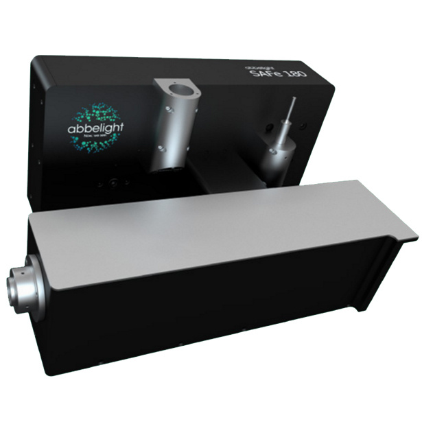
大视野单分子超分辨模块-SAFe 180 SAFe 180是法国abbelight公司推出的一款基于单分子定位技术的显微成像(SMLM)的超分辨模块。该设备具有高度灵活性,能够搭载在大多数的倒置显微镜上,并且仅仅需要使用一个C-mount(CCD或CMOS所连接的部位)接口,即可将您的倒置显微镜直接升为超分辨成像系统。并且改造过程不会破坏原有显微镜系统的光路和功能,不会与其它的显微镜改造相冲突。本设备既在配置上的选择也十分灵活。它既可以作为显微镜的一个升配件来改造您的显微镜,也拥有完整的超分辨系统。让用户在获得专业的图像质量的同时,获得经济合理的超分辨升方案。大视野单分子超分辨模块-SAFe 180 设备参数 + 模块化系统:可接到大多数倒置荧光显微镜+ 成像模式:PALM、STORM、smFRET、PAINT、SPT+ 光源模式:Epi、TIRF、HILO+ 超高分辨率:25 nm的XY轴分辨率,50nm的Z轴分辨率+ 超大视野:180 × 180 μm2的视野+ 全自动化控制+ 无需高功率激光光源加装TIRFPALMSTORMSPTsmFRET...... 兼容ConfocalSpinning-DeskWidefieldSIMSTED 测试数据 超大的视野神经元伪足小体模式生物微管蛋白 配套试剂 Smart kitCompatible dyes• 10 doses per box• 200 μL per dose• 30 sec prepartion• 2 months in a fridge• 2 weeks on sample• Atto 488, WGA-AF488• AF532, CF532, Cy3b• AF555, AF594, CF555, AF568, CF568, Cy5, MemBriteTM 568, TMR• AF647, CF647, AF680, CF680, MemBriteTM 640, Actin-stain 670, SiR647 发表文献列表 [1] Cabriel, Clément, et al. "Combining 3D single molecule localization strategies for reproducible bioimaging." Nature communications 10.1 (2019): 1980.[2] Woodhams, Stephen G., et al. "Cell type–specific super-resolution imaging reveals an increase in calcium-permeable AMPA receptors at spinal peptidergic terminals as an anatomical correlate of inflammatory pain." Pain 160.11 (2019): 2641-2650.[3] Belkahla, Hanen, et al. "Carbon dots, a powerful non-toxic support for bioimaging by fluorescence nanoscopy and eradication of bacteria by photothermia." Nanoscale Advances (2019).[4] Denis, Kevin, et al. "Targeting Type IV pili as an antivirulence strategy against invasive meningococcal disease." Nature microbiology 4.6 (2019): 972.[5] Szabo, Quentin, et al. "TADs are 3D structural units of higher-order chromosome organization in Drosophila." Science advances 4.2 (2018): eaar8082. [6] Boudjemaa, Rym, et al. "Impact of bacterial membrane fatty acid composition on the failure of daptomycin to kill Staphylococcus aureus." Antimicrobial agents and chemotherapy 62.7 (2018): e00023-18.[7] Culley, Sian, et al. "Quantitative mapping and minimization of super-resolution optical imaging artifacts." Nature methods 15.4 (2018): 263.[8] Berger, Stephen L., et al. "Localized myosin II activity regulates assembly and plasticity of the axon initial segment." Neuron 97.3 (2018): 555-570.[9] Cabriel, Clément, et al. "Aberration-accounting calibration for 3D single-molecule localization microscopy." Optics letters 43.2 (2018): 174-177. [10] Bouissou, Ana?s, et al. "Podosome force generation machinery: a local balance between protrusion at the core and traction at the ring." ACS nano 11.4 (2017): 4028-4040. [11] Sellés, Julien, et al. "Nuclear pore complex plasticity during developmental process as revealed by super-resolution microscopy." Scientific reports 7.1 (2017): 14732.[12] Bourg, Nicolas, et al. "Direct optical nanoscopy with axially localized detection." Nature Photonics 9.9 (2015): 587. 用户单位
 留言咨询
留言咨询
 400-860-5168转4170
 留言咨询
留言咨询
 400-860-5168转4170
 留言咨询
留言咨询
 400-803-3696
 留言咨询
留言咨询
 400-803-3696
 留言咨询
留言咨询
 400-803-3696
 留言咨询
留言咨询
 400-860-5168转2831
 留言咨询
留言咨询
 400-860-5168转0980
 留言咨询
留言咨询
 400-803-3696
 留言咨询
留言咨询

 留言咨询
留言咨询
 400-803-3696
 留言咨询
留言咨询

 留言咨询
留言咨询
 400-803-3696
 留言咨询
留言咨询
 400-803-3696
 留言咨询
留言咨询
 400-803-3696
 留言咨询
留言咨询
 400-803-3696
 留言咨询
留言咨询

 留言咨询
留言咨询

 留言咨询
留言咨询
 400-803-3696
 留言咨询
留言咨询
 400-803-3696
 留言咨询
留言咨询
 留言咨询
留言咨询

 400-803-3696
 留言咨询
留言咨询
 400-803-3696
 留言咨询
留言咨询
 400-803-3696
 留言咨询
留言咨询
 400-860-5168转3714
 留言咨询
留言咨询

 400-860-5168转5919
 留言咨询
留言咨询
 400-860-5168转3993
 留言咨询
留言咨询
 400-877-0075
 留言咨询
留言咨询
 留言咨询
留言咨询
 400-860-5168转3648
 留言咨询
留言咨询

 400-860-5168转1766
 留言咨询
留言咨询