阜外医院周洲团队Cell子刊:提出心脏流出道血管平滑肌细胞发育新模式
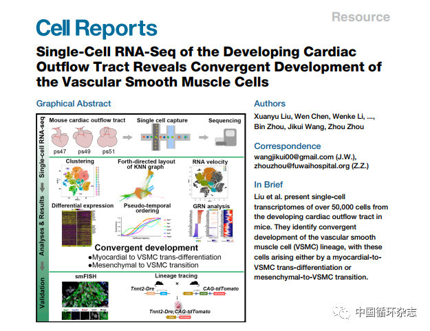
p style="text-align: justify text-indent: 2em line-height: 1.5em "span style="font-family: 楷体, 楷体_GB2312, SimKai "近日,中国医学科学院阜外医院周洲教授团队在Cell Reports发表的最新研究首次发现并证实了,心脏流出道发育过程中存在心肌细胞向血管平滑肌细胞的转分化(trans-differentiation)。/span/pp style="text-align: justify text-indent: 2em line-height: 1.5em "strong研究者就此提出了大动脉基部平滑肌汇聚发育(convergent development)的概念。/strong/pp style="text-align: justify text-indent: 2em line-height: 1.5em "其中,阜外医院实验诊断中心刘宣雨博士为论文第一作者,新乡医学院王计奎教授为共同通讯作者。/pp style="text-align: center"img style="max-width:100% max-height:100% " src="https://img1.17img.cn/17img/images/201908/uepic/5d0f19bf-17d9-40e8-be8b-89e6ba1b60f5.jpg" title="001.jpg" alt="001.jpg"//pp style="text-align: justify text-indent: 2em line-height: 1.5em "心脏流出道是先心病发病的热点部位,在这项研究中,研究者分析了来自小鼠流出道的3个连续发育阶段的共50,000多个细胞的单细胞转录组,同时结合单分子荧光原位杂交和基于Dre-Rox的谱系追踪技术进行了分析和验证。/pp style="text-align: justify text-indent: 2em line-height: 1.5em "研究发现,心脏流出道发育过程中涉及到6种细胞类型,共17个细胞亚群,研究者通过机器学习分类模型,为各种细胞类型及其亚群定义了分子特征(图1)。/pp style="text-align: center"img style="max-width:100% max-height:100% " src="https://img1.17img.cn/17img/images/201908/uepic/a1081aed-4d3e-411a-9b23-8ae01464bc9a.jpg" title="002.jpg" alt="002.jpg"//ppspan style="text-align: justify text-indent: 0em color: rgb(0, 112, 192) "注:A:17个细胞亚群;B:各种细胞类型及其比例;C:各种细胞类型及其亚群的分子特征/span/pp style="text-align: center line-height: 1.5em text-indent: 0em "span style="color: rgb(0, 112, 192) "图1 心脏流出道发育过程中的细胞亚群及其分子特征/span/pp/pp style="text-align: justify text-indent: 2em line-height: 1.5em " /pp style="text-align: justify text-indent: 2em line-height: 1.5em "为了分析细胞亚群间关系,研究者通过力导向的KNN图布局(force-directed layout of k-nearest-neighbor graph)在二维空间内更加准确反映数据结构(图2A),同时分析细胞状态随时间的变化动态(图2B)和细胞亚群的特异表达谱,发现了与平滑肌分化直接相关的细胞亚群(图2C)。/pp style="text-align: justify text-indent: 2em line-height: 1.5em "有趣的是,除了间充质细胞向平滑肌细胞转化外,一个可能向平滑肌细胞发生了转分化的心肌细胞中间态亚群c9“浮出水面”,研究者推测,流出道的平滑肌细胞可能存在汇聚发育模式。/pp style="text-align: center"img style="max-width:100% max-height:100% " src="https://img1.17img.cn/17img/images/201908/uepic/be4c2ffb-cb00-4c92-a03e-d1068157f53b.jpg" title="003.jpg" alt="003.jpg"//pp style="text-align: center "span style="text-align: justify text-indent: 2em color: rgb(0, 112, 192) "图2 心脏流出道平滑肌的汇聚发育模式/span/pp style="text-align: justify text-indent: 2em line-height: 1.5em "研究者进一步通过拟时间排序分析发现,在心肌向平滑肌转分化过程中,随着分化的进行,细胞的心肌标记的表达下调,平滑肌标记的表达上调(图3A)。/pp style="text-align: justify text-indent: 2em line-height: 1.5em "值得注意的是,Notch信号途径中的基因随着分化的进行逐渐上调(图3B)。通过基因调控网络打分分析,最终揭示出了心肌细胞向平滑肌细胞转分化过程中的关键转录因子(图3C),如Notch信号通路(已知在流出道的发育中扮演重要角色)的下游转录因子Heyl。/pp style="text-align: center"img style="max-width:100% max-height:100% " src="https://img1.17img.cn/17img/images/201908/uepic/9467d7f6-8f0b-4a78-9e53-39e72c304313.jpg" title="004.jpg" alt="004.jpg"//pp/pp style="text-align: center line-height: 1.5em text-indent: 0em "span style="color: rgb(0, 112, 192) font-size: 14px "图3 拟时间排序和基因调控网络分析揭示出心肌细胞向平滑肌细胞转分化过程中的关键转录因子/span/pp style="text-align: justify text-indent: 2em line-height: 1.5em "单分子荧光原位杂交结果显示,从近端到远端的流出道连续横截面可以观察到从心肌表型向平滑肌表型的过渡(图4A)。细胞共表达心肌的标记基因Myh7和流出道平滑肌的标记基因Cxcl12,为心肌细胞向平滑肌细胞转分化的存在提供了支持(图4B)。/pp style="text-align: center"img style="max-width:100% max-height:100% " src="https://img1.17img.cn/17img/images/201908/uepic/a1ccbd68-ea7d-4db2-ae6c-07344f4ead97.jpg" title="005.jpg" alt="005.jpg"/span style="color: rgb(0, 112, 192) text-align: justify text-indent: 0em font-size: 14px "图4 单分子荧光原位杂交共表达结果支持流出道发育过程中心肌细胞向平滑肌细胞转分化的存在/span/pp/pp style="text-align: justify text-indent: 2em line-height: 1.5em " /pp style="text-align: justify text-indent: 2em line-height: 1.5em "研究者利用可以特异性标记心肌细胞后代的小鼠胚胎模型Tnnt2-Dre CAG-tdTomato (图5A), 通过tdTomato和成熟平滑肌标记基因Myh11的共表达最终验证了流出道发育过程中心肌细胞向平滑肌细胞的转分化(图5B)。/pp style="text-align: center"img style="max-width:100% max-height:100% " src="https://img1.17img.cn/17img/images/201908/uepic/eeaf9199-00a4-47e6-9231-e6ee425dcd46.jpg" title="006.jpg" alt="006.jpg"//pp/pp style="text-align: center line-height: 1.5em text-indent: 0em "span style="color: rgb(0, 112, 192) "图5 谱系追踪验证了流出道发育过程中心肌细胞向平滑肌细胞的转分化/span/pp style="text-align: justify text-indent: 2em line-height: 1.5em "br//pp style="text-align: justify text-indent: 2em line-height: 1.5em "strongspan style="font-family: 楷体, 楷体_GB2312, SimKai color: rgb(127, 127, 127) "来源/span/strongspan style="font-family: 楷体, 楷体_GB2312, SimKai color: rgb(127, 127, 127) ":Xuanyu Liu, Wen Chen, Wenke Li, et al.Single-cell RNA-seq of the developing cardiac outflow tract reveals convergent development of the vascular smooth muscle cells. Cell Reports, 2019, 28: 1-16.DOI:10.1016/j.celrep.2019.06.092./span/pp style="text-align: center "span style="background-color: rgb(255, 255, 0) "strong扫码关注【3i生仪社】获取生命科学最新资讯/strong/spanbr//ppspan style="background-color: rgb(255, 255, 0) "strong/strong/span/pp style="text-align: center "img style="max-width:100% max-height:100% " src="https://img1.17img.cn/17img/images/201908/uepic/28979e25-472a-42e2-b206-56e81b58ea60.jpg" title="小icon.jpg" alt="小icon.jpg"//p


 我要推广仪器
 下载APP
下载APP