大家好,新手科研小白,想求助大家SEC分离外泌体需要采购的仪器有哪些,暂定是用血清外泌体
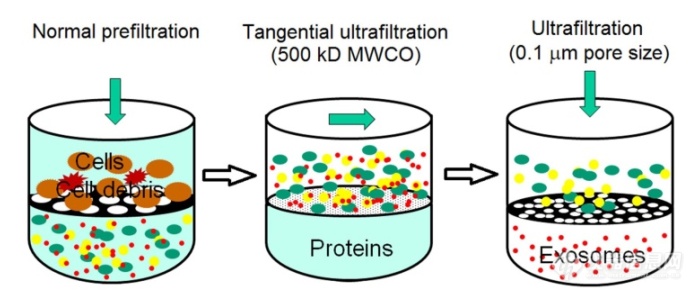
[align=center][font='times new roman'][size=18px][color=#000000]新型[/color][/size][/font][font='times new roman'][size=18px][color=#000000]外泌体分离方法[/color][/size][/font][/align][align=left][font='times new roman'][size=16px]肿瘤细胞来源的外泌体在分子水平上促进肿瘤的进展、侵袭和转移。因此,在探索细胞间信号传导,分析功能分子成分(蛋白质、[/size][/font][font='times new roman'][size=16px]mRNA[/size][/font][font='times new roman'][size=16px]和[/size][/font][font='times new roman'][size=16px]microRNA[/size][/font][font='times new roman'][size=16px])前需要有效的检测和分离肿瘤源性外泌体的能力,这可能为癌症诊断和预后提供关键信息。[/size][/font][/align][align=left][font='times new roman'][color=#000000]1[/color][/font][font='times new roman'][color=#000000]基于尺寸排阻的外泌体分离技术[/color][/font][/align][align=left][font='times new roman'][size=16px]外泌体是直径在[/size][/font][font='times new roman'][size=16px]30-200 nm[/size][/font][font='times new roman'][size=16px]的囊泡,其尺寸小于绝大部分的细胞外囊泡,因此,基于这一特性,可利用具有限制相对分子量或大小的过滤器来分离外泌体。目前,最常用的基于尺寸的外泌体分离技术就是超滤离心法。该方法是一种基于悬浮颗粒或聚合物大小的外泌体分离技术,小于膜孔径的物质会通过过滤膜,大于膜孔径的物质被截留在膜上。超滤法比[/size][/font][font='times new roman'][size=16px]UC[/size][/font][font='times new roman'][size=16px]速度更快,且不需要特殊的设备,已有研究表明该方法可以成功从[/size][/font][font='times new roman'][size=16px]0.5 mL[/size][/font][font='times new roman'][size=16px]尿液中分离外泌体。[/size][/font][/align][font='times new roman'][size=16px]目前已经开发了一种适合无细胞样品的商用外泌体分离试剂盒,兼具外泌体分离和[/size][/font][font='times new roman'][size=16px]RNA[/size][/font][font='times new roman'][size=16px]提取的功能。如图所示,该试剂盒利用注射过滤器双层膜结构,当样品通过两层膜时,较大的细胞外囊泡(如凋亡小体和微囊泡)被保留在上层膜上,而外泌体捕获在下层膜上。与[/size][/font][font='times new roman'][size=16px]UC[/size][/font][font='times new roman'][size=16px]和外泌体沉淀法相比,超滤法从尿液中获得的外泌体[/size][/font][font='times new roman'][size=16px]RNA[/size][/font][font='times new roman'][size=16px]产量最高。该方法的主要缺点在于分离的外泌体容易堵塞过滤膜,导致分离效率下降。此外,该方法可能会导致囊泡的变形和破裂,影响下游分析的结果。[/size][/font][font='times new roman'][size=16px]另一种基于尺寸的外泌体分离方法是尺寸排除色谱法[/size][/font][font='times new roman'][size=16px]([/size][/font][font='times new roman'][size=16px]SEC[/size][/font][font='times new roman'][size=16px])[/size][/font][font='times new roman'][size=16px]。该方法利用多孔固定相将悬浮颗粒和聚合物按照大小进行分类[/size][/font][font='times new roman'][size=16px],[/size][/font][font='times new roman'][size=16px]流体动力半径小的物质能够通过孔隙,而流体动力半径较大的物质会被截留在孔隙上。[/size][/font][font='times new roman'][size=16px]此外,[/size][/font][font='times new roman'][size=16px]该方法结合其他方法使用可取得更好的效果[/size][/font][font='times new roman'][size=16px]。[/size][/font][font='times new roman'][size=16px]例如,与单纯的超滤法或[/size][/font][font='times new roman'][size=16px]UC[/size][/font][font='times new roman'][size=16px]相比,该方法分离的外泌体[/size][/font][font='times new roman'][size=16px]结合[/size][/font][font='times new roman'][size=16px]后续超速离心可以[/size][/font][font='times new roman'][size=16px]提高[/size][/font][font='times new roman'][size=16px]尿外泌体[/size][/font][font='times new roman'][size=16px]的捕获效率[/size][/font][font='times new roman'][size=16px],从而有利于寻找肾脏疾病生物标志物。该方法分离外泌体[/size][/font][font='times new roman'][size=16px]主要[/size][/font][font='times new roman'][size=16px]缺点在于干扰物多[/size][/font][font='times new roman'][size=16px]、[/size][/font][font='times new roman'][size=16px]孔隙极易堵塞,导致色谱柱重复率低,分离效率较低。[/size][/font][align=left][img]https://ng1.17img.cn/bbsfiles/images/2021/08/202108012209419773_3887_5111497_3.png[/img][/align][align=center][font='times new roman']图[/font][font='times new roman']1-3[/font][font='times new roman'] [/font][font='times new roman']连续过滤原理图[/font][font='times new roman'][size=13px][68][/size][/font][/align][align=center][font='times new roman']Figure [/font][font='times new roman']1-[/font][font='times new roman']3[/font][font='times new roman'] [/font][font='times new roman']Schematic illustration of sequential filtration[/font][font='times new roman'][size=13px][68][/size][/font][/align][align=center][/align][font='times new roman'][color=#000000]2[/color][/font][font='times new roman'][color=#000000]基于聚合物沉淀的分离技术[/color][/font][font='times new roman'][size=16px]聚合物沉淀[/size][/font][font='times new roman'][size=16px]技术是通过添加水性聚合物使外泌体溶解度或分散性改变,减少外泌体的水合作用,使外泌体沉淀以达到分离的技术。通常使用分子量为[/size][/font][font='times new roman'][size=16px]8000 Da[/size][/font][font='times new roman'][size=16px]的聚乙二醇([/size][/font][font='times new roman'][size=16px]PEG[/size][/font][font='times new roman'][size=16px])与样品共孵育,[/size][/font][font='times new roman'][size=16px]4[/size][/font][font='times new roman'][size=16px]℃过夜后,用低速离心或过滤法分离含有外泌体的沉淀物。目前,已开发了一系列聚合物沉淀试剂盒可用于体液和培养基中外泌体的分离。聚合物沉淀分离外泌体的方法易于使用、回收率高,且不需要专门的设备。该方法的主要缺点在于容易引入蛋白质和聚合物材料等其他污染物,使得提取的外泌体纯度较低。[/size][/font][font='times new roman'][size=14px][color=#000000]3[/color][/size][/font][font='times new roman'][size=14px][color=#000000] [/color][/size][/font][font='times new roman'][size=14px][color=#000000]基于免疫亲和的分离技术[/color][/size][/font][font='times new roman'][size=16px]外泌体磷脂双层膜中含有丰富的蛋白质和受体,如[/size][/font][font='times new roman'][size=16px]CD81[/size][/font][font='times new roman'][size=16px]、[/size][/font][font='times new roman'][size=16px]CD63[/size][/font][font='times new roman'][size=16px]、[/size][/font][font='times new roman'][size=16px]TSG101[/size][/font][font='times new roman'][size=16px]、上皮细胞粘附分子等,利用这些受体与配体之间的相互作用,使外泌体与特殊设计的磁性颗粒之间建立免疫亲和作用,可用于外泌体的分离富集。例如,[/size][/font][font='times new roman'][size=16px]Zarovni[/size][/font][font='times new roman'][size=16px]等报道了一种基于微孔板的酶联免疫吸附试验([/size][/font][font='times new roman'][size=16px]ELISA[/size][/font][font='times new roman'][size=16px])用于捕获和定量检测外泌体。尽管与[/size][/font][font='times new roman'][size=16px]UC[/size][/font][font='times new roman'][size=16px]产量相当,但是该方法具有快速、易于使用和与常规设备兼容的优势。该报道继续开发了一种基于磁免疫捕获的外泌体分离试剂盒用于从细胞培养基和生物液中分离外泌体,其质量和纯度均优于其他技术。此外,这种方法对样品的初始体积没有要求,可以很容易地缩小或增大样品容量。而该技术主要缺点在于缺乏最佳的外泌体标志物。此外,随着肿瘤的进展,肿瘤抗原表达和调节的异质性可能导致低估和假阴性,并且有些抗原表位可能被阻断或掩蔽。[/size][/font]
[align=center][font='times new roman'][size=18px][color=#000000]外[/color][/size][/font][font='times new roman'][size=18px][color=#000000]泌[/color][/size][/font][font='times new roman'][size=18px][color=#000000]体蛋白质的提取[/color][/size][/font][/align][font='times new roman'][size=16px][color=#000000]([/color][/size][/font][font='times new roman'][size=16px][color=#000000]1[/color][/size][/font][font='times new roman'][size=16px][color=#000000])[/color][/size][/font][font='times new roman'][size=16px][color=#000000]RIPA[/color][/size][/font][font='times new roman'][size=16px][color=#000000]裂解液的配置:正常购买的裂解液中分别加入[/color][/size][/font][font='times new roman'][size=16px][color=#000000]0.1%[/color][/size][/font][font='times new roman'][size=16px][color=#000000]、[/color][/size][/font][font='times new roman'][size=16px][color=#000000]2%[/color][/size][/font][font='times new roman'][size=16px][color=#000000]、[/color][/size][/font][font='times new roman'][size=16px][color=#000000]4%[/color][/size][/font][font='times new roman'][size=16px][color=#000000]和[/color][/size][/font][font='times new roman'][size=16px][color=#000000]8%[/color][/size][/font][font='times new roman'][size=16px][color=#000000]的[/color][/size][/font][font='times new roman'][size=16px][color=#000000]SDS[/color][/size][/font][font='times new roman'][size=16px][color=#000000],配置系列[/color][/size][/font][font='times new roman'][size=16px][color=#000000]RIPA[/color][/size][/font][font='times new roman'][size=16px][color=#000000]裂解液。[/color][/size][/font][font='times new roman'][size=16px][color=#000000]([/color][/size][/font][font='times new roman'][size=16px][color=#000000]2[/color][/size][/font][font='times new roman'][size=16px][color=#000000])将提取的外[/color][/size][/font][font='times new roman'][size=16px][color=#000000]泌[/color][/size][/font][font='times new roman'][size=16px][color=#000000]体溶液加入等体积的蛋白裂解液在[/color][/size][/font][font='times new roman'][size=16px][color=#000000]冰浴[/color][/size][/font][font='times new roman'][size=16px][color=#000000]上[/color][/size][/font][font='times new roman'][size=16px][color=#000000]进行裂解。为了使其充分裂解,每[/color][/size][/font][font='times new roman'][size=16px][color=#000000]5 min[/color][/size][/font][font='times new roman'][size=16px][color=#000000]振荡一次,共裂解[/color][/size][/font][font='times new roman'][size=16px][color=#000000]50 min[/color][/size][/font][font='times new roman'][size=16px][color=#000000]。随后加入[/color][/size][/font][font='times new roman'][size=16px][color=#000000]4[/color][/size][/font][font='times new roman'][size=16px][color=#000000]×[/color][/size][/font][font='times new roman'][size=16px][color=#000000]蛋白变性缓冲液[/color][/size][/font][font='times new roman'][size=16px][color=#000000],[/color][/size][/font][font='times new roman'][size=16px][color=#000000]100[/color][/size][/font][font='times new roman'][size=16px][color=#000000]℃煮沸[/color][/size][/font][font='times new roman'][size=16px][color=#000000]5 min[/color][/size][/font][font='times new roman'][size=16px][color=#000000],将变性的蛋白样品放入[/color][/size][/font][font='times new roman'][size=16px][color=#000000]-20[/color][/size][/font][font='times new roman'][size=16px][color=#000000]℃[/color][/size][/font][font='times new roman'][size=16px][color=#000000]冰箱中保存备用。[/color][/size][/font][font='times new roman'][size=18px][color=#000000]蛋白浓度的测定[/color][/size][/font][font='times new roman'][size=16px][color=#000000]首先,将上述外[/color][/size][/font][font='times new roman'][size=16px][color=#000000]泌[/color][/size][/font][font='times new roman'][size=16px][color=#000000]体裂解后的蛋白原液于[/color][/size][/font][font='times new roman'][size=16px][color=#000000]4[/color][/size][/font][font='times new roman'][size=16px][color=#000000]℃以[/color][/size][/font][font='times new roman'][size=16px][color=#000000]12000[/color][/size][/font][font='times new roman'][size=16px][color=#000000] [/color][/size][/font][font='times new roman'][size=16px][color=#000000]rp[/color][/size][/font][font='times new roman'][size=16px][color=#000000]m[/color][/size][/font][font='times new roman'][size=16px][color=#000000]离心[/color][/size][/font][font='times new roman'][size=16px][color=#000000]15[/color][/size][/font][font='times new roman'][size=16px][color=#000000] [/color][/size][/font][font='times new roman'][size=16px][color=#000000]min[/color][/size][/font][font='times new roman'][size=16px][color=#000000],弃去沉淀。取[/color][/size][/font][font='times new roman'][size=16px][color=#000000]15[/color][/size][/font][font='times new roman'][size=16px][color=#000000] [/color][/size][/font][font='times new roman'][size=16px]μ[/size][/font][font='times new roman'][size=16px]L[/size][/font][font='times new roman'][size=16px][color=#000000]上清液与[/color][/size][/font][font='times new roman'][size=16px][color=#000000]45[/color][/size][/font][font='times new roman'][size=16px][color=#000000] [/color][/size][/font][font='times new roman'][size=16px]μL[/size][/font][font='times new roman'][size=16px] PBS[/size][/font][font='times new roman'][size=16px]缓冲液混合([/size][/font][font='times new roman'][size=16px][color=#000000]稀释[/color][/size][/font][font='times new roman'][size=16px][color=#000000]4[/color][/size][/font][font='times new roman'][size=16px][color=#000000]倍[/color][/size][/font][font='times new roman'][size=16px])配置蛋白稀释液[/size][/font][font='times new roman'][size=16px][color=#000000]。然后用[/color][/size][/font][font='times new roman'][size=16px][color=#000000]PBS[/color][/size][/font][font='times new roman'][size=16px][color=#000000]缓冲液稀释[/color][/size][/font][font='times new roman'][size=16px][color=#000000]BSA[/color][/size][/font][font='times new roman'][size=16px][color=#000000]标准蛋白溶液,配制终浓度为[/color][/size][/font][font='times new roman'][size=16px][color=#000000]0[/color][/size][/font][font='times new roman'][size=16px][color=#000000] [/color][/size][/font][font='times new roman'][size=16px][color=#000000]mg[/color][/size][/font][font='times new roman'][size=16px][color=#000000]/mL[/color][/size][/font][font='times new roman'][size=16px][color=#000000]、[/color][/size][/font][font='times new roman'][size=16px][color=#000000]0.025[/color][/size][/font][font='times new roman'][size=16px][color=#000000] [/color][/size][/font][font='times new roman'][size=16px][color=#000000]mg[/color][/size][/font][font='times new roman'][size=16px][color=#000000]/mL[/color][/size][/font][font='times new roman'][size=16px][color=#000000]、[/color][/size][/font][font='times new roman'][size=16px][color=#000000]0.05[/color][/size][/font][font='times new roman'][size=16px][color=#000000] [/color][/size][/font][font='times new roman'][size=16px][color=#000000]mg[/color][/size][/font][font='times new roman'][size=16px][color=#000000]/mL[/color][/size][/font][font='times new roman'][size=16px][color=#000000]、[/color][/size][/font][font='times new roman'][size=16px][color=#000000]0.1[/color][/size][/font][font='times new roman'][size=16px][color=#000000] [/color][/size][/font][font='times new roman'][size=16px][color=#000000]mg[/color][/size][/font][font='times new roman'][size=16px][color=#000000]/mL[/color][/size][/font][font='times new roman'][size=16px][color=#000000]、[/color][/size][/font][font='times new roman'][size=16px][color=#000000]0.2[/color][/size][/font][font='times new roman'][size=16px][color=#000000] [/color][/size][/font][font='times new roman'][size=16px][color=#000000]mg[/color][/size][/font][font='times new roman'][size=16px][color=#000000]/mL[/color][/size][/font][font='times new roman'][size=16px][color=#000000]、[/color][/size][/font][font='times new roman'][size=16px][color=#000000]0.3[/color][/size][/font][font='times new roman'][size=16px][color=#000000] [/color][/size][/font][font='times new roman'][size=16px][color=#000000]mg[/color][/size][/font][font='times new roman'][size=16px][color=#000000] /mL[/color][/size][/font][font='times new roman'][size=16px][color=#000000]、[/color][/size][/font][font='times new roman'][size=16px][color=#000000]0.4[/color][/size][/font][font='times new roman'][size=16px][color=#000000] [/color][/size][/font][font='times new roman'][size=16px][color=#000000]mg[/color][/size][/font][font='times new roman'][size=16px][color=#000000]/mL[/color][/size][/font][font='times new roman'][size=16px][color=#000000]、[/color][/size][/font][font='times new roman'][size=16px][color=#000000]0.5mg[/color][/size][/font][font='times new roman'][size=16px][color=#000000] /mL[/color][/size][/font][font='times new roman'][size=16px][color=#000000]的蛋白[/color][/size][/font][font='times new roman'][size=16px][color=#000000]溶液[/color][/size][/font][font='times new roman'][size=16px][color=#000000]。以[/color][/size][/font][font='times new roman'][size=16px][color=#000000]A[/color][/size][/font][font='times new roman'][size=16px][color=#000000]液:[/color][/size][/font][font='times new roman'][size=16px][color=#000000]B[/color][/size][/font][font='times new roman'][size=16px][color=#000000]液[/color][/size][/font][font='times new roman'][size=16px][color=#000000]=50[/color][/size][/font][font='times new roman'][size=16px][color=#000000]:[/color][/size][/font][font='times new roman'][size=16px][color=#000000]1[/color][/size][/font][font='times new roman'][size=16px][color=#000000]的浓度配制工作液。最后,将上述[/color][/size][/font][font='times new roman'][size=16px][color=#000000]配制好的标准蛋白和蛋白稀释液[/color][/size][/font][font='times new roman'][size=16px][color=#000000]2[/color][/size][/font][font='times new roman'][size=16px][color=#000000]0 [/color][/size][/font][font='times new roman'][size=16px][color=#000000]μ[/color][/size][/font][font='times new roman'][size=16px][color=#000000]L[/color][/size][/font][font='times new roman'][size=16px][color=#000000]加入[/color][/size][/font][font='times new roman'][size=16px][color=#000000]96[/color][/size][/font][font='times new roman'][size=16px][color=#000000]孔板中,每孔加入[/color][/size][/font][font='times new roman'][size=16px][color=#000000]200[/color][/size][/font][font='times new roman'][size=16px][color=#000000] [/color][/size][/font][font='times new roman'][size=16px][color=#000000]μ[/color][/size][/font][font='times new roman'][size=16px][color=#000000]L[/color][/size][/font][font='times new roman'][size=16px][color=#000000]工作液,设置[/color][/size][/font][font='times new roman'][size=16px][color=#000000]3[/color][/size][/font][font='times new roman'][size=16px][color=#000000]个复孔,吹打混匀,避免出现气泡。将锡箔纸覆盖的[/color][/size][/font][font='times new roman'][size=16px][color=#000000]96[/color][/size][/font][font='times new roman'][size=16px][color=#000000]孔板放入[/color][/size][/font][font='times new roman'][size=16px][color=#000000]37[/color][/size][/font][font='times new roman'][size=16px][color=#000000]℃恒温箱孵育[/color][/size][/font][font='times new roman'][size=16px][color=#000000]30[/color][/size][/font][font='times new roman'][size=16px][color=#000000] [/color][/size][/font][font='times new roman'][size=16px][color=#000000]min[/color][/size][/font][font='times new roman'][size=16px][color=#000000]。酶标仪的检测[/color][/size][/font][font='times new roman'][size=16px][color=#000000]波长设置为[/color][/size][/font][font='times new roman'][size=16px][color=#000000]562[/color][/size][/font][font='times new roman'][size=16px][color=#000000] [/color][/size][/font][font='times new roman'][size=16px][color=#000000]nm[/color][/size][/font][font='times new roman'][size=16px][color=#000000],检测各孔的[/color][/size][/font][font='times new roman'][size=16px][color=#000000]OD[/color][/size][/font][font='times new roman'][size=16px][color=#000000]值。根据酶标仪算出的标准蛋白拟合曲线计算蛋白原液的浓度。[/color][/size][/font][align=left][font='times new roman'][size=18px][color=#000000]Western blotting[/color][/size][/font][/align][font='times new roman'][size=16px][color=#000000]([/color][/size][/font][font='times new roman'][size=16px][color=#000000]1[/color][/size][/font][font='times new roman'][size=16px][color=#000000])[/color][/size][/font][font='times new roman'][size=16px][color=#000000]10%[/color][/size][/font][font='times new roman'][size=16px][color=#000000]分离胶的配置[/color][/size][/font][font='times new roman'][size=16px][color=#000000]将玻璃板清洗干净后组装起来,加入适当的三蒸水,[/color][/size][/font][font='times new roman'][size=16px][color=#000000]20 min[/color][/size][/font][font='times new roman'][size=16px][color=#000000]后,观察液面是否下降,来检查装置是否紧密。待玻璃板干燥后按比例配置分离胶:[/color][/size][/font][font='times new roman'][size=16px][color=#000000]4[/color][/size][/font][font='times new roman'][size=16px][color=#000000] [/color][/size][/font][font='times new roman'][size=16px][color=#000000]mL[/color][/size][/font][font='times new roman'][size=16px][color=#000000]去离子水;[/color][/size][/font][font='times new roman'][size=16px][color=#000000]3.3[/color][/size][/font][font='times new roman'][size=16px][color=#000000] [/color][/size][/font][font='times new roman'][size=16px][color=#000000]mL[/color][/size][/font][font='times new roman'][size=16px][color=#000000] [/color][/size][/font][font='times new roman'][size=16px][color=#000000]30%[/color][/size][/font][font='times new roman'][size=16px][color=#000000]丙烯酰胺;[/color][/size][/font][font='times new roman'][size=16px][color=#000000]2.5[/color][/size][/font][font='times new roman'][size=16px][color=#000000] [/color][/size][/font][font='times new roman'][size=16px][color=#000000]mL [/color][/size][/font][font='times new roman'][size=16px][color=#000000]p[/color][/size][/font][font='times new roman'][size=16px][color=#000000]H=8.8 Tris-HC[/color][/size][/font][font='times new roman'][size=16px][color=#000000]l[/color][/size][/font][font='times new roman'][size=16px][color=#000000];[/color][/size][/font][font='times new roman'][size=16px][color=#000000]100[/color][/size][/font][font='times new roman'][size=16px][color=#000000] [/color][/size][/font][font='times new roman'][size=16px][color=#000000]μ[/color][/size][/font][font='times new roman'][size=16px][color=#000000]L[/color][/size][/font][font='times new roman'][size=16px][color=#000000] [/color][/size][/font][font='times new roman'][size=16px][color=#000000]10[/color][/size][/font][font='times new roman'][size=16px][color=#000000] [/color][/size][/font][font='times new roman'][size=16px][color=#000000]%[/color][/size][/font][font='times new roman'][size=16px][color=#000000] [/color][/size][/font][font='times new roman'][size=16px][color=#000000]SDS[/color][/size][/font][font='times new roman'][size=16px][color=#000000];[/color][/size][/font][font='times new roman'][size=16px][color=#000000]100[/color][/size][/font][font='times new roman'][size=16px][color=#000000] [/color][/size][/font][font='times new roman'][size=16px][color=#000000]μ[/color][/size][/font][font='times new roman'][size=16px][color=#000000]L[/color][/size][/font][font='times new roman'][size=16px][color=#000000] 10%[/color][/size][/font][font='times new roman'][size=16px][color=#000000] [/color][/size][/font][font='times new roman'][size=16px][color=#000000]APS[/color][/size][/font][font='times new roman'][size=16px][color=#000000];[/color][/size][/font][font='times new roman'][size=16px][color=#000000]4[/color][/size][/font][font='times new roman'][size=16px][color=#000000] [/color][/size][/font][font='times new roman'][size=16px][color=#000000]μ[/color][/size][/font][font='times new roman'][size=16px][color=#000000]L[/color][/size][/font][font='times new roman'][size=16px][color=#000000] TEMED[/color][/size][/font][font='times new roman'][size=16px][color=#000000]。将分离胶混匀后加入玻璃板中,在该过程中应避免产生气泡,随后在玻璃板中加入适量的酒精压平界面,待[/color][/size][/font][font='times new roman'][size=16px][color=#000000]30-40 min[/color][/size][/font][font='times new roman'][size=16px][color=#000000]后,分离胶凝固,倒掉上层酒精。[/color][/size][/font][font='times new roman'][size=16px][color=#000000]([/color][/size][/font][font='times new roman'][size=16px][color=#000000]2[/color][/size][/font][font='times new roman'][size=16px][color=#000000])基层胶的配置[/color][/size][/font][font='times new roman'][size=16px][color=#000000]待玻璃板干燥后加入[/color][/size][/font][font='times new roman'][size=16px][color=#000000]已[/color][/size][/font][font='times new roman'][size=16px][color=#000000]配置好的基层胶([/color][/size][/font][font='times new roman'][size=16px][color=#000000]2.7 mL[/color][/size][/font][font='times new roman'][size=16px][color=#000000]去离子水;[/color][/size][/font][font='times new roman'][size=16px][color=#000000]670[/color][/size][/font][font='times new roman'][size=16px][color=#000000] [/color][/size][/font][font='times new roman'][size=16px][color=#000000]μ[/color][/size][/font][font='times new roman'][size=16px][color=#000000]L[/color][/size][/font][font='times new roman'][size=16px][color=#000000] 30% PAGE[/color][/size][/font][font='times new roman'][size=16px][color=#000000];[/color][/size][/font][font='times new roman'][size=16px][color=#000000]500 [/color][/size][/font][font='times new roman'][size=16px][color=#000000]μ[/color][/size][/font][font='times new roman'][size=16px][color=#000000]L[/color][/size][/font][font='times new roman'][size=16px][color=#000000] [/color][/size][/font][font='times new roman'][size=16px][color=#000000]p[/color][/size][/font][font='times new roman'][size=16px][color=#000000]H=6.8 Tris-HC[/color][/size][/font][font='times new roman'][size=16px][color=#000000]l[/color][/size][/font][font='times new roman'][size=16px][color=#000000];[/color][/size][/font][font='times new roman'][size=16px][color=#000000]40 [/color][/size][/font][font='times new roman'][size=16px][color=#000000]μ[/color][/size][/font][font='times new roman'][size=16px][color=#000000]L[/color][/size][/font][font='times new roman'][size=16px][color=#000000] 10%[/color][/size][/font][font='times new roman'][size=16px][color=#000000] [/color][/size][/font][font='times new roman'][size=16px][color=#000000]SDS[/color][/size][/font][font='times new roman'][size=16px][color=#000000];[/color][/size][/font][font='times new roman'][size=16px][color=#000000]40 [/color][/size][/font][font='times new roman'][size=16px][color=#000000]μL[/color][/size][/font][font='times new roman'][size=16px][color=#000000] 10% APS[/color][/size][/font][font='times new roman'][size=16px][color=#000000];[/color][/size][/font][font='times new roman'][size=16px][color=#000000]4[/color][/size][/font][font='times new roman'][size=16px][color=#000000] [/color][/size][/font][font='times new roman'][size=16px][color=#000000]μL[/color][/size][/font][font='times new roman'][size=16px][color=#000000] TEMED[/color][/size][/font][font='times new roman'][size=16px][color=#000000]),立刻插入梳子,避免产生气泡,等待基层胶凝固。[/color][/size][/font][font='times new roman'][size=16px][color=#000000]([/color][/size][/font][font='times new roman'][size=16px][color=#000000]3[/color][/size][/font][font='times new roman'][size=16px][color=#000000])[/color][/size][/font][font='times new roman'][size=16px][color=#000000]电泳[/color][/size][/font][font='times new roman'][size=16px][color=#000000]清洗并组装电泳装置,固定[/color][/size][/font][font='times new roman'][size=16px][color=#000000]SDS-PAGE[/color][/size][/font][font='times new roman'][size=16px][color=#000000]凝胶,倒入配置好的[/color][/size][/font][font='times new roman'][size=16px][color=#000000]1[/color][/size][/font][font='times new roman'][size=16px][color=#000000]×电泳缓冲液[/color][/size][/font][font='times new roman'][size=16px][color=#000000]([/color][/size][/font][font='times new roman'][size=16px][color=#000000]14.4 g [/color][/size][/font][font='times new roman'][size=16px][color=#000000]甘氨酸,[/color][/size][/font][font='times new roman'][size=16px][color=#000000]3 g Tris[/color][/size][/font][font='times new roman'][size=16px][color=#000000],[/color][/size][/font][font='times new roman'][size=16px][color=#000000]1 g SDS[/color][/size][/font][font='times new roman'][size=16px][color=#000000],加入去离子水,搅拌溶解,[/color][/size][/font][font='times new roman'][size=16px][color=#000000]定容至[/color][/size][/font][font='times new roman'][size=16px][color=#000000]1000 mL[/color][/size][/font][font='times new roman'][size=16px][color=#000000])。拔出梳子,用[/color][/size][/font][font='times new roman'][size=16px][color=#000000]20[/color][/size][/font][font='times new roman'][size=16px][color=#000000] [/color][/size][/font][font='times new roman'][size=16px][color=#000000]μ[/color][/size][/font][font='times new roman'][size=16px][color=#000000]L[/color][/size][/font][font='times new roman'][size=16px][color=#000000][url=https://insevent.instrument.com.cn/t/9p][color=#3333ff][url=https://insevent.instrument.com.cn/t/9p][color=#3333ff]移液枪[/color][/url][/color][/url][/color][/size][/font][font='times new roman'][size=16px][color=#000000]吹打每个[/color][/size][/font][font='times new roman'][size=16px][color=#000000]加样孔以[/color][/size][/font][font='times new roman'][size=16px][color=#000000]去除杂质,用[/color][/size][/font][font='times new roman'][size=16px][color=#000000]40 V[/color][/size][/font][font='times new roman'][size=16px][color=#000000]电压电泳[/color][/size][/font][font='times new roman'][size=16px][color=#000000]20 min[/color][/size][/font][font='times new roman'][size=16px][color=#000000]疏通每个泳道,关闭仪器。然后按照试验要求加入变性后的目的蛋白和[/color][/size][/font][font='times new roman'][size=16px][color=#000000]蛋白[/color][/size][/font][font='times new roman'][size=16px][color=#000000]Marker[/color][/size][/font][font='times new roman'][size=16px][color=#000000],样品的两边可以加入[/color][/size][/font][font='times new roman'][size=16px][color=#000000]1[/color][/size][/font][font='times new roman'][size=16px][color=#000000]×[/color][/size][/font][font='times new roman'][size=16px][color=#000000]上样缓冲[/color][/size][/font][font='times new roman'][size=16px][color=#000000]液进[/color][/size][/font][font='times new roman'][size=16px][color=#000000]行补齐。打开仪器开关,将电压调至[/color][/size][/font][font='times new roman'][size=16px][color=#000000]60 V[/color][/size][/font][font='times new roman'][size=16px][color=#000000]进行电泳,当溴酚蓝到达基层胶和分离胶的连接处,将电压调整到[/color][/size][/font][font='times new roman'][size=16px][color=#000000]90 V[/color][/size][/font][font='times new roman'][size=16px][color=#000000]继续电泳,直至溴酚蓝跑到接近底部。电泳结束后,拆除装置,取出凝胶,切除多余的部分备用。如需考马斯亮蓝染色,将凝胶放入考马斯亮蓝染色液中,摇床振荡[/color][/size][/font][font='times new roman'][size=16px][color=#000000]30 min[/color][/size][/font][font='times new roman'][size=16px][color=#000000]后,用蒸馏水洗涤[/color][/size][/font][font='times new roman'][size=16px][color=#000000]3[/color][/size][/font][font='times new roman'][size=16px][color=#000000]次后放入洗脱液中进行洗脱。[/color][/size][/font][font='times new roman'][size=16px][color=#000000]([/color][/size][/font][font='times new roman'][size=16px][color=#000000]4[/color][/size][/font][font='times new roman'][size=16px][color=#000000])[/color][/size][/font][font='times new roman'][size=16px][color=#000000]转膜[/color][/size][/font][font='times new roman'][size=16px][color=#000000]配置[/color][/size][/font][font='times new roman'][size=16px][color=#000000]1[/color][/size][/font][font='times new roman'][size=16px][color=#000000]×[/color][/size][/font][font='times new roman'][size=16px][color=#000000]转膜缓冲[/color][/size][/font][font='times new roman'][size=16px][color=#000000]液([/color][/size][/font][font='times new roman'][size=16px][color=#000000]14.4 g [/color][/size][/font][font='times new roman'][size=16px][color=#000000]甘氨酸,[/color][/size][/font][font='times new roman'][size=16px][color=#000000]3 g Tris[/color][/size][/font][font='times new roman'][size=16px][color=#000000],[/color][/size][/font][font='times new roman'][size=16px][color=#000000]150 mL [/color][/size][/font][font='times new roman'][size=16px][color=#000000]甲醇,加入去离子水,搅拌溶解,[/color][/size][/font][font='times new roman'][size=16px][color=#000000]定容至[/color][/size][/font][font='times new roman'][size=16px][color=#000000]1000 mL[/color][/size][/font][font='times new roman'][size=16px][color=#000000])。将适当大小的[/color][/size][/font][font='times new roman'][size=16px][color=#000000]PVDF[/color][/size][/font][font='times new roman'][size=16px][color=#000000]膜放入甲醇中激活[/color][/size][/font][font='times new roman'][size=16px][color=#000000]5 min[/color][/size][/font][font='times new roman'][size=16px][color=#000000],随后放入[/color][/size][/font][font='times new roman'][size=16px][color=#000000]转膜液[/color][/size][/font][font='times new roman'][size=16px][color=#000000]中。[/color][/size][/font][font='times new roman'][size=16px][color=#000000]转膜夹[/color][/size][/font][font='times new roman'][size=16px][color=#000000]白色面朝下放入[/color][/size][/font][font='times new roman'][size=16px][color=#000000]转膜液[/color][/size][/font][font='times new roman'][size=16px][color=#000000]中,然后放入泡沫层、[/color][/size][/font][font='times new roman'][size=16px][color=#000000]3[/color][/size][/font][font='times new roman'][size=16px][color=#000000]层滤纸、激活后的[/color][/size][/font][font='times new roman'][size=16px][color=#000000]PVDF[/color][/size][/font][font='times new roman'][size=16px][color=#000000]膜、切好的胶、三层滤纸和最后一个泡沫层。在上述过程中应保证每层均无气泡存在。将装置[/color][/size][/font][font='times new roman'][size=16px][color=#000000]放入转膜槽中[/color][/size][/font][font='times new roman'][size=16px][color=#000000],[/color][/size][/font][font='times new roman'][size=16px][color=#000000]转膜液[/color][/size][/font][font='times new roman'][size=16px][color=#000000]中可以加入小冰袋,防止[/color][/size][/font][font='times new roman'][size=16px][color=#000000]转膜过程[/color][/size][/font][font='times new roman'][size=16px][color=#000000]中温度过高。将装置放入冰盒,恒流[/color][/size][/font][font='times new roman'][size=16px][color=#000000]200 mA[/color][/size][/font][font='times new roman'][size=16px][color=#000000]进行转膜,[/color][/size][/font][font='times new roman'][size=16px][color=#000000]转膜时间[/color][/size][/font][font='times new roman'][size=16px][color=#000000]由目的蛋白的分子量所决定。[/color][/size][/font][font='times new roman'][size=16px][color=#000000]([/color][/size][/font][font='times new roman'][size=16px][color=#000000]5[/color][/size][/font][font='times new roman'][size=16px][color=#000000])[/color][/size][/font][font='times new roman'][size=16px][color=#000000]封闭和免疫反应[/color][/size][/font][font='times new roman'][size=16px][color=#000000]转膜结束[/color][/size][/font][font='times new roman'][size=16px][color=#000000]后,将膜和含[/color][/size][/font][font='times new roman'][size=16px][color=#000000]5% BSA[/color][/size][/font][font='times new roman'][size=16px][color=#000000]的[/color][/size][/font][font='times new roman'][size=16px][color=#000000]TBST[/color][/size][/font][font='times new roman'][size=16px][color=#000000]溶液放进封闭盒中,室温[/color][/size][/font][font='times new roman'][size=16px][color=#000000]摇床慢摇[/color][/size][/font][font='times new roman'][size=16px][color=#000000]2 h[/color][/size][/font][font='times new roman'][size=16px][color=#000000]。随后[/color][/size][/font][font='times new roman'][size=16px][color=#000000]TBST[/color][/size][/font][font='times new roman'][size=16px][color=#000000]洗[/color][/size][/font][font='times new roman'][size=16px][color=#000000]3[/color][/size][/font][font='times new roman'][size=16px][color=#000000]次,每次[/color][/size][/font][font='times new roman'][size=16px][color=#000000]10 min[/color][/size][/font][font='times new roman'][size=16px][color=#000000]。配置适合比例的[/color][/size][/font][font='times new roman'][size=16px][color=#000000]一[/color][/size][/font][font='times new roman'][size=16px][color=#000000]抗溶液,将膜放入其中,[/color][/size][/font][font='times new roman'][size=16px][color=#000000]4[/color][/size][/font][font='times new roman'][size=16px][color=#000000]℃摇床孵育过夜。第二天,[/color][/size][/font][font='times new roman'][size=16px][color=#000000]TBST[/color][/size][/font][font='times new roman'][size=16px][color=#000000]洗[/color][/size][/font][font='times new roman'][size=16px][color=#000000]3[/color][/size][/font][font='times new roman'][size=16px][color=#000000]次,每次[/color][/size][/font][font='times new roman'][size=16px][color=#000000]10 min[/color][/size][/font][font='times new roman'][size=16px][color=#000000],在室温下摇床[/color][/size][/font][font='times new roman'][size=16px][color=#000000]孵育二抗[/color][/size][/font][font='times new roman'][size=16px][color=#000000]1 h[/color][/size][/font][font='times new roman'][size=16px][color=#000000],随后[/color][/size][/font][font='times new roman'][size=16px][color=#000000]TBST[/color][/size][/font][font='times new roman'][size=16px][color=#000000]洗[/color][/size][/font][font='times new roman'][size=16px][color=#000000]3[/color][/size][/font][font='times new roman'][size=16px][color=#000000]次,每次[/color][/size][/font][font='times new roman'][size=16px][color=#000000]10 min[/color][/size][/font][font='times new roman'][size=16px][color=#000000]。[/color][/size][/font][font='times new roman'][size=16px][color=#000000]([/color][/size][/font][font='times new roman'][size=16px][color=#000000]6[/color][/size][/font][font='times new roman'][size=16px][color=#000000])[/color][/size][/font][font='times new roman'][size=16px][color=#000000]显影[/color][/size][/font][font='times new roman'][size=16px][color=#000000]避光配置[/color][/size][/font][font='times new roman'][size=16px][color=#000000]ECL[/color][/size][/font][font='times new roman'][size=16px][color=#000000]发光液。将发光液滴加到膜上,避光孵育[/color][/size][/font][font='times new roman'][size=16px][color=#000000]3 min[/color][/size][/font][font='times new roman'][size=16px][color=#000000],放入预冷后的发光板上,调整膜的位置,用[/color][/size][/font][font='times new roman'][size=16px][color=#000000]BIO-RAD[/color][/size][/font][font='times new roman'][size=16px][color=#000000]凝胶成像仪显影并拍照。[/color][/size][/font][font='times new roman'][size=16px]外[/size][/font][font='times new roman'][size=16px]泌[/size][/font][font='times new roman'][size=16px]体的成功捕获进一步通过[/size][/font][font='times new roman'][size=16px]Western blotting[/size][/font][font='times new roman'][size=16px]进行验证。[/size][/font][font='times new roman'][size=16px]HSP70[/size][/font][font='times new roman'][size=16px]、[/size][/font][font='times new roman'][size=16px]CD63[/size][/font][font='times new roman'][size=16px]、[/size][/font][font='times new roman'][size=16px]TSG101[/size][/font][font='times new roman'][size=16px]和[/size][/font][font='times new roman'][size=16px]CD81[/size][/font][font='times new roman'][size=16px]是四种典型的普遍存在的外[/size][/font][font='times new roman'][size=16px]泌[/size][/font][font='times new roman'][size=16px]体标记蛋白。通常这些蛋白在整个细胞上清裂解液中的[/size][/font][font='times new roman'][size=16px]表达水[/size][/font][font='times new roman'][size=16px]平极低。然而,通过所开发的[/size][/font][font='times new roman'][size=16px]Tim4@ILI-01[/size][/font][font='times new roman'][size=16px]免疫亲和材料富集后,这四种蛋白质均可以被显著识别(图[/size][/font][font='times new roman'][size=16px]3-7[/size][/font][font='times new roman'][size=16px])。以上结果进一步表明所开发的[/size][/font][font='times new roman'][size=16px]Tim4@ILI-01[/size][/font][font='times new roman'][size=16px]免疫亲和材料可用于成功捕获外[/size][/font][font='times new roman'][size=16px]泌[/size][/font][font='times new roman'][size=16px]体。[/size][/font][table][tr][td][align=center][img]https://ng1.17img.cn/bbsfiles/images/2021/08/202108012201079391_8222_5111497_3.jpeg[/img][/align][/td][/tr][/table][align=center][font='times new roman']图[/font][font='times new roman']1[/font][font='times new roman'] [/font][font='times new roman']培养基中外[/font][font='times new roman']泌[/font][font='times new roman']体特异性蛋白质[/font][font='times new roman']HSP70[/font][font='times new roman']、[/font][font='times new roman']CD63[/font][font='times new roman']、[/font][font='times new roman']TSG101[/font][font='times new roman']和[/font][font='times new roman']CD81[/font][font='times new roman']的[/font][font='times new roman']Western blotting[/font][font='times new roman']结果[/font][/align]
[align=center][size=16px]外泌体综述[/size][/align] 外泌体是直径30-150纳米之间的细胞外囊泡,在疾病发生和进展中起着重要作用。因此,外泌体在早期诊断、靶向治疗等方面均具有很大的潜力。 外泌体生物发生及作用 外泌体的生物发生 1967年,Wolf在人血浆中首次发现一种来源于血小板膜泡的物质,并将其称之为“血小板尘埃”。之后,所有的生物体液以及体外培养的细胞上清中都被检测到含有囊泡。外泌体的生物发生涉及多种机制,这些机制有助于蛋白质和RNA等在细胞间的传递,从而生成具有源细胞特定成分的外泌体。多囊泡体(MVBs)的极限膜向内萌芽形成腔内囊泡(ILVs),等到晚期内体成熟后,MVBs可以与质膜融合,在细胞外空间中释放封闭的ILVs,被释放出去的ILVs称为外泌体。外泌体主要通过两种不同的机制释放,即跨反式高尔基网络释放和诱导释放。Rab家族蛋白,如Rab27a和Rab27b,是外泌体分泌的关键调节剂。除了Rab27a和27b外,其他Rab家族成员Rab35和Rab11也已被证明通过与GTPase激活蛋白TBC1结构域家族成员10A-C(TBC1D10A-C)相互作用来调节外泌体的分泌。研究还表明,癌症抑制蛋白p53能通过调节各种基因的转录(如TSAP6和CHMP4C)来刺激和增加外泌体分泌的速率。 外泌体的作用 外泌体是由细胞释放的纳米级囊泡,存在于不同的生物体液中,如血液、唾液和尿液。这些囊泡携带丰富的“货物”,包括蛋白质、信使RNA(mRNA)和microRNA(miRNA)。1983年,Pan在大鼠网织红细胞中首次观察到内吞囊泡的分泌。1987年,科学家Johnstone将这类囊泡定义为“外泌体”。最初,科学家认为外泌体是由细胞产生的代谢废物。然而,随着对外泌体的研究更加深入,人们逐渐抛弃这一误解。越来越多的研究表明,外泌体参与细胞间通信,是细胞微环境和旁分泌信号的重要组成部分。1998年,L.Zitvogel等人发表了一项关于树突细胞(DCcell)能产生有抗原提呈能力的外泌体的研究,阐明了外泌体含有功能性的MHC-I类II类分子和共刺激因子。2007年,H.Valadi等人的研究证实,细胞之间可以利用外泌体RNA交换遗传物质。这说明了细胞之间可以通过外泌体互相影响,甚至可以将一个细胞的基因强加到另外一个细胞上。2013年,美国科学家JamesE.Rothman、RandyW.Schekman及德国科学家ThomasC.Südhof共同获得当年诺贝尔生理医学奖,以表彰他们发现并阐明了细胞囊泡运输系统及其调控机制。来自癌细胞的外泌体已被证实可以调节癌症细胞生长、增殖、迁移过程,还能影响癌症的化疗耐药。因此,外泌体是理想的可作为非侵入性诊断和预后的生物标志物。 外泌体的分离分析方法 基于对外泌体研究的需要,科学界对外泌体的高效分离、定量和分析方法也在不断尝试和深入。由于样品基质和外泌体理化性质的复杂性,从体液中准确分离外泌体仍存在重大挑战。在过去的几十年里,研究主要使用差分和密度梯度离心、超滤和免疫分离等方法。目前,已有商业外泌体分离试剂盒投入使用。商业试剂盒通过用聚乙二醇或类似成分沉淀囊泡来减少耗时,但是存在非囊泡与外泌体一起共沉淀的弊端,外泌体的常规检测方法仍需向快速、高效、可重复和低成本的方向改进。近年来,基于研究和临床需要,越来越多的分析方法已经被用来分析外泌体。例如,酶联免疫吸附测定(ELISA)、纳米颗粒跟踪分析技术(NTA)、流式细胞术和荧光活化细胞分选(FACS)已成功开发用于外泌体定量。 蛋白质谱分析在外泌体中的应用 外泌体蛋白质组学是对外泌体中的蛋白质进行全面分析,以了解其生物学功能和疾病相关性。外泌体蛋白质组学分析涉及到外泌体的分离纯化、鉴定、数据分析等过程。蛋白质谱是外泌体蛋白质组学研究的手段之一,通过质谱可以获得蛋白质的名称、组成、表达量等信息,进而找到与疾病相关的蛋白质,探索可以用于疾病早期诊断和预后评估的生物标志物。质谱分析具有灵敏度高、通用性强、准确性高等优点,在研究中发挥了重要作用。
各位观众将在这个视频中看到如何在chemstation工作站下对提取出的紫外光谱图标注最大波长方便贴到文章,ppt,报告中
[size=16px]BSA[/size][size=16px]和外泌体简介[/size][size=16px]牛血清白蛋白([/size][size=16px]BSA[/size][size=16px]),[/size][size=16px]BSA[/size][size=16px]:是牛血清中的一种球蛋白,包含[/size][size=16px]607[/size][size=16px]个氨基酸残基,[/size][font='calibri'][size=16px][back=#ffff00]分子量为[/back][/size][/font][font='calibri'][size=16px][back=#ffff00]66.446KDa[/back][/size][/font][size=16px],等电点为[/size][size=16px]4.7[/size][size=16px]。牛血清白蛋白在生化实验中有广泛的应用[/size][size=16px]。粗糙的[/size][size=16px]BSA[/size][size=16px]可以用来配置[/size][size=16px]western blot[/size][size=16px]的封闭液。另一个重要的用途就是用来测蛋白浓度配置标准曲线。[/size][size=16px]β[/size][size=16px]-Actin[/size][size=16px]抗体:β[/size][size=16px]-actin[/size][size=16px]由[/size][size=16px]375[/size][size=16px]个氨基酸组成,分子量大小为[/size][size=16px]42-43kDa[/size][size=16px]左右。[/size][font='calibri'][size=16px][back=#ffff00]β[/back][/size][/font][font='calibri'][size=16px][back=#ffff00]-actin[/back][/size][/font][font='calibri'][size=16px][back=#ffff00]作为内参是得到了公认的[/back][/size][/font][size=16px],这是针对大多数组织和细胞来说的,它广泛分布于细胞浆内,表达量非常丰富,其含量占所有细胞总蛋白的[/size][size=16px]50%[/size][size=16px]。但是在一些少量特殊的情况下,如脂肪组织或细胞内,β[/size][size=16px]-Actin[/size][size=16px]的表达量就很少。[/size][size=16px]外[/size][size=16px]泌[/size][size=16px]体:电子显微镜下呈现[/size][font='calibri'][size=16px][back=#ffff00]杯状结构(可用于验证)[/back][/size][/font][size=16px],外[/size][size=16px]泌体广泛[/size][size=16px]存在于各种生物体液中,如[/size][font='calibri'][size=16px][back=#ffff00]血液、唾液、尿液、乳汁以及细胞培养液中(可作为后续的[/back][/size][/font][font='calibri'][size=16px][back=#ffff00]实际[/back][/size][/font][font='calibri'][size=16px][back=#ffff00]分离样品)[/back][/size][/font][size=16px]。外[/size][size=16px]泌[/size][size=16px]体携带的蛋白质一般包括([/size][size=16px]i[/size][size=16px])跨[/size][size=16px]膜四分子[/size][size=16px]交联体蛋白质(例如[/size][size=16px] CD9[/size][size=16px]、[/size][size=16px]CD63[/size][size=16px]、[/size][size=16px]CD81[/size][size=16px]);([/size][size=16px]ii[/size][size=16px])[/size][size=16px]MVB [/size][size=16px]生物蛋白质(例如[/size][size=16px] TSG101[/size][size=16px]和[/size][size=16px] [/size][size=16px]Alix[/size][size=16px]);([/size][size=16px]iii[/size][size=16px])热休克蛋白质(例如[/size][size=16px] HSC70[/size][size=16px]),常被视为[/size][font='calibri'][size=16px][back=#ffff00]外[/back][/size][/font][font='calibri'][size=16px][back=#ffff00]泌[/back][/size][/font][font='calibri'][size=16px][back=#ffff00]体检测时的标志性蛋白质(用于[/back][/size][/font][font='calibri'][size=16px][back=#ffff00]WB[/back][/size][/font][font='calibri'][size=16px][back=#ffff00]抗体选择)[/back][/size][/font][size=16px]。[/size][size=16px]外[/size][size=16px]泌体参与[/size][size=16px]细胞间信号交流与传递,且在各种病理和生理过程中起着至关重要的作用,如[/size][font='calibri'][size=16px][back=#ffff00]信号转导、免疫应答、肿瘤发生、侵袭和转移等[/back][/size][/font][font='calibri'][size=16px][back=#ffff00](用于后期的分子及细胞水平的作用解释)[/back][/size][/font][size=16px]。优点:外[/size][size=16px]泌体成为[/size][size=16px]液体活检重要的生物标志物之一,其在癌症检测中具有微创、可重复采样、实时监测等显著优势。[/size][font='calibri'][size=16px][back=#ffff00]外[/back][/size][/font][font='calibri'][size=16px][back=#ffff00]泌[/back][/size][/font][font='calibri'][size=16px][back=#ffff00]体的提取和富集可参考[/back][/size][/font][font='calibri'][size=16px][back=#ffff00]CTC[/back][/size][/font][font='calibri'][size=16px][back=#ffff00]细胞[/back][/size][/font][size=16px]。[/size][size=16px]Exosome[/size][size=16px]在癌症[/size][font='calibri'][size=16px][back=#ffff00]靶向治疗[/back][/size][/font][font='calibri'][size=16px][back=#ffff00](载药)[/back][/size][/font][size=16px]中具有巨大的应用潜力[/size][size=16px]。[/size][size=16px]可以将其作为微[/size][size=16px]创疾病[/size][size=16px]生物标志物用于癌症患者的诊断、预后和治疗反应评估等方面[/size][size=16px]。[/size][size=16px]miRNA[/size][size=16px] [/size][size=16px]也是构成外[/size][size=16px]泌[/size][size=16px]体载物的关键成分,而外[/size][size=16px]泌[/size][size=16px]体传递的肿瘤[/size][size=16px] [/size][size=16px]miRNA[/size][size=16px] [/size][size=16px]通过调控微环境、血管生成、免疫逃避以及转移参与肿瘤进程[/size][size=16px]([/size][font='calibri'][size=16px][back=#ffff00]后期[/back][/size][/font][font='calibri'][size=16px][back=#ffff00]RNA[/back][/size][/font][font='calibri'][size=16px][back=#ffff00]检测用于外[/back][/size][/font][font='calibri'][size=16px][back=#ffff00]泌[/back][/size][/font][font='calibri'][size=16px][back=#ffff00]体的验证[/back][/size][/font][font='calibri'][size=16px][back=#ffff00]及分子水平的解释[/back][/size][/font][size=16px])。[/size]感谢仪器信息网提供平台
因为找不到HPLC——紫外检测器检测蜂蜜中磺胺类药物的,能用水产品的代替么?不过我有找到国标配荧光检测器检测蜂蜜中磺胺类药物的,我能用这个提取方法,但是用紫外检测么?
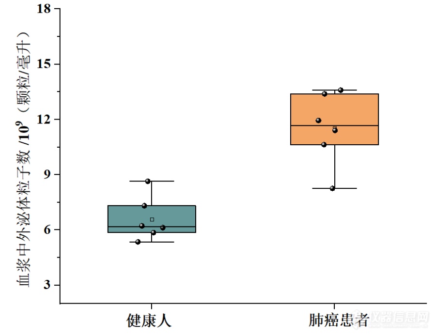
[align=center][font='times new roman'][size=16px]血浆外泌体[/size][/font][font='times new roman'][size=16px]在肺癌诊断中的应用[/size][/font][/align][font='times new roman'][size=16px]引言[/size][/font][font='宋体']外泌体作为重要的信息载体,参与肿瘤的发生、进展、转移和化疗耐药等过程,外泌体携带大量的遗传物质,已成为液体活检最理想的分析目标。外泌体也是目前最有前途的非侵入性诊断和预后的生物标志物之一。目前,超速离心法是外泌体分离的金标准。为了实现外泌体的临床价值,本研究以超速离心法富集了人血浆中的外泌体,并对健康人及肺腺癌患者的血浆样品做了区分,用于下游的肺癌诊断[/font][font='宋体']。[/font][align=left][font='times new roman'][size=16px]血浆外泌体用于肺癌诊断的策略[/size][/font][/align]肺癌仍然是世界范围内癌症相关死亡的主要诱因,因为大多数患者被诊断时均为晚期。因此,我们初步评估了超速离心法富集外泌体用于肺癌诊断的潜在适用性。我们收集了6名健康捐赠者和6名肺癌患者的血浆样本。利用超速离心法富集1 mL血浆样本中的外泌体,对比健康捐赠者和肺癌患者的血浆外泌体,结合数据分析,评估血浆外泌体在监测肺癌诊断方面潜在的适用性。[align=left][font='times new roman'][size=16px][color=#000000] [/color][/size][/font][font='times new roman'][size=16px]血浆中外泌体表征[/size][/font][/align]测试了超速离心法对复杂血浆样品的适用性。首先,利用纳米颗粒示踪分析对超速离心法得到的血浆外泌体进行了表征。如图(a)所示, 纳米颗粒示踪分析显示分离的外泌体粒径分布同样较窄,平均粒径为128.8 nm。与细胞上清样品类似,Western blot方法验证了分离外泌体的有效性。如图(b)所示,经富集后,典型的低丰度外泌体标记物同样得到了有效检测。以上结果证明,超速离心法能[img]" style="max-width: 100% max-height: 100% [/img]够成功的用于血浆中外泌体的富集。[align=center][font='黑体'][size=14px]图[/size][/font][font='黑体'][size=14px] (a)重悬液中血浆外泌体的[/size][/font][font='黑体'][size=14px]纳米颗粒示踪分析技术[/size][/font][font='黑体'][size=14px]表征,(b)[/size][/font][font='黑体'][size=14px]Western blot方法[/size][/font][font='黑体'][size=14px]表征血浆[/size][/font][font='黑体'][size=14px]外泌体标记物HSC70、TSG101、CD63和CD9[/size][/font][font='黑体'][size=14px]蛋白条带[/size][/font][/align][align=left][font='times new roman'][size=16px]血浆外泌体用于肺癌[/size][/font][font='times new roman'][size=16px]的[/size][/font][font='times new roman'][size=16px]诊断[/size][/font][/align][font='宋体']我[/font]们收集了6名健康捐赠者和6名肺癌患者的血浆样本。利用超速离心法富集血浆样本中的外泌体。采用纳米颗粒示踪分析技术对血浆外泌体标进行表征。如图所示,与健康对照组相比,肺癌患者的血浆外泌体显著上调。对比健康捐赠者和肺癌患者的血浆外泌体,结合数据分析,证明血浆外泌体在监测肺癌诊断方面具有潜在的适用性。[align=center][img]https://ng1.17img.cn/bbsfiles/images/2023/10/202310171534194891_9083_6197575_3.png[/img][font='黑体'][size=14px]图[/size][/font][font='黑体'][size=14px] [/size][/font][font='黑体'][size=14px]健康人与肺癌患者血浆中外泌体的[/size][/font][font='黑体'][size=14px]纳米颗粒示踪分析[/size][/font][font='黑体'][size=14px]技术[/size][/font][font='黑体'][size=14px]表征[/size][/font][/align][align=left][font='黑体'][size=18px]小结[/size][/font][/align]超速离心法可以高效、特异性地从人血浆中富集、纯化外泌体,在此我们评估了超速离心法在复杂血浆样品中的适用性。肺癌是全球癌症相关死亡的主要原因,因为在原发癌症扩散之前进行早期诊断具有挑战性。以6例肺癌患者(6例健康供者为对照)的人血浆为样本,进一步研究超速离心法富集外泌体在肺癌筛查和诊断中的应用。这些结果证实了血浆外泌体在肺癌筛查和监测中的应用潜力。
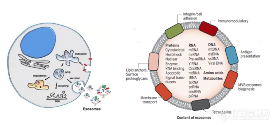
[align=center][font='times new roman'][size=16px]外[/size][/font][font='times new roman'][size=16px]泌[/size][/font][font='times new roman'][size=16px]体[/size][/font][font='times new roman'][size=16px]富集及在肿瘤诊断中的作用[/size][/font][/align]外泌体是直径30-200 nm,由细胞内陷产生的具有磷脂双分子层结构的细胞外小囊泡,是细胞间通讯的重要介质。此外,由于其易于获取、高度稳定且在体液中广泛分布,外泌体已成为液体活检中最有前途的生物标志物[font='宋体']。[/font][align=left][font='times new roman'][size=16px]外[/size][/font][font='times new roman'][size=16px]泌[/size][/font][font='times new roman'][size=16px]体的生发与分布[/size][/font][/align]外泌体是具有磷脂双分子层结构的细胞外小囊泡,它携带原始母细胞重要的物质信息,能够反应原始细胞的一些重要信息。随着研究的深入,已有研究证实外泌体是通过细胞内陷产生的胞外囊泡,如图1-1所示,它是起源于细胞内吞系统中的多囊泡内体,通过出芽、内陷、多泡体形成和分泌等形成的一种具有磷脂双分子层结构的小泡,形态呈球形、扁形或杯状小体,直径为30-200 nm。此外,外泌体的分布非常广泛,几乎分布在所有的体液中,如血液,尿液,乳汁,汗液等。携带活性生物分子(蛋白质、核酸和脂质等)和他们的母细胞有相似的特征,这可能适用于正常细胞和病理细胞的鉴别。特别是,外泌体膜蛋白的水平与癌症的动态密切相关,从而为癌症诊断和治疗提供了新的机会预后。[img]https://ng1.17img.cn/bbsfiles/images/2023/10/202310171529195086_7813_6197418_3.png[/img][align=center][font='黑体'][size=14px]图1.[/size][/font][font='黑体'][size=14px]1 外[/size][/font][font='黑体'][size=14px]泌[/size][/font][font='黑体'][size=14px]体的生发与组成[/size][/font][/align][align=left][font='times new roman'][size=16px]外[/size][/font][font='times new roman'][size=16px]泌[/size][/font][font='times new roman'][size=16px]体的通讯作用[/size][/font][/align]外泌体含有多种生物活性物质,这些被包裹的生物大分子可以在靶细胞中发挥一系列作用,是细胞间接通讯的重要媒介。外泌体与靶细胞间的信息传递主要通过三条途径实现:受体-配体相互作用;质膜直接融合;吞噬作用内吞。外泌体的生物学功能包括物质传递、信息交流、细胞增殖分化、血管生成、免疫调节等,在肿瘤发生发展、侵袭、转移、预后等过程中起重要的调控作用。肿瘤细胞较正常细胞分泌更多的外泌体,来源母细胞不同,所分泌的外泌体量与成分也不同。其次,由于其具有磷脂双分子层结构,生物学相对稳定,难于降解。外泌体的一个关键功能是将其内含物从供体细胞转运到受体细胞,使受体细胞的基因和表型修饰[font='宋体']。[/font][align=left][font='times new roman'][size=16px]外[/size][/font][font='times new roman'][size=16px]泌[/size][/font][font='times new roman'][size=16px]体的富集方法[/size][/font][/align]由于外泌体丰度相对较低且大分子干扰,高质量外泌体的分离仍然具有挑战性。基于不同的分离机制,已经提了各种分离方法。作为金标准的超速离心法在外泌体的分离中使用最广泛。其他策略,如免疫亲和的富集方法,微流控芯片等,这些方法大多依赖于抗体和适配体。分离的外泌体的质量和纯度受限于外泌体表面抗原等活性分子的缺失,失活和降解,并且不适合大规模捕获应用。[align=left][font='times new roman'][size=16px]外[/size][/font][font='times new roman'][size=16px]泌[/size][/font][font='times new roman'][size=16px]体与肿瘤[/size][/font][/align]肿瘤发病机制复杂,早期诊断困难,病程进展快,是严重威胁人类生命和社会发展的重大公共卫生问题。外泌体是由细胞分泌产生的纳米级囊泡,在肿瘤的发生发展、诊断和治疗方面发挥着重要作用。外泌体携带多种肿瘤相关因子,可以作为治疗靶点。外泌体以其天然的物质转运特性、相对较小的分子结构、优良的生物相容性及免疫原性低可避免被单核吞噬系统吞噬,可作为优良的药物载体,允许它们穿过生理病理屏障将其携带的物质递送到下面的组织。基于这些特性,越来越多的人关注外泌体在疾病特别是靶向治疗领域的研究[font='宋体']。[/font]
[align=center][font='宋体'][size=21px]以肝癌组织单外泌体表面蛋白组为基础的肝癌预后模型构建[/size][/font][/align]1、 [font='宋体']:[/font][align=left][size=16px]研究内容[/size][/align][align=left][size=16px]从肝细胞肝癌和癌旁组织中提取外泌体,深入探讨肝癌组织外泌体和癌旁组织外泌体的差异,通过生物信息学手段发现关键分子,并通过实验验证关键分子的作用,[/size][size=16px]结合临床信息构建肝癌预后模型,[/size][size=16px]具体研究内容为:[/size][/align][align=left][font='calibri'][size=16px][color=#000000]1[/color][/size][/font][font='calibri'][size=16px][color=#000000])临床样本收集:收集肝细胞肝癌和癌旁组织[/color][/size][/font][font='calibri'][size=16px][color=#000000]20[/color][/size][/font][font='calibri'][size=16px][color=#000000]例,分别提取外泌体;后期验证收集肝细胞肝癌和癌旁组织[/color][/size][/font][font='calibri'][size=16px][color=#000000]80[/color][/size][/font][font='calibri'][size=16px][color=#000000]例。[/color][/size][/font][/align][align=left][font='calibri'][size=16px][color=#000000]([/color][/size][/font][font='calibri'][size=16px][color=#000000]2[/color][/size][/font][font='calibri'][size=16px][color=#000000])外泌体表面蛋白测定及关键分子检测:前期采[/color][/size][/font][font='calibri'][size=16px][color=#000000]?[/color][/size][/font][font='calibri'][size=16px][color=#000000]邻近编码技术([/color][/size][/font][font='calibri'][size=16px][color=#000000]Proximity Barcoding Assay, PBA[/color][/size][/font][font='calibri'][size=16px][color=#000000])对单外泌体进行分析,对样本中的外泌体进行高通量、单颗粒、多种蛋白的检测;[/color][/size][/font][font='calibri'][size=16px][color=#000000]miRNA[/color][/size][/font][font='calibri'][size=16px][color=#000000]组学分析:与疾病发生相关的外泌体源功能分子;差异分子的功能、通路的富集与注释;组织外泌体验证关键分子;关键分子上下游通路预测、功能富集、表型预测;[/color][/size][/font][/align][font='calibri'][size=16px][color=#000000]([/color][/size][/font][font='calibri'][size=16px][color=#000000]3[/color][/size][/font][font='calibri'][size=16px][color=#000000])功能机制研究:获得或缺失关键分子后的外泌体处理肿瘤细胞,检测干性表型;体内实验验证[/color][/size][/font][font='calibri'][size=16px][color=#000000]其对小鼠成瘤的影响。[/color][/size][/font]1、 [font='宋体']研究方案:[/font][font='宋体'][size=16px]1.研究方案(有关方法、技术路线、实验手段、关键技术等说明,项目可行性分析)[/size][/font][font='宋体'][size=16px]研究方法[/size][/font][font='宋体'][size=16px](1)临床样本收集:收集肝细胞肝癌和癌旁组织20例,分别提取外泌体;[/size][/font][font='宋体'][size=16px](2)[/size][/font][font='宋体'][size=16px]外泌体表面蛋白测定及关键分子检测:[/size][/font][font='宋体'][size=16px]前期采用[/size][/font][font='宋体'][size=16px]邻近编码技术([/size][/font][size=16px]Proximity Barcoding Assay[/size][size=16px],[/size][size=16px] [/size][size=16px]PBA[/size][font='宋体'][size=16px])对单外泌体进行分析,[/size][/font][font='宋体'][size=16px]对样本中的外泌体进行高[/size][/font][font='宋体'][size=16px]通量、单颗粒、多种[/size][/font][font='宋体'][size=16px]蛋白[/size][/font][font='宋体'][size=16px]的检测;[/size][/font][size=16px]miRNA[/size][font='宋体'][size=16px]组学分析:与疾病发生相关的[/size][/font][font='宋体'][size=16px]外泌体[/size][/font][font='宋体'][size=16px]源功能分子;差异分子的功能、通路的富集与注释;组织外泌体验证关键分子;关键分子上下游通路预测、功能富集、表型预测[/size][/font][font='宋体'][size=16px];[/size][/font][font='宋体'][size=16px](3)功能机制研究:获得或缺失关键分子后的外泌体处理肿瘤细胞,检测干[/size][/font][font='宋体'][size=16px]性表型;体内实验验证其对小鼠成瘤的影响;[/size][/font][font='宋体'][size=16px]([/size][/font][font='宋体'][size=16px]4[/size][/font][font='宋体'][size=16px])预后模型的构建:将临床信息与关键分子信息相结合,构建肝癌预后模型。[/size][/font][font='宋体'][size=16px]实验方案[/size][/font][font='宋体'][size=16px]一、组织外泌体的提取[/size][/font][font='宋体'][size=16px]1.组织切片解离,获得[/size][/font][font='宋体'][size=16px]外泌体[/size][/font][font='宋体'][size=16px]悬液[/size][/font][size=16px]([/size][size=16px]1[/size][size=16px])冰冻切片机对组织进行切片处理[/size][size=16px], [/size][size=16px]([/size][size=16px]2[/size][size=16px])将提前配好的消化酶转移到[/size][size=16px]50[/size][size=16px] [/size][size=16px]ml[/size][size=16px]的离心管中。[/size][size=16px] [/size][size=16px]([/size][size=16px]3[/size][size=16px])[/size][font='宋体'][size=16px]将[/size][/font][size=16px]装有切片组织的离心管转移至于[/size][size=16px]37℃[/size][size=16px]水浴锅中水浴孵育[/size][size=16px] 10[/size][size=16px]-[/size][size=16px]15[/size][size=16px] [/size][size=16px]min[/size][size=16px],过程每隔[/size][size=16px] 5[/size][size=16px] [/size][size=16px]min[/size][size=16px]充分混合一次观察组织形态消化情况。[/size][size=16px] [/size][size=16px]([/size][size=16px]4[/size][size=16px])[/size][size=16px]37℃[/size][size=16px]孵育结束后将组织消化混合液放置在冰上,用[/size][size=16px]70[/size][size=16px] [/size][size=16px]μm[/size][size=16px]滤膜过滤上述溶液到新的[/size][size=16px]15[/size][size=16px] [/size][size=16px]ml[/size][size=16px]的离心管中。[/size][size=16px] [/size][size=16px]([/size][size=16px]5[/size][size=16px])向每个[/size][size=16px]15[/size][size=16px] [/size][size=16px]ml[/size][size=16px]的离心管中加入[/size][size=16px] 25[/size][size=16px] [/size][size=16px]ul [/size][size=16px]蛋白酶和磷酸酶抑制剂[/size][size=16px]([/size][size=16px]1:100[/size][size=16px])[/size][size=16px]。[/size][size=16px] [/size][size=16px]2.[/size][size=16px]外泌体[/size][size=16px]悬液差速离心[/size][size=16px] [/size][size=16px]([/size][size=16px]1[/size][size=16px])[/size][size=16px]4℃[/size][size=16px],[/size][size=16px]300×g[/size][size=16px],离心[/size][size=16px] 10 min[/size][size=16px],转移上清到新的[/size][size=16px] 15[/size][size=16px] [/size][size=16px]ml [/size][size=16px]离心管中。[/size][size=16px] [/size][size=16px]([/size][size=16px]2[/size][size=16px])[/size][size=16px]4℃[/size][size=16px],[/size][size=16px]2000×g[/size][size=16px],离心[/size][size=16px] 10 min[/size][size=16px],转移上清到新的[/size][size=16px] 15[/size][size=16px] [/size][size=16px]ml [/size][size=16px]离心管中。[/size][size=16px] [/size][size=16px]([/size][size=16px]3[/size][size=16px])[/size][size=16px]4℃[/size][size=16px],[/size][size=16px]10, 000×g[/size][size=16px],离心[/size][size=16px] 20 min[/size][size=16px],上清转移到新的[/size][size=16px] 15[/size][size=16px] [/size][size=16px]ml [/size][size=16px]离心管中。[/size][size=16px] [/size][size=16px]([/size][size=16px]4[/size][size=16px])步骤([/size][size=16px]3[/size][size=16px])得到的上清液用[/size][size=16px] 10[/size][size=16px] [/size][size=16px]ml [/size][size=16px]注射器通过[/size][size=16px] 0.22[/size][size=16px] [/size][size=16px]μm [/size][size=16px]滤膜过滤进入大超离管中。[/size][size=16px] [/size][size=16px]([/size][size=16px]5[/size][size=16px])[/size][size=16px]4℃[/size][size=16px],[/size][size=16px]150,000[/size][size=16px] [/size][size=16px]×g[/size][size=16px],超离[/size][size=16px]2 h[/size][size=16px]。[/size][size=16px] [/size][font='宋体'][size=16px]([/size][/font][size=16px]6[/size][size=16px])超离结束后,弃上清,沉淀以[/size][size=16px]0.4 ml+0.4 ml[/size][size=16px]遇冷的[/size][size=16px]PBS[/size][size=16px](含有蛋白酶和磷酸酶抑制剂)溶解后,获得小囊泡。[/size][size=16px] [/size][size=16px]3. SEC [/size][size=16px]排阻[/size][size=16px]+[/size][size=16px]超滤[/size][size=16px] [/size][size=16px]([/size][size=16px]1[/size][size=16px])将超离后[/size][size=16px]1[/size][size=16px] [/size][size=16px]ml[/size][size=16px]的[/size][size=16px]PBS[/size][size=16px]洗涤的外泌体加入到排阻柱中,待液面下降至上筛板时,依次加入[/size][size=16px]PBS[/size][size=16px],并同时[/size][size=16px]([/size][size=16px]2[/size][size=16px])收集对应馏分加入[/size][size=16px] 100kd [/size][size=16px]超滤管中。[/size][size=16px] [/size][size=16px]([/size][size=16px]3[/size][size=16px])[/size][size=16px]4℃[/size][size=16px],[/size][size=16px]4000×g[/size][size=16px],离心[/size][size=16px] 1min[/size][size=16px]。[/size][size=16px]([/size][size=16px]4[/size][size=16px])超滤至合适体积后,对着滤膜反复吹打,吸出[/size][size=16px]200[/size][size=16px] [/size][size=16px]ul[/size][size=16px]后进行[/size][size=16px]BCA[/size][size=16px]蛋白浓度检测。[/size][size=16px] [/size][size=16px]4. BCA[/size][size=16px]检测[/size][size=16px] [/size][size=16px]将超滤得到的外泌体进行[/size][size=16px]BCA[/size][size=16px]蛋白浓度检测。[/size][size=16px]二、外泌体表面蛋白及[/size][size=16px]miRNA[/size][size=16px]组学分析[/size][size=16px]1.[/size][size=16px] [/size][size=16px]与疾病发生相关的[/size][size=16px]外泌体[/size][size=16px]源功能分子,差异分子的功能、通路的富集和注释。[/size][size=16px]2.[/size][size=16px] [/size][size=16px]组织外泌体验证关键分子,供体细胞的推断。[/size][size=16px]3.[/size][size=16px] [/size][size=16px]关键分子上下游通路预测,功能富集,表型预测。[/size][size=16px]4.[/size][size=16px] [/size][size=16px]预后模型的构建:将临床信息与关键分子信息相结合,构建肝癌预后模型。[/size][size=16px]三、功能机制研究[/size][size=16px]1.[/size][size=16px] [/size][size=16px]获得或缺失关键分子后的外泌体处理肿瘤细胞,检测干性表型。[/size][size=16px]2.[/size][size=16px] [/size][size=16px]体内实验验证其对小鼠成瘤的影响:经溯源分析得出不同亚群外泌体的来源,将特定来源的外泌体注入小鼠体内,研究其对小鼠成瘤的影响。[/size][size=16px]可行性分析[/size][size=16px]本课题组前期进行了外泌体提取的相关研究,已取得学术成果。[/size][size=16px]1[/size][size=16px])科研基础扎实:申请者研究团队从事相关基础研究多年,近年在国内外期刊上发表论文多篇,具备高质量完成项目的能力。[/size][size=16px]2[/size][size=16px])研究目标切实:本课题密切联系外泌体研究现状,旨在研究组织外泌体与疾病关系,并为疾病的诊断和治疗提供切实合理的方案。[/size][size=16px]3[/size][size=16px])技术平台与硬件设施完善:申请者所在单位的分子生物学及生物信息学各项研究方法比较成熟,具备完成实验所需的全套设备,并且已熟练掌握上述技术路线涉及的各种实验方法。[/size][size=16px]4[/size][size=16px])项目组成员近年来连续从事外泌体捕获及分离方法研究,熟悉该研究领域的动态前沿,有一定的研究功底。[/size][size=16px]5[/size][size=16px])本项目的研究团队人员配置合理,骨干成员均为年富力强的中青年科学工作者和技术骨干,有较强科研履约能力和良好履约记录。[/size]创新点:[size=16px]目前对于外泌体的研究大多局限于体液外泌体,体液来源的[/size][size=16px]外泌体[/size][size=16px]在疾病研究和早诊方面存在一定的局限性。首先,体液[/size][size=16px]外泌体[/size][size=16px]来源于机体内的各种细胞、组织导致其组成复杂,鉴定出的标志物是否为肿瘤所特异并无清晰阐述。其次,组织微环境中包括肿瘤细胞和其他各种细胞的复杂作用。本研究直接从组织中提取的[/size][size=16px]外泌体([/size][size=16px]Ti-EVs[/size][size=16px])[/size][size=16px]具有组织特异性、准确反映组织微环境以及携带更丰富的信息等优点。[/size]
[align=center][font='times new roman'][size=16px]超速离心法[/size][/font][font='times new roman'][size=16px]富集[/size][/font][font='times new roman'][size=16px]细胞分泌的外[/size][/font][font='times new roman'][size=16px]泌[/size][/font][font='times new roman'][size=16px]体[/size][/font][/align][align=left][font='times new roman'][size=16px]引言[/size][/font][/align]本章利用超速离心法富集细胞分泌的外泌体。采用透射电子显微镜,Western Blot实验,纳米颗粒示踪分析等表征了外泌体的形态结构及分布特征。最后,用增殖实验和划痕实验测试了富集得到的外泌体的生物完整性[font='宋体']。[/font][align=left][font='times new roman'][size=16px]细胞分泌的[/size][/font][font='times new roman'][size=16px]外[/size][/font][font='times new roman'][size=16px]泌[/size][/font][font='times new roman'][size=16px]体表征[/size][/font][/align][img]" style="max-width: 100% max-height: 100% [/img]利用透射电子显微镜对超速离心法得到的外泌体进行了表征。如图(a)所示,洗脱后的外泌体具有典型的杯状结构。纳米颗粒示踪分析技术显示富集的外泌体粒径分布较窄,平均粒径为115.3 nm(图 b)。Western blot方法验证了富集方法的有效性。如图(c)所示,经超速离心法富集后,典型的低丰度外泌体标记物HSC70、TSG101、CD63和CD9可得到有效检测。以上结果证明,基于超速离心法对外泌体的富集可行性。[align=center][font='黑体'][size=14px]图[/size][/font][font='黑体'][size=14px] (a)重悬液中外[/size][/font][font='黑体'][size=14px]泌[/size][/font][font='黑体'][size=14px]体的透射电子显微镜表征,(b)[/size][/font][font='黑体'][size=14px]纳米颗粒示踪分析技术[/size][/font][font='黑体'][size=14px]表征外[/size][/font][font='黑体'][size=14px]泌[/size][/font][font='黑体'][size=14px]体的尺寸分布,(c)[/size][/font][font='黑体'][size=14px]Western blot方法[/size][/font][font='黑体'][size=14px]表征[/size][/font][font='黑体'][size=14px]外[/size][/font][font='黑体'][size=14px]泌[/size][/font][font='黑体'][size=14px]体标记物HSC70、TSG101、CD63和CD9[/size][/font][font='黑体'][size=14px]蛋白条带[/size][/font][/align][align=left][font='times new roman'][size=16px][color=#000000] [/color][/size][/font][font='times new roman'][size=16px][color=#000000] [/color][/size][/font][font='times new roman'][size=16px]完整性验证[/size][/font][/align][img]" style="max-width: 100% max-height: 100% [/img]首先用细胞增殖用于评估超速离心法分离的外泌体的生物完整性。不同浓度(10[font='times new roman'][sup][size=16px]6[/size][/sup][/font]、10[font='times new roman'][sup][size=16px]8[/size][/sup][/font]、10[font='times new roman'][sup][size=16px]10[/size][/sup][/font]颗粒/孔)的H1299细胞外泌体分别与H1299细胞共培养24、48和72小时。如图(a)所示,H1299细胞来源的外泌体以剂量依赖性方式显著促进细胞增殖。此外,伤口愈合实验显示,H1299细胞来源的外泌体加速了伤口愈合速度,并以剂量和时间依赖的方式提高了伤口愈合质量(图 b)。上述结果表明,通过超速离心法富集的外泌体具有完整性,可以促进细胞增殖、分化和迁移。[align=center][font='黑体'][size=14px]图[/size][/font][font='黑体'][size=14px](a)H[/size][/font][font='黑体'][size=14px]1299[/size][/font][font='黑体'][size=14px]细胞来源的外[/size][/font][font='黑体'][size=14px]泌[/size][/font][font='黑体'][size=14px]体对细胞增殖的影响结果,(b)[/size][/font][font='黑体'][size=14px]H1299细胞来源的外[/size][/font][font='黑体'][size=14px]泌[/size][/font][font='黑体'][size=14px]体对细胞[/size][/font][font='黑体'][size=14px]划后伤口愈合[/size][/font][font='黑体'][size=14px]的影响结果[/size][/font][/align][align=left][font='times new roman'][size=16px]小结[/size][/font][/align]本章利用超速离心法富集了细胞来源的外泌体,并对富集得到的外泌体进行了一系列表征表征。通过透射电子显微镜和纳米颗粒示踪分析技术分析表明得到的外泌体具有典型的杯状结构和小的粒径分布,证明了富集方法的可行性。Western Blot结果表明,富集后得到的外泌体溶液经裂解后,特征蛋白HSC70、CD63、TSG101和CD81,其含量比富集前显著提高。利用细胞增殖实验和划痕实验证明超速离心法富集得到的外泌体具有良好的生物学活性,可应用于下游的实验研究。
[align=left][font='times new roman'][size=18px][color=#000000]外[/color][/size][/font][font='times new roman'][size=18px][color=#000000]泌[/color][/size][/font][font='times new roman'][size=18px][color=#000000]体作为癌症治疗的载体[/color][/size][/font][/align][font='times new roman'][size=16px]理想的治疗载体[/size][/font][font='times new roman'][size=16px]应作用[/size][/font][font='times new roman'][size=16px]于特定靶向部位而对其他器官具有低毒性,并具有高传递效率。此外,在循环过程中应有[/size][/font][font='times new roman'][size=16px]效[/size][/font][font='times new roman'][size=16px]保护负载物,并保持稳定的释放效率。近年来,开发了一种具有特定靶向分子的纳米载体,主要包括阳离子聚合物和脂质体等。其主要问题包括药物会优先在其他组织而非疾病部位积累释放。外[/size][/font][font='times new roman'][size=16px]泌[/size][/font][font='times new roman'][size=16px]体可以作为一种有效的药物传递手段用于癌症治疗。其主要优势在于,首先,外[/size][/font][font='times new roman'][size=16px]泌[/size][/font][font='times new roman'][size=16px]体在体液中较为稳定。其次,与其他纳米载体相比,外[/size][/font][font='times new roman'][size=16px]泌体更[/size][/font][font='times new roman'][size=16px]容易穿透肿瘤细胞,外[/size][/font][font='times new roman'][size=16px]泌[/size][/font][font='times new roman'][size=16px]体的毒性和免疫原性更低。再次,由于外[/size][/font][font='times new roman'][size=16px]泌体独特[/size][/font][font='times new roman'][size=16px]的膜蛋白和脂质可以与受体细胞的特定受体结合,使其可以携带药物运送到特定的受体细胞从而提高运输效率。最后,外[/size][/font][font='times new roman'][size=16px]泌[/size][/font][font='times new roman'][size=16px]体能够穿过血脑屏障,这也是药物输送中的一个主要挑战。[/size][/font][font='times new roman'][size=16px]研究人员已成功利用外[/size][/font][font='times new roman'][size=16px]泌[/size][/font][font='times new roman'][size=16px]体携带抗癌药物和功能性[/size][/font][font='times new roman'][size=16px]RNA[/size][/font][font='times new roman'][size=16px]递送给癌细胞,大大提高了药物的抗肿瘤作用。外[/size][/font][font='times new roman'][size=16px]泌[/size][/font][font='times new roman'][size=16px]体携带阿霉素和紫杉[/size][/font][font='times new roman'][size=16px]醇靶向[/size][/font][font='times new roman'][size=16px]给药可以有效抑制结肠直肠癌和乳腺癌异种移植瘤的体内增强作用。而且外[/size][/font][font='times new roman'][size=16px]泌[/size][/font][font='times new roman'][size=16px]体可以携带治疗性[/size][/font][font='times new roman'][size=16px]RNA[/size][/font][font='times new roman'][size=16px]如[/size][/font][font='times new roman'][size=16px]siRNA[/size][/font][font='times new roman'][size=16px]和[/size][/font][font='times new roman'][size=16px]miRNAs[/size][/font][font='times new roman'][size=16px]靶向递送给癌细胞。[/size][/font][font='times new roman'][size=16px]如[/size][/font][font='times new roman'][size=16px]miRNA-122[/size][/font][font='times new roman'][size=16px]会诱导肝癌的化疗敏感性。研究人员利用来自脂肪间充质干细胞的外[/size][/font][font='times new roman'][size=16px]泌[/size][/font][font='times new roman'][size=16px]体携带[/size][/font][font='times new roman'][size=16px]miRNA-122[/size][/font][font='times new roman'][size=16px]以提高肝癌细胞的药物敏感性。靶向[/size][/font][font='times new roman'][size=16px]RAD5[/size][/font][font='times new roman'][size=16px]1[/size][/font][font='times new roman'][size=16px]的[/size][/font][font='times new roman'][size=16px]siRNA[/size][/font][font='times new roman'][size=16px]通过外[/size][/font][font='times new roman'][size=16px]泌[/size][/font][font='times new roman'][size=16px]体传递到[/size][/font][font='times new roman'][size=16px]Hela[/size][/font][font='times new roman'][size=16px]、[/size][/font][font='times new roman'][size=16px]HT1080[/size][/font][font='times new roman'][size=16px]细胞导致受体癌细胞大量死亡。来自[/size][/font][font='times new roman'][size=16px]MSC[/size][/font][font='times new roman'][size=16px]和[/size][/font][font='times new roman'][size=16px]HEK293[/size][/font][font='times new roman'][size=16px]细胞的外[/size][/font][font='times new roman'][size=16px]泌[/size][/font][font='times new roman'][size=16px]体携带靶向[/size][/font][font='times new roman'][size=16px]polo[/size][/font][font='times new roman'][size=16px]样激酶[/size][/font][font='times new roman'][size=16px]1[/size][/font][font='times new roman'][size=16px]([/size][/font][font='times new roman'][size=16px]PLK-1[/size][/font][font='times new roman'][size=16px])的[/size][/font][font='times new roman'][size=16px]siRNA[/size][/font][font='times new roman'][size=16px],使得膀胱癌细胞中[/size][/font][font='times new roman'][size=16px]PKL-1[/size][/font][font='times new roman'][size=16px]的表达降低。实验证明,外[/size][/font][font='times new roman'][size=16px]泌[/size][/font][font='times new roman'][size=16px]体已经成功的将药物递送到了癌症病灶,起到治疗肿瘤的作用。[/size][/font]
[align=center][font='黑体'][size=21px]外泌体对肿瘤疾病靶向治疗的研究进展[/size][/font][/align][size=18px]1[/size][size=18px]外泌体在乳腺癌靶向治疗[/size][size=18px]中[/size][size=18px]的研究进展[/size][size=16px]乳腺癌是女性最常见的恶性肿瘤之一,居我国女性恶性肿瘤发病率的首位。中国女性乳腺癌的发病率呈逐年上升趋势,而且趋于年轻化。外泌体在乳腺癌治疗上也发挥着日益重要的作用,包括调控外泌体的表达、将外泌体作为药物运输载体、将外泌体进行工程化修饰用于靶向治疗等。[/size][size=16px]Limoni[/size][size=16px]等[/size][font='times new roman'][sup][size=16px][12][/size][/sup][/font][size=16px]通过特异性改造,将[/size][size=16px]siRNA[/size][size=16px]装载于[/size][size=16px]HEK293[/size][size=16px]细胞来源的外泌体中,改造后的外泌体显示出显著的[/size][size=16px]HER-2[/size][size=16px]靶向性,并成功敲[/size][size=16px]除[/size][size=16px]低乳腺癌细胞[/size][size=16px]TPD52[/size][size=16px]基因的表达。[/size][size=16px]Liang[/size][size=16px]等[/size][font='times new roman'][sup][size=16px][13][/size][/sup][/font][size=16px]将抗肿瘤药物[/size][size=16px]5-[/size][size=16px]氟尿嘧啶和[/size][size=16px]miRNA-21[/size][size=16px]寡核苷酸拮抗片段一同包裹在外泌体中,成功实现了[/size][size=16px]HER-2[/size][size=16px]阳性肿瘤细胞的靶向治疗。外泌体表面定向性修饰是增强外泌体靶向性的一种有效方式。[/size][size=16px]Yu[/size][size=16px]等[/size][font='times new roman'][sup][size=16px][14][/size][/sup][/font][size=16px]通过修饰改造,将化疗药物[/size][size=16px]Erastin[/size][size=16px]成功装载于叶酸标记的外泌体中,实现了乳腺癌的靶向治疗。[/size][size=16px]Li[/size][size=16px]等[/size][font='times new roman'][sup][size=16px][15][/size][/sup][/font][size=16px]通过对巨噬细胞来源的外泌体做[/size][size=16px]c-Met[/size][size=16px]修饰,成功实现了外泌体携带多西他滨对乳腺癌的靶向治疗。[/size][size=18px]2[/size][size=18px]外泌体在肝癌靶向治疗[/size][size=18px]中[/size][size=18px]的研究进展[/size][size=16px]肝癌是病死率较高的恶性肿瘤,中国是全球肝癌发病率最高的国家。[/size][size=16px]miR-122[/size][size=16px]是目前研究较多的与肝癌相关的外泌体[/size][size=16px]miRNA[/size][size=16px],它在正常肝组织中丰度最高,约占肝组织[/size][size=16px]miRNA[/size][size=16px]的[/size][size=16px]50%[/size][size=16px]。[/size][size=16px]miR-122[/size][size=16px]在肝癌组织中表达量明显降低,且在肝癌患者血清外泌体中的表达量也显著下降。[/size][size=16px]L[/size][size=16px]o[/size][size=16px]u[/size][size=16px]等[/size][font='times new roman'][sup][size=16px][16][/size][/sup][/font][size=16px]研究证实,将[/size][size=16px]miR-122[/size][size=16px]转染到脂肪组织来源的间充质干细胞中,待其产生大量富含[/size][size=16px]miR-122[/size][size=16px]的外泌体后,将外泌体运载到肝癌细胞中,能提高肝癌细胞对化疗的敏感性。鉴于[/size][size=16px]GPC3[/size][size=16px]在肝癌细胞外泌体分泌的特异性,也表明了[/size][size=16px]GPC3[/size][size=16px]有望成为肝癌的治疗靶点。[/size][size=16px]Zhang[/size][size=16px]等[/size][font='times new roman'][sup][size=16px][17][/size][/sup][/font][size=16px]将阿霉素或索拉菲尼装载至红细胞来源的外泌体中,该载药外泌体可明显抑制小鼠原位肝癌细胞的生长,并且其对肝癌的抑制作用强于传统化疗药物给药方式及剂量所诱导的肝癌抑制作用。[/size][size=18px]3[/size][size=18px]外泌体在胃癌靶向治疗中的研究进展[/size][size=16px]胃癌是起源于胃黏膜上皮的恶性肿瘤,可发生于胃的任何部位,我国是胃癌发病大国,好发于[/size][size=16px]50[/size][size=16px]岁[/size][size=16px]以上[/size][size=16px]的中老年人,其早期发现困难,极易误诊,患者预后差,生存期短。因此对于胃癌的早期诊断以及靶向性治疗研究成为亟待解决的[/size][size=16px]热点问题。在研究[/size][size=16px]miRNA[/size][size=16px]靶点或制定[/size][size=16px]miRNA[/size][size=16px]靶向治疗策略时,发现外泌体可研发成新型[/size][size=16px]miRNA[/size][size=16px]的纳米载体,能够调控某些[/size][size=16px]miRNA[/size][size=16px]的表达,最终抑制肿瘤的进展。[/size][size=16px]Z[/size][size=16px]hang[/size][size=16px]等[/size][font='times new roman'][sup][size=16px][18][/size][/sup][/font][size=16px]研究发现外泌体可以包裹肝细胞生长因子小干扰[/size][size=16px]RNA[/size][size=16px]([/size][size=16px]HGF siRNA[/size][size=16px]),并将其转运到胃癌细胞中,负向调控[/size][size=16px]HGF[/size][size=16px]表达,从而抑制胃癌细胞的增殖和迁移,[/size][size=16px]HGF siRNA[/size][size=16px]在胃癌的靶向治疗中具有潜在的应用前景。外泌体在胃癌中[/size][size=16px]也[/size][size=16px]可作为治疗靶点。服用[/size][size=16px]PPI[/size][size=16px]抑制剂已被证明是可以减少胃酸产生和促进抗癌作用的药物。[/size][size=16px]Guan[/size][size=16px]等[/size][font='times new roman'][sup][size=16px][19][/size][/sup][/font][size=16px]人最近证明了[/size][size=16px]PPI[/size][size=16px]抑制剂可能抑制外泌体释放作为胃癌治疗的一种潜在的治疗工具。[/size][size=18px]4[/size][size=18px]外泌体在卵巢癌靶向治疗中的研究进展[/size][size=16px]卵巢癌是影响全世界众多女性的最致命的妇科恶性肿瘤,其早期症状隐匿,临床诊断时往往已为中晚期。靶向杀灭恶性肿瘤细胞是一个高效的方式,外泌体和外泌体模拟物在靶向药物的治疗剂递送中的新兴作用已得到广泛认可[/size][size=16px]。[/size][size=16px]卵泡刺激素受体[/size][size=16px]β[/size][size=16px]([/size][size=16px]FSHβ[/size][size=16px])链特定的氨基酸片段能够特异性的识别[/size][size=16px]FSHβ[/size][size=16px]阳性的卵巢癌细胞,锚定[/size][size=16px]FSHβ[/size][size=16px]的外泌体通过其表面的特异表达分子诱导外周血[/size][size=16px]T[/size][size=16px]淋巴细胞的增值效应从而激发其抗肿瘤效应,负载外泌体[/size][size=16px]/FSHβ[/size][size=16px]的树突状细胞能显著激活[/size][size=16px]T[/size][size=16px]细胞的卵巢癌细胞杀伤力,展示了外泌体装载靶向肽的潜力[/size][font='times new roman'][sup][size=16px][20][/size][/sup][/font][size=16px]。外泌体[/size][size=16px]miR-21-5p[/size][size=16px]在肿瘤患者中高表达,研究表明其可促进腹膜间皮细胞间皮[/size][size=16px]-[/size][size=16px]间质转化,促进肿瘤细胞腹腔转移,外泌体[/size][size=16px]miR-21-5p[/size][size=16px]可能成为腹腔转移的新型治疗靶点[/size][font='times new roman'][sup][size=16px][21][/size][/sup][/font][size=16px]。此外[/size][size=16px]miR-1246[/size][size=16px]在卵巢癌外泌体中高表达,[/size][size=16px]Kanlikilicer[/size][size=16px]等[/size][font='times new roman'][sup][size=16px][22][/size][/sup][/font][size=16px]发现其表达水平是来源细胞的几百倍,抑制[/size][size=16px]miR-1246[/size][size=16px]的表达,可显著降低肿瘤负荷。[/size][size=18px]5[/size][size=18px]外泌体在肺癌靶向治疗中的研究进展[/size][size=16px]近年来外泌体在肺癌治疗方面的研究越来越多,认为外泌体在肺癌的治疗领域中有望为新的治疗靶点。可通过减少外泌体含量、调控特异性[/size][size=16px]miRNA[/size][size=16px]的表达,增强肿瘤细胞对药物敏感性、提高抗肿瘤免疫等途径促进肿瘤细胞凋亡,抑制肿瘤细胞增殖、侵袭及转移。[/size][size=16px]L[/size][size=16px]i[/size][size=16px]等[/size][font='times new roman'][sup][size=16px][23][/size][/sup][/font][size=16px]证实耐紫杉醇肺腺癌细胞([/size][size=16px]A549/PTX[/size][size=16px])、顺铂耐药肺癌细胞([/size][size=16px]A549/PTX[/size][size=16px])中[/size][size=16px]miR[/size][size=16px]-181a[/size][size=16px]过度表达,促进肺腺癌细胞([/size][size=16px]A549[/size][size=16px])细胞上皮间充质转化([/size][size=16px]EMT[/size][size=16px]),而抑制[/size][size=16px]miR[/size][size=16px]-181a[/size][size=16px]表达,可逆转[/size][size=16px]A549/PTX[/size][size=16px]和[/size][size=16px]A549/PTX[/size][size=16px]细胞[/size][size=16px]EMT[/size][size=16px]表型,并增强肺腺癌细胞对紫杉醇和铂类化疗敏感性。上调外泌体[/size][size=16px] mi[/size][size=16px]R[/size][size=16px]-630[/size][size=16px]在[/size][size=16px]NSCLC[/size][size=16px]细胞中的表达,通过靶向[/size][size=16px]LM03[/size][size=16px]蛋白,可抑制肿瘤[/size][size=16px]细胞的生长增殖及转移。[/size][size=16px]Kim[/size][size=16px]等[/size][font='times new roman'][sup][size=16px][24][/size][/sup][/font][size=16px]将紫杉醇载入外泌体中制成外泌体紫杉醇制剂,发现气道给予的外泌体能够将药物足量有效的运送至肺癌细胞,并且对耐药肺肿瘤有显著治疗效果。[/size]
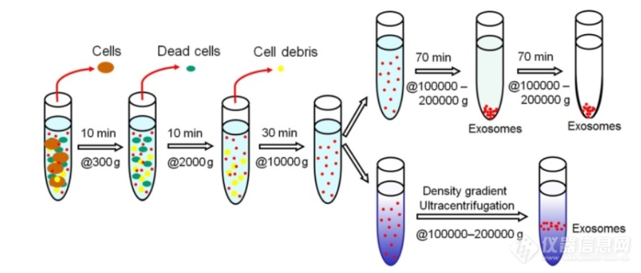
[font='times new roman'][size=18px][color=#000000]基于超速离心的外泌体分离技术[/color][/size][/font][align=left][font='times new roman'][size=16px]超速离心法([/size][/font][font='times new roman'][size=16px]UC[/size][/font][font='times new roman'][size=16px])是目前外泌体分离的“金标准”,大约[/size][/font][font='times new roman'][size=16px]56%[/size][/font][font='times new roman'][size=16px]的实验人员使用这种技术分离外泌体。目前[/size][/font][font='times new roman'][size=16px]UC[/size][/font][font='times new roman'][size=16px]包括差速超速离心和密度梯度超速离心。差速超速离心分离外泌体的方法主要受颗粒的大小、密度和形状的影响,基于颗粒的沉降速率不同,通过施加离心力,样品可以根据它们的物理性质被分离。在相同的颗粒密度下,大颗粒的沉积速度比小颗粒快,因此,更小的颗粒,如外泌体,可以通过一系列连续增加的旋转速度分离出来,具体步骤如图所示。首先用[/size][/font][font='times new roman'][size=16px]300 g[/size][/font][font='times new roman'][size=16px],[/size][/font][font='times new roman'][size=16px]2000 g[/size][/font][font='times new roman'][size=16px],[/size][/font][font='times new roman'][size=16px]10000 g[/size][/font][font='times new roman'][size=16px]的转速分别去除培养基中的细胞、坏死细胞和细胞碎片,上清液继续进行[/size][/font][font='times new roman'][size=16px]100,000 g 70[/size][/font][font='times new roman'][size=16px]分钟的超速离心,沉淀部分重悬在磷酸盐([/size][/font][font='times new roman'][size=16px]PBS[/size][/font][font='times new roman'][size=16px])缓冲液中进行另一轮[/size][/font][font='times new roman'][size=16px]100,000 g[/size][/font][font='times new roman'][size=16px]超速离心,最后,将得到的外泌体重悬于[/size][/font][font='times new roman'][size=16px]PBS[/size][/font][font='times new roman'][size=16px]缓冲液中以作下一步分析。[/size][/font][/align][align=left][font='times new roman'][size=16px]密度梯度离心[/size][/font][font='times new roman'][size=16px]法将待测生物样品添加到自上而下密度逐步增大的溶液中,在超速离心之后,这些外泌体就会移动到对应密度梯度层的底部(外泌体的密度介于[/size][/font][font='times new roman'][size=16px]1.10-1.21 g/mL[/size][/font][font='times new roman'][size=16px])。密度梯度离心法获得的外泌体具有更好的完整性和生物活性。此外,由于[/size][/font][font='times new roman'][size=16px]外泌体[/size][/font][font='times new roman'][size=16px]与胞外囊泡的大小存在重叠且外泌体存在异质性,差速超速离心分离得到的外泌体纯度和效率均较低,而密度梯度离心法使密度相对较低的外泌体漂浮,进一步净化了外泌体。[/size][/font][/align][font='times new roman'][size=16px]虽然[/size][/font][font='times new roman'][size=16px]UC[/size][/font][font='times new roman'][size=16px]是目前最常用的方法,但它也存在一些缺点:它是一种劳动密集型、耗时的方法(通常持续[/size][/font][font='times new roman'][size=16px]5-10 h[/size][/font][font='times new roman'][size=16px]),需要大量的样品和昂贵的专用设备。聚集的蛋白质和核蛋白颗粒污染使得分离的外泌体的效率和纯度相对较低。此[/size][/font][font='times new roman'][size=16px][color=#000000]外,[/color][/size][/font][font='times new roman'][size=16px]分离过程中需要超高的离心力,这可能会导致外泌体的形态和组成发生变化。[/size][/font][align=center][img]https://ng1.17img.cn/bbsfiles/images/2021/08/202108012208553147_7986_5111497_3.png[/img][/align][align=center][font='times new roman']图[/font][font='times new roman'] 1 [/font][font='times new roman']用差速超离心法分离外泌体示意图[/font][/align]
用GC7890-5975MSD检测经索氏萃取后的十溴联苯醚标准样品,除了有十溴联苯醚的提取离子外,还有647.5的(它不是一溴到九溴联苯醚的提取离子),不知道它是什么物质的
何为细胞外泌体? 外泌体最早发现于体外培养的绵羊红细胞上清液中,是细胞主动分泌的大小较为均一,直径为40~100纳米,密度1.10~1.18 g/ml的囊泡样小体。细胞外泌体携带多种蛋白质、mRNA、miRNA,参与细胞通讯、细胞迁移、血管新生和肿瘤细胞生长等过程并且有可能成为药物的天然载体,应用于临床治疗。 然而,测量技术手段的局限限制了外泌体在这些领域的研究进展。所以,在这篇文章中,作者总结了外泌体的纯化方法(离心法、过滤离心法、密度梯度离心法、免疫磁珠法以及色谱法),比较了现存各种外泌体测量技术(电子显微镜、动态光散射技术及纳米微粒追踪分析术)在外泌体尺寸和表征研究中的应用。原文点击——综述:细胞外泌体颗粒表征测量技术新进展

黄芩提取物-黄芩苷的测定http://ng1.17img.cn/bbsfiles/images/2012/12/201212091347_411149_2369266_3.jpg http://ng1.17img.cn/bbsfiles/images/2012/12/201212091349_411153_2369266_3.jpg 黄芩茎叶 http://ng1.17img.cn/bbsfiles/images/2012/12/201212091348_411151_2369266_3.jpg http://ng1.17img.cn/bbsfiles/images/2012/12/201212091350_411154_2369266_3.jpg黄芩根部http://ng1.17img.cn/bbsfiles/images/2012/12/201212091350_411155_2369266_3.jpg黄芩提取物-黄芩苷黄芩提取物-黄芩苷C21H18O11446.37黄芩苷药理作用:⑴抗炎抗变态反应,黄芩甙、黄芩甙对豚鼠离体气管过敏性收缩及整体动物过敏性气喘,均有缓解作用;⑵抗病毒、抗微生物(菌)作用;⑶镇静、解热、解痉作用;⑷抗癌、降压、利尿作用;⑸对血脂及血糖上升的作用;⑹利胆、保肝作用;⑺可降低乙醇所致的甘油三酸酯水平等。实验室测定方法名称:黄芩提取物-黄芩苷的测定-高效液相色谱法应用范围:本方法采用高效液相色谱法测定黄芩提取物中黄芩苷的含量。本方法适用于黄芩经加工制成的提取物。方法原理:本品加甲醇溶解、稀释,摇匀,滤过,续滤液进入高效液相色谱仪进行色谱分离,用紫外吸收检测器,于波长280nm处检测黄芩苷的吸收值,计算出其含量。试剂:1.甲醇2.磷酸仪器设备:高效液相色谱仪(配带紫外检测器)色谱条件:1.色谱柱:Xtimate C18柱2.流动相:甲醇 水 磷酸 =47 53 0.23.流速:1.0ml/min4.检测波长:280nm5.柱温:室温样品的制备:1.对照品溶液的制备精密称取黄芩苷对照品适量
[font='times new roman'][size=16px]荧光表征外泌体捕获和释放[/size][/font][font='times new roman'][size=16px]为了进一步阐明外泌体的捕获和释放过程,采用[/size][/font][font='times new roman'][size=16px]Tim4@ILI-01[/size][/font][font='times new roman'][size=16px]材料[/size][/font][font='times new roman'][size=16px]富集外泌体后,分别加入[/size][/font][font='times new roman'][size=16px]CD63[/size][/font][font='times new roman'][size=16px]和[/size][/font][font='times new roman'][size=16px]TSG101[/size][/font][font='times new roman'][size=16px]抗体进行孵育,经过[/size][/font][font='times new roman'][size=16px]3[/size][/font][font='times new roman'][size=16px]次洗涤后,加入[/size][/font][font='times new roman'][size=16px]FITC[/size][/font][font='times new roman'][size=16px]标记的山羊抗兔[/size][/font][font='times new roman'][size=16px]IgG[/size][/font][font='times new roman'][size=16px]进行避光孵育。通过免疫荧光实验对捕获的外泌体进行特异性识别。如图所示,[/size][/font][font='times new roman'][size=16px]Tim4@ILI-01[/size][/font][font='times new roman'][size=16px]材料[/size][/font][font='times new roman'][size=16px]经过后续的免疫反应未见荧光信号([/size][/font][font='times new roman'][size=16px]图[/size][/font][font='times new roman'][size=16px]A[/size][/font][font='times new roman'][size=16px],[/size][/font][font='times new roman'][size=16px]D[/size][/font][font='times new roman'][size=16px])[/size][/font][font='times new roman'][size=16px]。[/size][/font][font='times new roman'][size=16px]捕获外泌体[/size][/font][font='times new roman'][size=16px]后[/size][/font][font='times new roman'][size=16px]的材料表面可[/size][/font][font='times new roman'][size=16px]观察[/size][/font][font='times new roman'][size=16px]到强的[/size][/font][font='times new roman'][size=16px]荧光信号[/size][/font][font='times new roman'][size=16px]([/size][/font][font='times new roman'][size=16px]图[/size][/font][font='times new roman'][size=16px]B[/size][/font][font='times new roman'][size=16px],[/size][/font][font='times new roman'][size=16px]E[/size][/font][font='times new roman'][size=16px])[/size][/font][font='times new roman'][size=16px]。添加[/size][/font][font='times new roman'][size=16px]EDTA[/size][/font][font='times new roman'][size=16px]螯合剂后[/size][/font][font='times new roman'][size=16px],外泌体与材料分离,[/size][/font][font='times new roman'][size=16px]洗脱液中存在荧光信号[/size][/font][font='times new roman'][size=16px]([/size][/font][font='times new roman'][size=16px]图[/size][/font][font='times new roman'][size=16px] C[/size][/font][font='times new roman'][size=16px],[/size][/font][font='times new roman'][size=16px]F[/size][/font][font='times new roman'][size=16px])[/size][/font][font='times new roman'][size=16px],表明捕获的外泌体已成功从[/size][/font][font='times new roman'][size=16px]Tim4@ILI-01[/size][/font][font='times new roman'][size=16px]免疫亲和材料中释放出来[/size][/font][font='times new roman'][size=16px]。[/size][/font][table][tr][td][align=center][img]https://ng1.17img.cn/bbsfiles/images/2023/06/202306302129594249_5113_5389809_3.jpeg[/img][/align][/td][/tr][/table][align=center][font='times new roman']图[/font][font='times new roman']外泌体捕获和释放过程的荧光图像:([/font][font='times new roman']A[/font][font='times new roman'],[/font][font='times new roman']D[/font][font='times new roman'])[/font][font='times new roman']Tim4@ILI-01[/font][font='times new roman']免疫亲和材料([/font][font='times new roman']B[/font][font='times new roman'],[/font][font='times new roman']E[/font][font='times new roman'])捕获外泌体后的[/font][font='times new roman']Tim4@ILI-01[/font][font='times new roman']免疫亲和材料([/font][font='times new roman']C[/font][font='times new roman'],[/font][font='times new roman']F[/font][font='times new roman'])洗脱液中释放的外泌体。比例尺:[/font][font='times new roman']1 [/font][font='times new roman']μ[/font][font='times new roman']m[/font][/align]
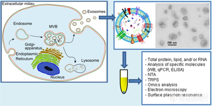
[align=center][font='times new roman'][size=20px][color=#000000]外泌体[/color][/size][/font][font='times new roman'][size=20px][color=#000000]的发生及组成[/color][/size][/font][/align][font='times new roman'][size=16px]1967[/size][/font][font='times new roman'][size=16px]年,[/size][/font][font='times new roman'][size=16px]Wolf[/size][/font][font='times new roman'][size=16px]在人血浆中首次发现一种来源于血小板膜泡的物质,并将其称之为“血小板尘埃”[/size][/font][font='times new roman'][size=16px][1][/size][/font][font='times new roman'][size=16px]。之后,所有的生物体液以及体外培养的细胞上清中都被检测到含有囊泡[/size][/font][font='times new roman'][size=16px][2-3][/size][/font][font='times new roman'][size=16px]。[/size][/font][font='times new roman'][size=16px]1983[/size][/font][font='times new roman'][size=16px]年,[/size][/font][font='times new roman'][size=16px]Pan[/size][/font][font='times new roman'][size=16px]首次在大鼠网织红细胞中观察到其分泌这类[/size][/font][font='times new roman'][size=16px][color=#191919]内吞囊泡[/color][/size][/font][font='times new roman'][size=16px][color=#191919][[/color][/size][/font][font='times new roman'][size=16px][color=#191919]4[/color][/size][/font][font='times new roman'][size=16px][color=#191919]][/color][/size][/font][font='times new roman'][size=16px]。几年后,该领域的先驱[/size][/font][font='times new roman'][size=16px]Johnstone[/size][/font][font='times new roman'][size=16px]将这种囊泡命名为“外泌体”[/size][/font][font='times new roman'][size=16px][5-6][/size][/font][font='times new roman'][size=16px]。最初,外泌体[/size][/font][font='times new roman'][size=16px]被认为是[/size][/font][font='times new roman'][size=16px]细胞产生的代谢废物。然而,最近的研究表明外泌体参与细胞间通讯,[/size][/font][font='times new roman'][size=16px]是细胞微环境和旁分泌信号系统的重要组成部分[/size][/font][font='times new roman'][size=16px][7-8][/size][/font][font='times new roman'][size=16px]。来自癌细胞的外泌体已被[/size][/font][font='times new roman'][size=16px]证实[/size][/font][font='times new roman'][size=16px]可以促进[/size][/font][font='times new roman'][size=16px]肿瘤发生、进展、转移、迁移和化疗耐药[/size][/font][font='times new roman'][size=16px][9][/size][/font][font='times new roman'][size=16px]。[/size][/font][font='times new roman'][size=16px]因此,外泌体是理想的非侵入性诊断和预后的生物标志物[/size][/font][align=left][font='times new roman'][size=18px][color=#000000]1[/color][/size][/font][font='times new roman'][size=18px][color=#000000] [/color][/size][/font][font='times new roman'][size=18px][color=#000000]外泌体的[/color][/size][/font][font='times new roman'][size=18px][color=#000000]发生[/color][/size][/font][/align][font='times new roman'][size=16px]根据产生机制和大小的不同,细胞外囊泡主要分为三种类型[/size][/font][font='times new roman'][size=16px]: [/size][/font][font='times new roman'][size=16px]外泌体、微泡颗粒和凋亡小体。不同于微泡颗粒和凋亡小体从质膜上直接释放的过程,[/size][/font][font='times new roman'][size=16px]外泌体的形成[/size][/font][font='times new roman'][size=16px]类似于反向内吞作用[/size][/font][font='times new roman'][size=16px][10-11][/size][/font][font='times new roman'][size=16px]。近年来,科学家进一步研究了外泌体的释放过程,虽然具体的调控机制还不是很清楚,但是[/size][/font][font='times new roman'][size=16px]大体由几个步骤组成:晚期核内体[/size][/font][font='times new roman'][size=16px]/[/size][/font][font='times new roman'][size=16px]多泡体[/size][/font][font='times new roman'][size=16px]([/size][/font][font='times new roman'][size=16px]MVBs[/size][/font][font='times new roman'][size=16px])腔内囊泡[/size][/font][font='times new roman'][size=16px]([/size][/font][font='times new roman'][size=16px]ILVs[/size][/font][font='times new roman'][size=16px])的形成、[/size][/font][font='times new roman'][size=16px]MVBs[/size][/font][font='times new roman'][size=16px]向细胞膜的转运以及[/size][/font][font='times new roman'][size=16px]MVBs[/size][/font][font='times new roman'][size=16px]与细胞膜的融合[/size][/font][font='times new roman'][size=16px][12][/size][/font][font='times new roman'][size=16px]。外泌体的形成过程如图[/size][/font][font='times new roman'][size=16px]1-1[/size][/font][font='times new roman'][size=16px]所示,首先细胞膜通过向内凹陷形成早期核内体;核内体膜向内出芽导致腔内囊泡在大的多泡体内逐渐积累;随后,细胞内多泡体可以运输到溶酶体内降解,或者,也可以与细胞膜特定部位融合释放其内部的小囊泡到胞外形成外泌体[/size][/font][font='times new roman'][size=16px][13-15][/size][/font][font='times new roman'][size=16px]。内吞体分选复合物([/size][/font][font='times new roman'][size=16px]Endosomal Sorting complex for Transport, ESCRTs[/size][/font][font='times new roman'][size=16px])是一种调控多泡体的生物发生[/size][/font][font='times new roman'][size=16px]/[/size][/font][font='times new roman'][size=16px]降解机制的多蛋白复合物。[/size][/font][font='times new roman'][size=16px]ESCRT[/size][/font][font='times new roman'][size=16px]多蛋白复合物组分、磷脂和泛素之间的联系为多泡体的生物发生和转运提供新的见解。而且[/size][/font][font='times new roman'][size=16px]ILVs[/size][/font][font='times new roman'][size=16px]中蛋白质的组成也受转运机制所需的[/size][/font][font='times new roman'][size=16px]ESCRTs[/size][/font][font='times new roman'][size=16px]的调节。研究表明外泌体的释放速度和蛋白组成与特定的[/size][/font][font='times new roman'][size=16px]ESCRT[/size][/font][font='times new roman'][size=16px]蛋白的缺失有关[/size][/font][font='times new roman'][size=16px][16-19][/size][/font][font='times new roman'][size=16px]。目前为止,外泌体是唯一一种起源于细胞内的膜泡。[/size][/font][align=left][img]https://ng1.17img.cn/bbsfiles/images/2021/08/202108012204290582_7776_5111497_3.png[/img][/align][align=center][font='times new roman']图[/font][font='times new roman']1[/font][font='times new roman']外泌体分泌及其结构示意图[/font][font='times new roman'][size=13px][12][/size][/font][/align][align=center][font='times new roman']Figure [/font][font='times new roman']1 The exosome secretion and its structure diagram[/font][font='times new roman'][size=13px][12][/size][/font][/align][align=center][/align][font='times new roman'][size=18px][color=#000000]2 [/color][/size][/font][font='times new roman'][size=18px][color=#000000]外泌体的组成[/color][/size][/font][align=left][font='times new roman'][size=16px]外泌体的生物组成不仅反映了母细胞的组成,而且反映了其受调节的分选机制[/size][/font][font='times new roman'][size=16px][20][/size][/font][font='times new roman'][size=16px]。外泌体主要包含各种受体、转录因子、酶、细胞外基质蛋白、脂质、外泌体内部和表面的核酸([/size][/font][font='times new roman'][size=16px]DNA[/size][/font][font='times new roman'][size=16px]、[/size][/font][font='times new roman'][size=16px]mRNA[/size][/font][font='times new roman'][size=16px]和[/size][/font][font='times new roman'][size=16px]miRNA[/size][/font][font='times new roman'][size=16px])在内的各种蛋白质的复合体[/size][/font][font='times new roman'][size=16px][21-22][/size][/font][font='times new roman'][size=16px]。对外泌体蛋白质组成的分析表明,有些蛋白质特异性地起源于细胞和组织,例如粘附分子、整合素[/size][/font][font='times new roman'][size=16px][23][/size][/font][font='times new roman'][size=16px];有些则是所有外泌体共有的,包括一系列融合和转移蛋白,如[/size][/font][font='times new roman'][size=16px]Rab2[/size][/font][font='times new roman'][size=16px]、[/size][/font][font='times new roman'][size=16px]Rab7[/size][/font][font='times new roman'][size=16px]、[/size][/font][font='times new roman'][size=16px]flotillin[/size][/font][font='times new roman'][size=16px]和[/size][/font][font='times new roman'][size=16px]annexin[/size][/font][font='times new roman'][size=16px],热休克蛋白,如[/size][/font][font='times new roman'][size=16px]Hsc70[/size][/font][font='times new roman'][size=16px]和[/size][/font][font='times new roman'][size=16px]Hsc90[/size][/font][font='times new roman'][size=16px],以及介导多泡体形成的蛋白,如[/size][/font][font='times new roman'][size=16px]Alix[/size][/font][font='times new roman'][size=16px][24][/size][/font][font='times new roman'][size=16px]。[/size][/font][/align][align=left][font='times new roman'][size=16px]此外,[/size][/font][font='times new roman'][size=16px]外泌体还包含多种类型的脂质。[/size][/font][font='times new roman'][size=16px]脂类不仅在保护外泌体形状方面发挥重要作用,还参与外泌体的生物发生和受体细胞内的稳态调节[/size][/font][font='times new roman'][size=16px][25-27][/size][/font][font='times new roman'][size=16px]。多泡体内膜中溶双磷脂酸([/size][/font][font='times new roman'][size=16px]LBPA[/size][/font][font='times new roman'][size=16px])等高密度脂质促进腔内囊泡的形成[/size][/font][font='times new roman'][size=16px][28][/size][/font][font='times new roman'][size=16px]。[/size][/font][font='times new roman'][size=16px]LBPA[/size][/font][font='times new roman'][size=16px]与[/size][/font][font='times new roman'][size=16px]Alix[/size][/font][font='times new roman'][size=16px]的相互作用促进了多泡体膜向内出芽[/size][/font][font='times new roman'][size=16px][29][/size][/font][font='times new roman'][size=16px]。鞘磷脂、磷脂酰胆碱和[/size][/font][font='times new roman'][size=16px]BMP[/size][/font][font='times new roman'][size=16px]是区分囊泡类型的主要因素。不同类型的微[/size][/font][font='times new roman'][size=16px][color=#000000]囊[/color][/size][/font][font='times new roman'][size=16px]泡磷脂酰胆碱含量相近,而鞘磷脂在外泌体中浓度会偏高,[/size][/font][font='times new roman'][size=16px]BMP[/size][/font][font='times new roman'][size=16px]是外泌体所独有的脂质成分[/size][/font][font='times new roman'][size=16px][30-31][/size][/font][font='times new roman'][size=16px]。此外,研究表明,转移到靶细胞的外泌体可以改变受体细胞的脂质组成,特别是胆固醇和鞘磷脂,从而影响细胞内稳态。[/size][/font][/align][font='times new roman'][size=16px]外泌体中存在大量的核酸,调节着受体细胞的活动[/size][/font][font='times new roman'][size=16px][32][/size][/font][font='times new roman'][size=16px]。[/size][/font][font='times new roman'][size=16px][color=#000000]外泌体携带的[/color][/size][/font][font='times new roman'][size=16px][color=#000000]RNA[/color][/size][/font][font='times new roman'][size=16px][color=#000000]可以在多种细胞间转移,[/color][/size][/font][font='times new roman'][size=16px][color=#000000]为受体靶点提供生理和病理功能,[/color][/size][/font][font='times new roman'][size=16px][color=#000000]因此被称为[/color][/size][/font][font='times new roman'][size=16px][color=#000000]“[/color][/size][/font][font='times new roman'][size=16px][color=#000000]外泌体穿梭[/color][/size][/font][font='times new roman'][size=16px][color=#000000]RNA[/color][/size][/font][font='times new roman'][size=16px][color=#000000]”[/color][/size][/font][font='times new roman'][size=16px][color=#000000]。近年来,通过广泛的研究,已在不同物种组织的纳米囊泡中发现了[/color][/size][/font][font='times new roman'][size=16px][color=#000000]764[/color][/size][/font][font='times new roman'][size=16px][color=#000000]个[/color][/size][/font][font='times new roman'][size=16px][color=#000000]miRNAs[/color][/size][/font][font='times new roman'][size=16px][color=#000000]和[/color][/size][/font][font='times new roman'][size=16px][color=#000000]1639[/color][/size][/font][font='times new roman'][size=16px][color=#000000]个[/color][/size][/font][font='times new roman'][size=16px][color=#000000]mRNA[/color][/size][/font][font='times new roman'][size=16px][color=#000000][33][/color][/size][/font][font='times new roman'][size=16px][color=#000000]。外泌体的组成因不同病理生理状态和起源细胞的不同而不同。[/color][/size][/font][font='times new roman'][size=16px][color=#000000]外泌体作为遗传信息的载体和传递者在细胞生物学中具有重要意[/color][/size][/font][font='times new roman'][size=16px][color=#000000]义。[/color][/size][/font][font='times new roman'][size=21px][color=#000000]主要[/color][/size][/font][font='times new roman'][size=21px][color=#000000]参考文献[/color][/size][/font][1] [font='times new roman'][color=#000000]Wolf P. The Nature and Significance of Platelet Products in Human Plasma[J]. [/color][/font][font='times new roman'][color=#000000]British Journal of Haematology,[/color][/font][font='times new roman'][color=#000000] 1967, 13(3):269-288.[/color][/font][2] [font='times new roman'][color=#000000]Raposo G, Stoorvogel W. Extracellular vesicles: Exosomes, microvesicles, and friends[J]. Journal of Cell Biology, 2013, 200(4):373-383.[/color][/font][3] [font='times new roman'][color=#000000]Yanez-Mo M, Siljander P[/color][/font][font='times new roman'][color=#000000] [/color][/font][font='times new roman'][color=#000000]R, Andreu Z[/color][/font][font='times new roman'][color=#000000], et al. [/color][/font][font='times new roman'][color=#000000]Biological properties of extracellular vesicles and their physiological functions[/color][/font][font='times new roman'][color=#000000][J]. J Extracell Vesicles, 2015, 4:27066.[/color][/font][4] [font='times new roman'][color=#000000]Harding C, Heuser J, Stahl P[/color][/font][font='times new roman'][color=#000000].[/color][/font][font='times new roman'][color=#000000] Receptor-mediated endocytosis of transferrin and recycling of the transferrin receptor in rat reticulocytes.[J]. The Journal of Cell Biology, 1983, 97(2):329-339.[/color][/font][5] [font='times new roman'][color=#000000]Johnstone R M. Revisiting the road to the discovery of exosomes[J]. Blood Cells Molecules & Diseases, 2005, 34(3):214-219.[/color][/font][6] [font='times new roman'][color=#000000]Johnstone R M, Adam M, Hammond J R, et al. [/color][/font][font='times new roman'][color=#000000]Vesicle formation during reticulocyte maturation. Association of plasma membrane activities with released vesicles (exosomes)[/color][/font][font='times new roman'][color=#000000][J]. Journal of Biological Chemistry, 1987, 262[/color][/font][font='times new roman'][color=#000000](19):9412-9420[/color][/font][font='times new roman'][color=#000000].[/color][/font][7] [font='times new roman'][color=#000000]Mathivanan S, Ji H, Simpson R J . Exosomes: extracellular organelles important in intercellular communication[J]. Journal of Proteomics, 2010, 73(10):1907-1920.[/color][/font][8] [font='times new roman'][color=#000000]Record M, Carayon K, Poirot M, et al. Exosomes as new vesicular lipid transporters involved in cell-cell communication and various pathophysiologies[J]. Biochimica et Biophysica Acta, 2014, 1841(1):108-120.[/color][/font][9] [font='times new roman'][color=#000000]Becker A, Thakur B K , Weiss J M, et al. Extracellular Vesicles in Cancer: Cell-to-Cell Mediators of Metastasis[J]. Cancer Cell, 2016, 30(6):836-848.[/color][/font][10] [font='times new roman'][color=#000000]Pan B T, Teng K, Wu C, et al. Electron microscopic evidence for externalization of the transferrin receptor in vesicular form in sheep reticulocytes[J]. Journal of Cell Biology, 1985, 101(3):942-948.[/color][/font]
方法:将富集过后的外泌体与底衬材料一起放在酶标板里,顶读模式,孵育CD63荧光一抗,定量抗体响应量,作为外泌体的定量标准
[align=center][size=18px]外[/size][size=18px]泌[/size][size=18px]体[/size][size=18px]在[/size][size=18px]肝癌的预后及靶[/size][size=18px]向治疗[/size][size=18px]中的[/size][size=18px]作用[/size][/align][size=16px]肝细胞癌([/size][size=16px]HCC[/size][size=16px])是世界上第三大常见恶性肿瘤,占原发性肝癌的[/size][size=16px]85%-90%[/size][font='times new roman'][sup][size=16px][1-3][/size][/sup][/font][size=16px]。肝细胞癌新发病例估计约为[/size][size=16px]841[/size][size=16px],[/size][size=16px]000[/size][size=16px]例[/size][font='times new roman'][sup][size=16px][4][/size][/sup][/font][size=16px]。乙型肝炎病毒([/size][size=16px]HBV[/size][size=16px])感染被认为是中国发生肝细胞癌的主要危险因素[/size][font='times new roman'][sup][size=16px][5][/size][/sup][/font][size=16px]。尽管影像技术、化疗、介入放射学、外科技术和肝移植近年取得了新的突破,晚期肝癌患者预后依然很差,至今没有有效的治疗[/size][font='times new roman'][sup][size=16px][6-8][/size][/sup][/font][size=16px]。目前,肝细胞癌的[/size][size=16px]5[/size][size=16px]年生存率不超过[/size][size=16px]20%[/size][font='times new roman'][sup][size=16px][9[/size][/sup][/font][font='times new roman'][sup][size=16px],[/size][/sup][/font][font='times new roman'][sup][size=16px]10][/size][/sup][/font][size=16px],[/size][size=16px]然而,[/size][size=16px]早期诊断可显著降低肝癌患者的死亡率[/size][font='times new roman'][sup][size=16px][11][/size][/sup][/font][size=16px]。因此,提高肝细胞癌的总生存率依赖于对肝细胞癌的生物学行为机制的深入研究和新治疗靶点的开发。[/size][size=16px]外[/size][size=16px]泌[/size][size=16px]体是小的纳米级[/size][size=16px]细胞外[/size][size=16px]囊泡,[/size][size=16px]是细胞间通讯的重要介质[/size][size=16px],通过多种生物分子(如蛋白质、[/size][size=16px]RNA[/size][size=16px]和[/size][size=16px]DNA[/size][size=16px])调节细胞之间的微环境和免疫系统[/size][font='times new roman'][sup][size=16px][4[/size][/sup][/font][font='times new roman'][sup][size=16px], [/size][/sup][/font][font='times new roman'][sup][size=16px]12[/size][/sup][/font][font='times new roman'][sup][size=16px]-[/size][/sup][/font][font='times new roman'][sup][size=16px]13][/size][/sup][/font][size=16px]以及细胞的遗传和表观遗传机制[/size][font='times new roman'][sup][size=16px][14][/size][/sup][/font][size=16px]。研究表明,外[/size][size=16px]泌体不仅[/size][size=16px]将下游信号传递给靶细胞,还将遗传物质转移到下游细胞,为细胞间通讯提供了新的机制[/size][font='times new roman'][sup][size=16px][4][/size][/sup][/font][size=16px]。[/size][size=16px]已有的研究结果表明[/size][size=16px],[/size][size=16px]Vesiclepedia[/size][size=16px]、[/size][size=16px]EVpedia[/size][size=16px]、[/size][size=16px]Exocarta[/size][size=16px]等数据库提供的不同类型的外[/size][size=16px]泌[/size][size=16px]体成分存在于生理或病理条件相似的不同细胞中[/size][font='times new roman'][sup][size=16px][15-17][/size][/sup][/font][size=16px]。此外,外[/size][size=16px]泌[/size][size=16px]体还参与血管生成、肿瘤细胞转移和正常细胞向肿瘤细胞的转化[/size][font='times new roman'][sup][size=16px][18][/size][/sup][/font][size=16px]。许多类型的细胞,如间充质细胞、免疫细胞和肿瘤细胞可以诱导外[/size][size=16px]泌[/size][size=16px]体的释放[/size][size=16px],[/size][size=16px]外[/size][size=16px]泌[/size][size=16px]体在这些细胞中[/size][size=16px]上调[/size][size=16px],[/size][size=16px]间接[/size][size=16px]表明外[/size][size=16px]泌体参与[/size][size=16px]肿瘤的发生、发展、转移、免疫逃逸和耐药性。此外,肿瘤来源的外[/size][size=16px]泌[/size][size=16px]体中含有大量的肿瘤相关[/size][size=16px]的生物[/size][size=16px]标志物[/size][size=16px]([/size][size=16px]如[/size][size=16px]miRNAs[/size][size=16px])[/size][size=16px],可用于检测早期[/size][size=16px]HCC[/size][font='times new roman'][sup][size=16px][20][/size][/sup][/font][size=16px]。因此[/size][size=16px],[/size][size=16px]外[/size][size=16px]泌[/size][size=16px]体对肝癌的预后及靶[/size][size=16px]向治疗[/size][size=16px]可能发挥了重要作用[/size][size=16px]由于外[/size][size=16px]泌[/size][size=16px]体携带的生物分子与来源细胞具有一致性,近年来,肿瘤源性外[/size][size=16px]泌[/size][size=16px]体([/size][size=16px]Tumor-derived exosomes[/size][size=16px],[/size][size=16px]TDE[/size][size=16px])作为肿瘤分子标志物在肿瘤诊治中的应用得到了越来越多的关注。研究发现,与健康对照组比较,[/size][size=16px]HCC [/size][size=16px]患者血清中有[/size][size=16px]19[/size][size=16px]种[/size][size=16px]miRNAs[/size][size=16px]显著增加。其中,[/size][size=16px]miR-210[/size][size=16px]分泌由[/size][size=16px]HCC[/size][size=16px]细胞来源的外[/size][size=16px]泌[/size][size=16px]体[/size][size=16px]介[/size][size=16px]导,因此,[/size][size=16px]miR-210[/size][size=16px]也可作为[/size][size=16px]HCC[/size][size=16px]诊断的生物学标志物之一[/size][font='times new roman'][sup][size=16px][[/size][/sup][/font][font='times new roman'][sup][size=16px]21[/size][/sup][/font][font='times new roman'][sup][size=16px]][/size][/sup][/font][size=16px]。[/size][size=16px]Pu[/size][size=16px]等[/size][font='times new roman'][sup][size=16px][22][/size][/sup][/font][size=16px]研究发现,[/size][size=16px]HCC[/size][size=16px]患者血清中外[/size][size=16px]泌[/size][size=16px]体[/size][size=16px]has-miR-21-5p[/size][size=16px]和[/size][size=16px]has-miR-144-3p[/size][size=16px]表达水平显著高于慢性乙型肝炎[/size][size=16px]([/size][size=16px]CHB[/size][size=16px])[/size][size=16px]患者,[/size][size=16px]ROC[/size][size=16px]曲线显示,两者在[/size][size=16px]HCC[/size][size=16px]诊断上的表现优于甲胎蛋白,敏感度和特异性更高,作为肿瘤分子标志物具有良好的临床应用前景。肿瘤微环境是由肿瘤细胞、血管内皮细胞、成纤维细胞、抗原递呈细胞以及细胞外基质、生长因子、炎性细胞因子等组成。不同细胞外[/size][size=16px]泌[/size][size=16px]体通过携带不同信号分子发挥细胞通讯作用,参与肝癌的发生、转移、血管生成以及免疫抑制型微环境构建。当癌基因表达过度活化或抑癌基因表达过度受抑时,细胞表现为增殖能力的过度增强和凋亡能力的过度削弱,进而导致肿瘤的发生。[/size]
[align=left][font='calibri'][size=16px]外泌体[/size][/font][font='calibri'][size=16px]作为标志物用于肿瘤的早期诊断[/size][/font][/align][font='times new roman'][size=16px]外泌体是一种磷脂双层膜包裹的生物纳米颗粒,直径为[/size][/font][font='times new roman'][size=16px]30-200 nm[/size][/font][font='times new roman'][size=16px]。近年来,由于外泌体携带大量遗传物质,在介导细胞[/size][/font][font='times new roman'][size=16px]-[/size][/font][font='times new roman'][size=16px]细胞间通信中发挥重要作用,已成为液体活检最理想的分析目标。[/size][/font][font='times new roman'][size=16px]肿瘤生物标志物的检测可以协助肿瘤的早期诊断和治疗。近年来,外泌体在肿瘤的发生发展过程中显示出重要的作用,逐渐成为重要的诊断生物标志物和治疗靶标。肿瘤来源的外泌体携带分子和基因载体,反映了肿瘤细胞的部分组成,广泛分布于血液和包括尿液在内的其他体液。这些外泌体可以作为一种微创的液体活组织检查,对肿瘤进行实时的非侵入性分析,帮助疾病的临床诊断。外泌体作为疾病诊断和预后的标志物,相比于传统的[/size][/font][font='times new roman'][size=16px]miRNA[/size][/font][font='times new roman'][size=16px]分子具有明显的优势。肿瘤细胞衍生的外泌体携带的[/size][/font][font='times new roman'][size=16px]mRNA[/size][/font][font='times new roman'][size=16px]、[/size][/font][font='times new roman'][size=16px]miRNAs[/size][/font][font='times new roman'][size=16px]和蛋白质等生物活性分子通常构成了肿瘤细胞的总蛋白质组或转录组的一部分,反映了亲本细胞的遗传信息。而且这些生物活性分子由于受到外泌体双层膜结构的保护使其在运输过程中不被降解。此外,这些外泌体在不同的储存情况下,包括在[/size][/font][font='times new roman'][size=16px]4[/size][/font][font='times new roman'][size=16px]℃下短期储存[/size][/font][font='times new roman'][size=16px]96 h[/size][/font][font='times new roman'][size=16px]或在[/size][/font][font='times new roman'][size=16px]-80[/size][/font][font='times new roman'][size=16px]℃下长期储存,都具有很强的活力。外泌体的这些特性使其成为癌症诊断、预后和治疗反应的重要监测指标。[/size][/font][font='times new roman'][size=16px]多项体外研究表明,通过应用蛋白质组学和转录组学的方法,使用外泌体及其内容物和表面蛋白可以更早地发现癌症,从而提高预后和生存率[/size][/font][font='times new roman'][size=16px][43][/size][/font][font='times new roman'][size=16px]。结肠癌患者血浆标本中外泌体水平的提高与肿瘤细胞的整体存活率的下降有相当大的联系[/size][/font][font='times new roman'][size=16px][32][/size][/font][font='times new roman'][size=16px]。另一项研究表明,外泌体上的[/size][/font][font='times new roman'][size=16px]CD24[/size][/font][font='times new roman'][size=16px]、[/size][/font][font='times new roman'][size=16px]EDIL3[/size][/font][font='times new roman'][size=16px]和纤维连接蛋白被认为是早期乳腺癌的标志物。此外,在非小细胞肺癌的外泌体中,研究人员发现表皮生长因子受体([/size][/font][font='times new roman'][size=16px]EGFR[/size][/font][font='times new roman'][size=16px])、胎盘碱性磷酸酶([/size][/font][font='times new roman'][size=16px]PLAP[/size][/font][font='times new roman'][size=16px])和富亮氨酸的α[/size][/font][font='times new roman'][size=16px]-2-[/size][/font][font='times new roman'][size=16px]糖蛋白[/size][/font][font='times new roman'][size=16px]1[/size][/font][font='times new roman'][size=16px]([/size][/font][font='times new roman'][size=16px]LRG1[/size][/font][font='times new roman'][size=16px])的过表达。最近的报告显示,使用无标记的液相色谱[/size][/font][font='times new roman'][size=16px]-[/size][/font][font='times new roman'][size=16px]串联质谱([/size][/font][font='times new roman'][size=16px][url=https://insevent.instrument.com.cn/t/Yp][color=#3333ff]LC-MS[/color][/url]/MS[/size][/font][font='times new roman'][size=16px])检测前列腺癌患者的尿液,研究人员发现存在于尿外泌体中的[/size][/font][font='times new roman'][size=16px]PCA3[/size][/font][font='times new roman'][size=16px]和[/size][/font][font='times new roman'][size=16px]TMPRSS2:ERG[/size][/font][font='times new roman'][size=16px]蛋白是潜在的生物标志物。这些发现表明从体液中分离得到的外泌体可能是肿瘤早期检测的重要非侵入性标记物。[/size][/font]
[align=center][font='times new roman'][size=18px][color=#000000]外[/color][/size][/font][font='times new roman'][size=18px][color=#000000]泌[/color][/size][/font][font='times new roman'][size=18px][color=#000000]体对耐药性的影响[/color][/size][/font][/align][font='times new roman'][size=16px][color=#000000]([/color][/size][/font][font='times new roman'][size=16px][color=#000000]1[/color][/size][/font][font='times new roman'][size=16px][color=#000000])分别用[/color][/size][/font][font='times new roman'][size=16px][color=#000000]0[/color][/size][/font][font='times new roman'][size=16px][color=#000000]、[/color][/size][/font][font='times new roman'][size=16px][color=#000000]1[/color][/size][/font][font='times new roman'][size=16px][color=#000000]、[/color][/size][/font][font='times new roman'][size=16px][color=#000000]10[/color][/size][/font][font='times new roman'][size=16px][color=#000000] [/color][/size][/font][font='times new roman'][size=16px][color=#000000]μ[/color][/size][/font][font='times new roman'][size=16px][color=#000000]M[/color][/size][/font][font='times new roman'][size=16px][color=#000000]的[/color][/size][/font][font='times new roman'][size=16px][color=#000000]含顺铂[/color][/size][/font][font='times new roman'][size=16px][color=#000000]培养基培养细胞,待[/color][/size][/font][font='times new roman'][size=16px][color=#000000]3[/color][/size][/font][font='times new roman'][size=16px][color=#000000]天后,收集各自培养上清,编号为[/color][/size][/font][font='times new roman'][size=16px][color=#000000]1[/color][/size][/font][font='times new roman'][size=16px][color=#000000]、[/color][/size][/font][font='times new roman'][size=16px][color=#000000]2[/color][/size][/font][font='times new roman'][size=16px][color=#000000]、[/color][/size][/font][font='times new roman'][size=16px][color=#000000]3[/color][/size][/font][font='times new roman'][size=16px][color=#000000]。[/color][/size][/font][font='times new roman'][size=16px][color=#000000]([/color][/size][/font][font='times new roman'][size=16px][color=#000000]2[/color][/size][/font][font='times new roman'][size=16px][color=#000000])经前期预处理后用[/color][/size][/font][font='times new roman'][size=16px]Tim4@ILI-01[/size][/font][font='times new roman'][size=16px]免疫亲和材料[/size][/font][font='times new roman'][size=16px]分别富集[/size][/font][font='times new roman'][size=16px]1[/size][/font][font='times new roman'][size=16px]、[/size][/font][font='times new roman'][size=16px]2[/size][/font][font='times new roman'][size=16px]、[/size][/font][font='times new roman'][size=16px]3[/size][/font][font='times new roman'][size=16px]培养基中的外[/size][/font][font='times new roman'][size=16px]泌[/size][/font][font='times new roman'][size=16px]体。[/size][/font][font='times new roman'][size=16px]([/size][/font][font='times new roman'][size=16px]3[/size][/font][font='times new roman'][size=16px])将另一瓶生长好的细胞消化处理计数,均匀的传到[/size][/font][font='times new roman'][size=16px]3[/size][/font][font='times new roman'][size=16px]个[/size][/font][font='times new roman'][size=16px]皿[/size][/font][font='times new roman'][size=16px]中。待细胞过夜后,将上述分离得到的外[/size][/font][font='times new roman'][size=16px]泌体加入[/size][/font][font='times new roman'][size=16px]这三个[/size][/font][font='times new roman'][size=16px]皿[/size][/font][font='times new roman'][size=16px]中培养[/size][/font][font='times new roman'][size=16px]3[/size][/font][font='times new roman'][size=16px]天。[/size][/font][font='times new roman'][size=16px]([/size][/font][font='times new roman'][size=16px]4[/size][/font][font='times new roman'][size=16px])分别将[/size][/font][font='times new roman'][size=16px]3[/size][/font][font='times new roman'][size=16px]个[/size][/font][font='times new roman'][size=16px]皿[/size][/font][font='times new roman'][size=16px]中的细胞按照上述步骤铺[/size][/font][font='times new roman'][size=16px]96[/size][/font][font='times new roman'][size=16px]孔板,检测用不同浓度[/size][/font][font='times new roman'][size=16px]顺铂处理[/size][/font][font='times new roman'][size=16px]细胞后,细胞分泌的外[/size][/font][font='times new roman'][size=16px]泌[/size][/font][font='times new roman'][size=16px]体对细胞耐药性的影响。[/size][/font][font='times new roman'][size=18px][color=#000000]顺铂处理[/color][/size][/font][font='times new roman'][size=18px][color=#000000]后细胞外[/color][/size][/font][font='times new roman'][size=18px][color=#000000]泌[/color][/size][/font][font='times new roman'][size=18px][color=#000000]体的分泌[/color][/size][/font][font='times new roman'][size=16px]为了[/size][/font][font='times new roman'][size=16px]进一步验证[/size][/font][font='times new roman'][size=16px]Tim4@ILI-01[/size][/font][font='times new roman'][size=16px]免疫亲和[/size][/font][font='times new roman'][size=16px]材料的捕获性能,[/size][/font][font='times new roman'][size=16px]采用[/size][/font][font='times new roman'][size=16px]不同浓度的[/size][/font][font='times new roman'][size=16px]顺铂处理[/size][/font][font='times new roman'][size=16px]H1299[/size][/font][font='times new roman'][size=16px]细胞来验证[/size][/font][font='times new roman'][size=16px]Tim4@ILI-01[/size][/font][font='times new roman'][size=16px]免疫亲和[/size][/font][font='times new roman'][size=16px]材料是否可以检测细胞培养上清液中外[/size][/font][font='times new roman'][size=16px]泌[/size][/font][font='times new roman'][size=16px]体分泌的变化。如图所示,[/size][/font][font='times new roman'][size=16px]随着顺铂浓度[/size][/font][font='times new roman'][size=16px]的增加,细胞活力[/size][/font][font='times new roman'][size=16px]持续下降[/size][/font][font='times new roman'][size=16px],[/size][/font][font='times new roman'][size=16px]细胞分泌[/size][/font][font='times new roman'][size=16px]外[/size][/font][font='times new roman'][size=16px]泌[/size][/font][font='times new roman'][size=16px]体[/size][/font][font='times new roman'][size=16px]的[/size][/font][font='times new roman'][size=16px]浓度随着药物浓度的增加呈现先小幅增加后持续下降的趋势。外[/size][/font][font='times new roman'][size=16px]泌[/size][/font][font='times new roman'][size=16px]体浓度的下降[/size][/font][font='times new roman'][size=16px]主要[/size][/font][font='times new roman'][size=16px]是由大多数细胞活性降低[/size][/font][font='times new roman'][size=16px]所致[/size][/font][font='times new roman'][size=16px]。活细胞数量及其形态变化进一步证实了[/size][/font][font='times new roman'][size=16px]顺铂仅[/size][/font][font='times new roman'][size=16px]在特定浓度下调控癌细胞外[/size][/font][font='times new roman'][size=16px]泌[/size][/font][font='times new roman'][size=16px]体的分泌[/size][/font][font='times new roman'][size=16px],[/size][/font][font='times new roman'][size=16px]顺铂诱导[/size][/font][font='times new roman'][size=16px]细胞毒性存在浓度依赖性机制[/size][/font][font='times new roman'][size=16px](图[/size][/font][font='times new roman'][size=16px]B[/size][/font][font='times new roman'][size=16px])[/size][/font][font='times new roman'][size=16px]。为了进一步验证外[/size][/font][font='times new roman'][size=16px]泌[/size][/font][font='times new roman'][size=16px]体是否能提高细胞的化疗耐药能力,我们用不同浓度的顺铂[/size][/font][font='times new roman'][size=16px]([/size][/font][font='times new roman'][size=16px]0 [/size][/font][font='times new roman'][size=16px]μM[/size][/font][font='times new roman'][size=16px],[/size][/font][font='times new roman'][size=16px]1[/size][/font][font='times new roman'][size=16px] [/size][/font][font='times new roman'][size=16px]μM[/size][/font][font='times new roman'][size=16px]和[/size][/font][font='times new roman'][size=16px]10[/size][/font][font='times new roman'][size=16px] [/size][/font][font='times new roman'][size=16px]μM[/size][/font][font='times new roman'][size=16px])[/size][/font][font='times new roman'][size=16px]处理细胞,[/size][/font][font='times new roman'][size=16px]其分泌的[/size][/font][font='times new roman'][size=16px]外[/size][/font][font='times new roman'][size=16px]泌[/size][/font][font='times new roman'][size=16px]体孵育[/size][/font][font='times new roman'][size=16px]下一代[/size][/font][font='times new roman'][size=16px]细胞后,细胞[/size][/font][font='times new roman'][size=16px]在相同刺激条件下[/size][/font][font='times new roman'][size=16px]存活率较对照组明显提高,以[/size][/font][font='times new roman'][size=16px]10[/size][/font][font='times new roman'][size=16px] [/size][/font][font='times new roman'][size=16px]μM[/size][/font][font='times new roman'][size=16px]浓度效果最好[/size][/font][font='times new roman'][size=16px](图[/size][/font][font='times new roman'][size=16px]C[/size][/font][font='times new roman'][size=16px])[/size][/font][font='times new roman'][size=16px]。[/size][/font][font='times new roman'][size=16px]实验[/size][/font][font='times new roman'][size=16px]表明,[/size][/font][font='times new roman'][size=16px]H1299[/size][/font][font='times new roman'][size=16px]细胞暴露于特定浓度[/size][/font][font='times new roman'][size=16px]的顺铂后[/size][/font][font='times new roman'][size=16px],外[/size][/font][font='times new roman'][size=16px]泌[/size][/font][font='times new roman'][size=16px]体的分泌[/size][/font][font='times new roman'][size=16px]量增加,并且这些[/size][/font][font='times new roman'][size=16px]外[/size][/font][font='times new roman'][size=16px]泌[/size][/font][font='times new roman'][size=16px]体[/size][/font][font='times new roman'][size=16px]可以[/size][/font][font='times new roman'][size=16px]降低其他[/size][/font][font='times new roman'][size=16px]H1299[/size][/font][font='times new roman'][size=16px]细胞[/size][/font][font='times new roman'][size=16px]对顺铂的[/size][/font][font='times new roman'][size=16px]敏感性,增加了它们[/size][/font][font='times new roman'][size=16px]对顺铂的[/size][/font][font='times new roman'][size=16px]耐药性。以上研究表明,来自肺腺癌的外[/size][/font][font='times new roman'][size=16px]泌[/size][/font][font='times new roman'][size=16px]体在肺癌化疗耐药中起着重要作用[/size][/font][font='times new roman'][size=16px],[/size][/font][font='times new roman'][size=16px]这个结果与之前的[/size][/font][font='times new roman'][size=16px]报道[/size][/font][font='times new roman'][size=16px]一致。[/size][/font][table][tr][td][img]https://ng1.17img.cn/bbsfiles/images/2021/08/202108012222057317_3122_5111497_3.jpeg[/img][/td][/tr][/table][align=center][font='times new roman']图[/font][font='times new roman']1[/font][font='times new roman']顺铂对[/font][font='times new roman']H1299[/font][font='times new roman']细胞外[/font][font='times new roman']泌[/font][font='times new roman']体分泌的影响:([/font][font='times new roman']A[/font][font='times new roman'])[/font][font='times new roman']H1299[/font][font='times new roman']细胞的剂量反应;([/font][font='times new roman']B[/font][font='times new roman'])不同浓度[/font][font='times new roman']顺铂[/font][font='times new roman']处理[/font][font='times new roman']H1299[/font][font='times new roman']细胞[/font][font='times new roman']3[/font][font='times new roman']天后的形态;([/font][font='times new roman']C[/font][font='times new roman'])外[/font][font='times new roman']泌[/font][font='times new roman']体作用后的[/font][font='times new roman']H1299[/font][font='times new roman']细胞[/font][font='times new roman']对顺铂的[/font][font='times new roman']剂量反应。[/font][font='times new roman'] [/font][font='times new roman']比例尺:[/font][font='times new roman']200 [/font][font='times new roman']μ[/font][font='times new roman']m[/font][/align]
[align=center][font='times new roman'][size=21px]肿瘤细胞分泌的外[/size][/font][font='times new roman'][size=21px]泌[/size][/font][font='times new roman'][size=21px]体在机体内的作用[/size][/font][/align][font='times new roman'][size=16px]摘要[/size][/font][font='times new roman'][size=16px] [/size][/font][font='times new roman'][size=16px]肿瘤细胞通过产生,释放和利用外[/size][/font][font='times new roman'][size=16px]泌[/size][/font][font='times new roman'][size=16px]体来促进肿瘤发生发展。肿瘤来源的外[/size][/font][font='times new roman'][size=16px]泌[/size][/font][font='times new roman'][size=16px]体在肿瘤中的作用机制以成为目前的研究热点。外[/size][/font][font='times new roman'][size=16px]泌[/size][/font][font='times new roman'][size=16px]体作为信息载体,可将遗传信息从肿瘤细胞传递到位于近处或远处的正常或其他异常细胞。所有体液中[/size][/font][font='times new roman'][size=16px]均[/size][/font][font='times new roman'][size=16px]发现了肿瘤来源的外[/size][/font][font='times new roman'][size=16px]泌[/size][/font][font='times new roman'][size=16px]体,与靶细胞接触后,[/size][/font][font='times new roman'][size=16px]外[/size][/font][font='times new roman'][size=16px]泌[/size][/font][font='times new roman'][size=16px]体[/size][/font][font='times new roman'][size=16px]会改变受体[/size][/font][font='times new roman'][size=16px]细胞[/size][/font][font='times new roman'][size=16px]的表型和功能属性,[/size][/font][font='times new roman'][size=16px]起到促进[/size][/font][font='times new roman'][size=16px]血管生成,血栓形成,免疫抑制,肿瘤转移和耐药的作用。[/size][/font][font='times new roman'][size=16px]外[/size][/font][font='times new roman'][size=16px]泌[/size][/font][font='times new roman'][size=16px]体[/size][/font][font='times new roman'][size=16px]在抑制宿主抗肿瘤反应和[/size][/font][font='times new roman'][size=16px]介导[/size][/font][font='times new roman'][size=16px]耐药中发挥重要作用。肿瘤来源的外[/size][/font][font='times new roman'][size=16px]泌[/size][/font][font='times new roman'][size=16px]体可能会干扰癌症患者的免疫治疗。它们在癌症进展以及癌症治疗中的生物学作用表明肿瘤来源的外[/size][/font][font='times new roman'][size=16px]泌[/size][/font][font='times new roman'][size=16px]体是致癌转化的关键组成部分。[/size][/font][font='times new roman'][size=16px]关键词[/size][/font][font='times new roman'][size=16px] [/size][/font][font='times new roman'][size=16px]外[/size][/font][font='times new roman'][size=16px]泌[/size][/font][font='times new roman'][size=16px]体;肿瘤转移;耐药;免疫抑制;血栓形成[/size][/font][font='times new roman'][size=16px]多细胞生物[/size][/font][font='times new roman'][size=16px]体[/size][/font][font='times new roman'][size=16px]中相邻细胞之间的通讯通常包括细胞内物质的交换和共享,这些[/size][/font][font='times new roman'][size=16px]过程[/size][/font][font='times new roman'][size=16px]是通过细胞间连接、突触或通过吞噬作用形成的,都需要细胞接触并且在短距离内进行。相反,外[/size][/font][font='times new roman'][size=16px]泌体代表[/size][/font][font='times new roman'][size=16px]了信息传递的独特形式,既可以在短距离传递,也可以在长距离下进行信息交流。肿瘤来源的外[/size][/font][font='times new roman'][size=16px]泌[/size][/font][font='times new roman'][size=16px]体可以将信号从肿瘤细胞转移到远端组织和器官。[/size][/font][font='times new roman'][size=16px]外[/size][/font][font='times new roman'][size=16px]泌[/size][/font][font='times new roman'][size=16px]体[/size][/font][font='times new roman'][size=16px]存在于机体循环中,并可以随时进入身体的各个部位。它们带有能够与内皮细胞接触并促进外[/size][/font][font='times new roman'][size=16px]泌[/size][/font][font='times new roman'][size=16px]体进入血管和组织的表面成分[/size][/font][font='times new roman'][size=16px][[/size][/font][font='times new roman'][size=16px]1,2[/size][/font][font='times new roman'][size=16px]][/size][/font][font='times new roman'][size=16px]。但是肿瘤来源的外[/size][/font][font='times new roman'][size=16px]泌[/size][/font][font='times new roman'][size=16px]体仅占患者血浆中总外[/size][/font][font='times new roman'][size=16px]泌[/size][/font][font='times new roman'][size=16px]体的[/size][/font][font='times new roman'][size=16px]一[/size][/font][font='times new roman'][size=16px]小部分,且该部分的含量可根据肿瘤进展而改变。[/size][/font]1. [font='times new roman'][size=16px]外[/size][/font][font='times new roman'][size=16px]泌[/size][/font][font='times new roman'][size=16px]体在肿瘤转移中的作用[/size][/font][font='times new roman'][size=16px]肿瘤[/size][/font][font='times new roman'][size=16px]细胞[/size][/font][font='times new roman'][size=16px]的转移过程[/size][/font][font='times new roman'][size=16px]开始[/size][/font][font='times new roman'][size=16px]于肿瘤细胞经历[/size][/font][font='times new roman'][size=16px]了[/size][/font][font='times new roman'][size=16px]上皮间质转化([/size][/font][font='times new roman'][size=16px]Epithelial-to-mesenchymal transition[/size][/font][font='times new roman'][size=16px],[/size][/font][font='times new roman'][size=16px]EMT[/size][/font][font='times new roman'][size=16px])。肿瘤细胞[/size][/font][font='times new roman'][size=16px]获得[/size][/font][font='times new roman'][size=16px]迁移[/size][/font][font='times new roman'][size=16px]能力,并[/size][/font][font='times new roman'][size=16px]进入血液[/size][/font][font='times new roman'][size=16px]和[/size][/font][font='times new roman'][size=16px]淋巴系统[/size][/font][font='times new roman'][size=16px],逐渐[/size][/font][font='times new roman'][size=16px]转移[/size][/font][font='times new roman'][size=16px]到其他组织[/size][/font][font='times new roman'][size=16px]。[/size][/font][font='times new roman'][size=16px]这些[/size][/font][font='times new roman'][size=16px]肿瘤细胞产生具有独特分子特征的外[/size][/font][font='times new roman'][size=16px]泌[/size][/font][font='times new roman'][size=16px]体,[/size][/font][font='times new roman'][size=16px]主要包含[/size][/font][font='times new roman'][size=16px]EMT[/size][/font][font='times new roman'][size=16px]相关的蛋白质与迁移和侵袭所需的分子,[/size][/font][font='times new roman'][size=16px]如[/size][/font][font='times new roman'][size=16px]前列腺癌释放的外[/size][/font][font='times new roman'][size=16px]泌[/size][/font][font='times new roman'][size=16px]体中的α[/size][/font][font='times new roman'][size=16px]v[/size][/font][font='times new roman'][size=16px]β[/size][/font][font='times new roman'][size=16px]6[/size][/font][font='times new roman'][size=16px]整联蛋白[/size][/font][font='times new roman'][size=16px][3][/size][/font][font='times new roman'][size=16px],白血病[/size][/font][font='times new roman'][size=16px]或[/size][/font][font='times new roman'][size=16px]乳腺癌衍生的外[/size][/font][font='times new roman'][size=16px]泌[/size][/font][font='times new roman'][size=16px]体的[/size][/font][font='times new roman'][size=16px]Wnt[/size][/font][font='times new roman'][size=16px]通路[/size][/font][font='times new roman'][size=16px]成分[/size][/font][font='times new roman'][size=16px][4,5][/size][/font][font='times new roman'][size=16px],以及胃肠道间质瘤([/size][/font][font='times new roman'][size=16px]GIST[/size][/font][font='times new roman'][size=16px])产生的外[/size][/font][font='times new roman'][size=16px]泌[/size][/font][font='times new roman'][size=16px]体中的[/size][/font][font='times new roman'][size=16px]KIT [/size][/font][font='times new roman'][size=16px][6][/size][/font][font='times new roman'][size=16px]。这些外[/size][/font][font='times new roman'][size=16px]泌[/size][/font][font='times new roman'][size=16px]体中缺氧诱导因子[/size][/font][font='times new roman'][size=16px]([/size][/font][font='times new roman'][size=16px]HIF[/size][/font][font='times new roman'][size=16px])[/size][/font][font='times new roman'][size=16px]的含量增加,[/size][/font][font='times new roman'][size=16px]促炎因子[/size][/font][font='times new roman'][size=16px]的含量也增加[/size][/font][font='times new roman'][size=16px][7][/size][/font][font='times new roman'][size=16px]。准备迁移的肿瘤细胞产生的外[/size][/font][font='times new roman'][size=16px]泌[/size][/font][font='times new roman'][size=16px]体可与机体的血管、基质成分和免疫细胞相互作用,完成转移前的准备[/size][/font][font='times new roman'][size=16px][8][/size][/font][font='times new roman'][size=16px]。黑色素瘤来源的外[/size][/font][font='times new roman'][size=16px]泌[/size][/font][font='times new roman'][size=16px]体显示在前哨淋巴结中积累,刺激血管生成,重塑细胞外基质并诱导黑色素瘤细胞富集到淋巴结中[/size][/font][font='times new roman'][size=16px][9][/size][/font][font='times new roman'][size=16px]。[/size][/font][font='times new roman'][size=16px]Peinado[/size][/font][font='times new roman'][size=16px]研究团队证明了,从高度转移的鼠类黑[/size][/font][font='times new roman'][size=16px]色素瘤细胞衍生的外[/size][/font][font='times new roman'][size=16px]泌[/size][/font][font='times new roman'][size=16px]体能够将骨髓重编程为转移前的生理状态。现在已有研究支持肿瘤细胞分泌的外[/size][/font][font='times new roman'][size=16px]泌[/size][/font][font='times new roman'][size=16px]体与高度侵袭性的黑色素瘤细胞的发展有关,与空白对照组相比,实验组小鼠先前外[/size][/font][font='times new roman'][size=16px]泌[/size][/font][font='times new roman'][size=16px]体进行过注射处理,其体内黑色素瘤细胞的增殖和转移速率明显提高[/size][/font][font='times new roman'][size=16px][10][/size][/font][font='times new roman'][size=16px]。在许多有关鼠类和人体肿瘤来源的外[/size][/font][font='times new roman'][size=16px]泌[/size][/font][font='times new roman'][size=16px]体的近期研究中,已证明这些外[/size][/font][font='times new roman'][size=16px]泌[/size][/font][font='times new roman'][size=16px]体还携带微小[/size][/font][font='times new roman'][size=16px]RNA[/size][/font][font='times new roman'][size=16px]分子,将其转移至正常细胞并诱导其遗传和蛋白质谱发生变化,从而有利于转移形成[/size][/font][font='times new roman'][size=16px][11,12][/size][/font][font='times new roman'][size=16px]。肿瘤来源的外[/size][/font][font='times new roman'][size=16px]泌[/size][/font][font='times new roman'][size=16px]体已被证明带有[/size][/font][font='times new roman'][size=16px]CD39[/size][/font][font='times new roman'][size=16px]和[/size][/font][font='times new roman'][size=16px]CD73[/size][/font][font='times new roman'][size=16px],它们是催化腺苷产生的外核苷酸酶[/size][/font][font='times new roman'][size=16px][13][/size][/font][font='times new roman'][size=16px]。腺苷在机体内可[/size][/font][font='times new roman'][size=16px]介[/size][/font][font='times new roman'][size=16px]导免疫抑制,发挥促进血管生成并驱动细胞基质重塑的重要作用,所有这些功能都促进肿瘤细胞迁移及其进入淋巴结。肿瘤外[/size][/font][font='times new roman'][size=16px]泌体支持[/size][/font][font='times new roman'][size=16px]转移的能力可以通过腺苷参与不同类别的分子途径[/size][/font][font='times new roman'][size=16px][14][/size][/font][font='times new roman'][size=16px]。[/size][/font]2. [font='times new roman'][size=16px]外[/size][/font][font='times new roman'][size=16px]泌[/size][/font][font='times new roman'][size=16px]体在肿瘤耐药性中的作用[/size][/font][font='times new roman'][size=16px]肿瘤对放射和化学药物的抵抗作用是肿瘤患者临床治疗中面对的严重问题,至今尚未得到解决。值得注意的是有研究指出肿瘤分泌的外[/size][/font][font='times new roman'][size=16px]泌[/size][/font][font='times new roman'][size=16px]体在肿瘤的耐药性中起重要作用;肿瘤来源的外[/size][/font][font='times new roman'][size=16px]泌[/size][/font][font='times new roman'][size=16px]体通过多种机制促进耐药性的发展,例如肿瘤细胞可以将化学治疗性药物(例如顺铂)浓缩并通过外[/size][/font][font='times new roman'][size=16px]泌[/size][/font][font='times new roman'][size=16px]体从细胞质中去除[/size][/font][font='times new roman'][size=16px][15][/size][/font][font='times new roman'][size=16px];此外肿瘤细胞还可以简单地将化疗药物包装到外[/size][/font][font='times new roman'][size=16px]泌[/size][/font][font='times new roman'][size=16px]体中以保护自己免受细胞毒性作用。耐药性肿瘤细胞可以通过外[/size][/font][font='times new roman'][size=16px]泌[/size][/font][font='times new roman'][size=16px]体将抗性传递给敏感细胞,从而产生新的耐药性肿瘤细胞株。例如,已显示某些[/size][/font][font='times new roman'][size=16px]RNA[/size][/font][font='times new roman'][size=16px]([/size][/font][font='times new roman'][size=16px]miR-100[/size][/font][font='times new roman'][size=16px],[/size][/font][font='times new roman'][size=16px]miR-222[/size][/font][font='times new roman'][size=16px],[/size][/font][font='times new roman'][size=16px]miR-30a[/size][/font][font='times new roman'][size=16px]和[/size][/font][font='times new roman'][size=16px]miR-17[/size][/font][font='times new roman'][size=16px])在外[/size][/font][font='times new roman'][size=16px]泌[/size][/font][font='times new roman'][size=16px]体中从抗阿霉素和多[/size][/font][font='times new roman'][size=16px]西他赛[/size][/font][font='times new roman'][size=16px]的乳腺癌耐药细胞系转移至敏感细胞[/size][/font][font='times new roman'][size=16px]系[/size][/font][font='times new roman'][size=16px]赋予抗药性[/size][/font][font='times new roman'][size=16px][16][/size][/font][font='times new roman'][size=16px]。有研究报道,在乳腺癌中,由[/size][/font][font='times new roman'][size=16px]HER2[/size][/font][font='times new roman'][size=16px]+[/size][/font][font='times new roman'][size=16px]细胞系或[/size][/font][font='times new roman'][size=16px]HER2[/size][/font][font='times new roman'][size=16px]+[/size][/font][font='times new roman'][size=16px]的肿瘤患者[/size][/font][font='times new roman'][size=16px]产生的[/size][/font][font='times new roman'][size=16px]携带[/size][/font][font='times new roman'][size=16px]HER2[/size][/font][font='times new roman'][size=16px]的外[/size][/font][font='times new roman'][size=16px]泌[/size][/font][font='times new roman'][size=16px]体[/size][/font][font='times new roman'][size=16px]可以[/size][/font][font='times new roman'][size=16px]清除特异性抗肿瘤药[/size][/font][font='times new roman'][size=16px]物曲妥珠单[/size][/font][font='times new roman'][size=16px]抗[/size][/font][font='times new roman'][size=16px][17,18][/size][/font][font='times new roman'][size=16px]。多[/size][/font][font='times new roman'][size=16px]西他赛[/size][/font][font='times new roman'][size=16px]耐药性已在前列腺癌中进行了研究,发现其抗药性是通过多药耐药蛋白[/size][/font][font='times new roman'][size=16px]-1[/size][/font][font='times new roman'][size=16px]([/size][/font][font='times new roman'][size=16px]MDR-1 / P-[/size][/font][font='times new roman'][size=16px]gp[/size][/font][font='times new roman'][size=16px])的外[/size][/font][font='times new roman'][size=16px]泌体转移[/size][/font][font='times new roman'][size=16px]而赋予的,多药耐药蛋白[/size][/font][font='times new roman'][size=16px]-1[/size][/font][font='times new roman'][size=16px]是一种[/size][/font][font='times new roman'][size=16px]P-[/size][/font][font='times new roman'][size=16px]糖蛋白转运蛋白,通常在耐药肿瘤中过表达[/size][/font][font='times new roman'][size=16px][19][/size][/font][font='times new roman'][size=16px]。[/size][/font][font='times new roman'][size=16px]顺铂耐药[/size][/font][font='times new roman'][size=16px]的卵巢癌产生的外[/size][/font][font='times new roman'][size=16px]泌[/size][/font][font='times new roman'][size=16px]体中富含其他转运蛋白,例如[/size][/font][font='times new roman'][size=16px]MDR-2[/size][/font][font='times new roman'][size=16px],[/size][/font][font='times new roman'][size=16px]ATP-7A[/size][/font][font='times new roman'][size=16px]和[/size][/font][font='times new roman'][size=16px]ATP-7B [/size][/font][font='times new roman'][size=16px][15][/size][/font][font='times new roman'][size=16px]。最近的研究表明,耐药性部分归因于外[/size][/font][font='times new roman'][size=16px]泌[/size][/font][font='times new roman'][size=16px]体中的[/size][/font][font='times new roman'][size=16px]miRNA[/size][/font][font='times new roman'][size=16px]从耐药性癌细胞向敏感性癌细胞的细胞间转移[/size][/font][font='times new roman'][size=16px][16][/size][/font][font='times new roman'][size=16px]。黑色素瘤动物模型的体内研究表明,质子泵抑制剂([/size][/font][font='times new roman'][size=16px]PPI[/size][/font][font='times new roman'][size=16px])和低[/size][/font][font='times new roman'][size=16px]pH[/size][/font][font='times new roman'][size=16px]剂的联合使用可有效降低外[/size][/font][font='times new roman'][size=16px]泌[/size][/font][font='times new roman'][size=16px]体[/size][/font][font='times new roman'][size=16px]对顺铂的[/size][/font][font='times new roman'][size=16px]耐药性[/size][/font][font='times new roman'][size=16px][20][/size][/font][font='times new roman'][size=16px]。尽管现有的研究表明外[/size][/font][font='times new roman'][size=16px]泌[/size][/font][font='times new roman'][size=16px]体与肿瘤的耐药[/size][/font][font='times new roman'][size=16px]性转移有关,但更详细的分子和遗传学分析对于确认上述研究并确定该过程中的潜在机制是十分重要的。[/size][/font]3. [font='times new roman'][size=16px]外[/size][/font][font='times new roman'][size=16px]泌[/size][/font][font='times new roman'][size=16px]体对宿主免疫功能的影响[/size][/font][font='times new roman'][size=16px]肿瘤来源的外[/size][/font][font='times new roman'][size=16px]泌[/size][/font][font='times new roman'][size=16px]体[/size][/font][font='times new roman'][size=16px]不仅仅在[/size][/font][font='times new roman'][size=16px]肿瘤微环境[/size][/font][font='times new roman'][size=16px]起[/size][/font][font='times new roman'][size=16px]免疫抑制或免疫刺激作用,与循环[/size][/font][font='times new roman'][size=16px]系统[/size][/font][font='times new roman'][size=16px]以及各种淋巴器官中的免疫细胞也[/size][/font][font='times new roman'][size=16px]可以[/size][/font][font='times new roman'][size=16px]相互[/size][/font][font='times new roman'][size=16px]作用[/size][/font][font='times new roman'][size=16px]。[/size][/font][font='times new roman'][size=16px]例如,[/size][/font][font='times new roman'][size=16px]白血病胚泡衍生的外[/size][/font][font='times new roman'][size=16px]泌[/size][/font][font='times new roman'][size=16px]体在血浆中[/size][/font][font='times new roman'][size=16px]聚集[/size][/font][font='times new roman'][size=16px]并直接与免疫细胞作用[/size][/font][font='times new roman'][size=16px][21][/size][/font][font='times new roman'][size=16px]。在肿瘤存在的情况下,外[/size][/font][font='times new roman'][size=16px]泌[/size][/font][font='times new roman'][size=16px]体与周围免疫细胞的相互作用会导致免疫抑制[/size][/font][font='times new roman'][size=16px][22][/size][/font][font='times new roman'][size=16px]。[/size][/font][font='times new roman'][size=16px]实验性小鼠模型的体内研究表明,注射肿瘤来源的外[/size][/font][font='times new roman'][size=16px]泌[/size][/font][font='times new roman'][size=16px]体后,外周免疫细胞的功能发生改变,这些改变[/size][/font][font='times new roman'][size=16px]导致[/size][/font][font='times new roman'][size=16px]肿瘤生长和更短的生长周期[/size][/font][font='times new roman'][size=16px][23][/size][/font][font='times new roman'][size=16px]。将离体分离的人[/size][/font][font='times new roman'][size=16px]T[/size][/font][font='times new roman'][size=16px]细胞、[/size][/font][font='times new roman'][size=16px]B[/size][/font][font='times new roman'][size=16px]细胞或[/size][/font][font='times new roman'][size=16px]NK[/size][/font][font='times new roman'][size=16px]细胞与肿瘤来源的外[/size][/font][font='times new roman'][size=16px]泌[/size][/font][font='times new roman'][size=16px]体共同孵育,导致[/size][/font][font='times new roman'][size=16px]其[/size][/font][font='times new roman'][size=16px]介[/size][/font][font='times new roman'][size=16px]导的抗肿瘤功能部分或完全丧失,其机制与上述中外[/size][/font][font='times new roman'][size=16px]泌[/size][/font][font='times new roman'][size=16px]体的机制相同。癌症患者血液和淋巴器官中常见免疫抑制因子,并且似乎与血浆中外[/size][/font][font='times new roman'][size=16px]泌[/size][/font][font='times new roman'][size=16px]体的水平相关。循环肿瘤源性外[/size][/font][font='times new roman'][size=16px]泌[/size][/font][font='times new roman'][size=16px]体的分子和遗传特征部分反映了在肿瘤细胞中发现的分子和遗传特征,这些特征正在作为鉴定癌症进展的非侵入性生物标志物的潜在方法被广泛研究[/size][/font][font='times new roman'][size=16px][24][/size][/font][font='times new roman'][size=16px]。[/size][/font][font='times new roman'][size=16px]肿瘤源性外[/size][/font][font='times new roman'][size=16px]泌[/size][/font][font='times new roman'][size=16px]体的免疫抑制机制之一是癌症患者循环中活化的[/size][/font][font='times new roman'][size=16px]CD8[/size][/font][font='times new roman'][size=16px]+[/size][/font][font='times new roman'][size=16px] T[/size][/font][font='times new roman'][size=16px]效应细胞的凋亡。癌症患者循环中几乎所有的[/size][/font][font='times new roman'][size=16px]CD8[/size][/font][font='times new roman'][size=16px]+[/size][/font][font='times new roman'][size=16px] T[/size][/font][font='times new roman'][size=16px]淋巴细胞都表达表面[/size][/font][font='times new roman'][size=16px]CD95[/size][/font][font='times new roman'][size=16px],同时有许多表达[/size][/font][font='times new roman'][size=16px]PD-1 [/size][/font][font='times new roman'][size=16px][25][/size][/font][font='times new roman'][size=16px]。因此,它们分别受到携带[/size][/font][font='times new roman'][size=16px]FasL[/size][/font][font='times new roman'][size=16px]膜形式[/size][/font][font='times new roman'][size=16px]的外[/size][/font][font='times new roman'][size=16px]泌[/size][/font][font='times new roman'][size=16px]体或携带[/size][/font][font='times new roman'][size=16px]PD-L1[/size][/font][font='times new roman'][size=16px]的外[/size][/font][font='times new roman'][size=16px]泌[/size][/font][font='times new roman'][size=16px]体的凋亡影响。这些凋亡诱导分子在外[/size][/font][font='times new roman'][size=16px]泌[/size][/font][font='times new roman'][size=16px]体中的表达水平与癌症患者循环中对细胞凋亡敏感的活化[/size][/font][font='times new roman'][size=16px]CD8[/size][/font][font='times new roman'][size=16px]+[/size][/font][font='times new roman'][size=16px] T[/size][/font][font='times new roman'][size=16px]细胞的频率相关。此外,循环中的[/size][/font][font='times new roman'][size=16px]CD8[/size][/font][font='times new roman'][size=16px]+[/size][/font][font='times new roman'][size=16px] T[/size][/font][font='times new roman'][size=16px]细胞的“自发凋亡”与疾病分期和预后之间存在显着相关性[/size][/font][font='times new roman'][size=16px][26][/size][/font][font='times new roman'][size=16px]。[/size][/font][font='times new roman'][size=16px]TEX[/size][/font][font='times new roman'][size=16px]介[/size][/font][font='times new roman'][size=16px]导的导致活化[/size][/font][font='times new roman'][size=16px]CD8[/size][/font][font='times new roman'][size=16px]+[/size][/font][font='times new roman'][size=16px] T[/size][/font][font='times new roman'][size=16px]细胞凋亡的信号与靶细胞中的早期膜变化([/size][/font][font='times new roman'][size=16px]即膜联蛋白[/size][/font][font='times new roman'][size=16px]V[/size][/font][font='times new roman'][size=16px]结合)、半[/size][/font][font='times new roman'][size=16px]胱天冬酶[/size][/font][font='times new roman'][size=16px]3[/size][/font][font='times new roman'][size=16px]裂解、线粒体细胞色素[/size][/font][font='times new roman'][size=16px]C[/size][/font][font='times new roman'][size=16px]释放、线粒体膜电位([/size][/font][font='times new roman'][size=16px]MMP[/size][/font][font='times new roman'][size=16px])的损失以及最后的[/size][/font][font='times new roman'][size=16px]DNA[/size][/font][font='times new roman'][size=16px]片段[/size][/font][font='times new roman'][size=16px]化有关[/size][/font][font='times new roman'][size=16px][27][/size][/font][font='times new roman'][size=16px]。[/size][/font][font='times new roman'][size=16px] PI3K / AKT[/size][/font][font='times new roman'][size=16px]途径成为活化的[/size][/font][font='times new roman'][size=16px]CD8[/size][/font][font='times new roman'][size=16px]+[/size][/font][font='times new roman'][size=16px] T[/size][/font][font='times new roman'][size=16px]细胞中肿瘤来源的外[/size][/font][font='times new roman'][size=16px]泌[/size][/font][font='times new roman'][size=16px]体的主要靶标。有研究发现调节[/size][/font][font='times new roman'][size=16px]PI3K-AKT[/size][/font][font='times new roman'][size=16px]信号的[/size][/font][font='times new roman'][size=16px]PTEN[/size][/font][font='times new roman'][size=16px]是外[/size][/font][font='times new roman'][size=16px]泌[/size][/font][font='times new roman'][size=16px]体的组成成分,并[/size][/font][font='times new roman'][size=16px]介[/size][/font][font='times new roman'][size=16px]导受体细胞中的磷酸酶活性。将活化的[/size][/font][font='times new roman'][size=16px]CD8[/size][/font][font='times new roman'][size=16px]+[/size][/font][font='times new roman'][size=16px] T[/size][/font][font='times new roman'][size=16px]细胞与肿瘤来源的外[/size][/font][font='times new roman'][size=16px]泌[/size][/font][font='times new roman'][size=16px]体共同孵育会导致严重的时间依赖性[/size][/font][font='times new roman'][size=16px]AKT[/size][/font][font='times new roman'][size=16px]去磷酸化,并同时下调抗凋亡蛋白[/size][/font][font='times new roman'][size=16px]Bcl-2[/size][/font][font='times new roman'][size=16px],[/size][/font][font='times new roman'][size=16px]Bcl-xL[/size][/font][font='times new roman'][size=16px]和[/size][/font][font='times new roman'][size=16px]MCl-1[/size][/font][font='times new roman'][size=16px]的表达水平,同时,肿瘤细胞分泌的外[/size][/font][font='times new roman'][size=16px]泌[/size][/font][font='times new roman'][size=16px]体会上调促凋亡蛋白[/size][/font][font='times new roman'][size=16px]Bax[/size][/font][font='times new roman'][size=16px][28][/size][/font][font='times new roman'][size=16px]。这些研究表明,肿瘤来源的外[/size][/font][font='times new roman'][size=16px]泌[/size][/font][font='times new roman'][size=16px]体通过参与外在和[/size][/font][font='times new roman'][size=16px]内在的凋亡途径来诱导活化的[/size][/font][font='times new roman'][size=16px]CD8[/size][/font][font='times new roman'][size=16px]+[/size][/font][font='times new roman'][size=16px] T[/size][/font][font='times new roman'][size=16px]细胞凋亡[/size][/font][font='times new roman'][size=16px][22][/size][/font][font='times new roman'][size=16px]。体外数据与癌症患者循环[/size][/font][font='times new roman'][size=16px]T[/size][/font][font='times new roman'][size=16px]细胞中促凋亡或抗凋亡家族成员表达的类似变化的报道一致[/size][/font][font='times new roman'][size=16px][7][/size][/font][font='times new roman'][size=16px]。[/size][/font][font='times new roman'][size=16px]肿瘤来源的外[/size][/font][font='times new roman'][size=16px]泌[/size][/font][font='times new roman'][size=16px]体可能会激活宿主的免疫系统。由于发现了肿瘤相关抗原([/size][/font][font='times new roman'][size=16px]TAA[/size][/font][font='times new roman'][size=16px])、[/size][/font][font='times new roman'][size=16px]MHC[/size][/font][font='times new roman'][size=16px]分子、伴侣蛋白(例如热休克蛋白[/size][/font][font='times new roman'][size=16px]HSP-70[/size][/font][font='times new roman'][size=16px]和[/size][/font][font='times new roman'][size=16px]HSP-90[/size][/font][font='times new roman'][size=16px])等,因此,研究人员对肿瘤衍生的外[/size][/font][font='times new roman'][size=16px]泌[/size][/font][font='times new roman'][size=16px]体的免疫刺激作用进行了详细的研究。实际上,肿瘤细胞释放并被免疫系统内化的外[/size][/font][font='times new roman'][size=16px]泌[/size][/font][font='times new roman'][size=16px]体是开发抗肿瘤疫苗中[/size][/font][font='times new roman'][size=16px]TAA[/size][/font][font='times new roman'][size=16px]的良好来源[/size][/font][font='times new roman'][size=16px][29,30][/size][/font][font='times new roman'][size=16px]。有研究报道在鼠类肿瘤模型中进行的疫苗接种研究证实,使用肿瘤衍生的外[/size][/font][font='times new roman'][size=16px]泌[/size][/font][font='times new roman'][size=16px]体进行有效的疫苗接种可诱导小鼠产生强大的抗肿瘤免疫力和肿瘤排斥反应[/size][/font][font='times new roman'][size=16px][31][/size][/font][font='times new roman'][size=16px]。基于这些报告,在人类临床试验中,肿瘤来源的外[/size][/font][font='times new roman'][size=16px]泌[/size][/font][font='times new roman'][size=16px]体分别被认为是疫苗佐剂和治疗性疫苗的设计的免疫激活剂和免疫原性抗原的贡献者。[/size][/font]4. [font='times new roman'][size=16px]外[/size][/font][font='times new roman'][size=16px]泌[/size][/font][font='times new roman'][size=16px]体在血栓形成过程中的作用[/size][/font][font='times new roman'][size=16px]晚期恶性肿瘤患者可能会产生足以危及生命的血栓。有研究指出,携带转移因子([/size][/font][font='times new roman'][size=16px]Tf[/size][/font][font='times new roman'][size=16px])的肿瘤来源外[/size][/font][font='times new roman'][size=16px]泌[/size][/font][font='times new roman'][size=16px]体可以诱导癌症相关的血栓形成[/size][/font][font='times new roman'][size=16px][32][/size][/font][font='times new roman'][size=16px]。[/size][/font][font='times new roman'][size=16px] [/size][/font][font='times new roman'][size=16px]Tf[/size][/font][font='times new roman'][size=16px]也被称为凝血因子,其在患者体内的过表达与肿瘤的进展和转移密切相关[/size][/font][font='times new roman'][size=16px][33][/size][/font][font='times new roman'][size=16px]。当癌细胞发生[/size][/font][font='times new roman'][size=16px]EMT[/size][/font][font='times new roman'][size=16px]过程时,它们开始释放含有高水平[/size][/font][font='times new roman'][size=16px]Tf[/size][/font][font='times new roman'][size=16px]的外[/size][/font][font='times new roman'][size=16px]泌[/size][/font][font='times new roman'][size=16px]体。这些富含[/size][/font][font='times new roman'][size=16px]Tf[/size][/font][font='times new roman'][size=16px]的外[/size][/font][font='times new roman'][size=16px]泌[/size][/font][font='times new roman'][size=16px]体可以被内皮细胞内化,并诱导其快速转化为促凝血表型。癌症患者血浆中存在大量促凝囊泡是一种不良预后因素[/size][/font][font='times new roman'][size=16px][32][/size][/font][font='times new roman'][size=16px]。但是,目前尚不清楚外[/size][/font][font='times new roman'][size=16px]泌体转移[/size][/font][font='times new roman'][size=16px]Tf[/size][/font][font='times new roman'][size=16px]及其促血栓作用如何促进癌症进展和转移形成。[/size][/font][font='times new roman'][size=16px]总结[/size][/font][font='times new roman'][size=16px]在过去的[/size][/font][font='times new roman'][size=16px]10[/size][/font][font='times new roman'][size=16px]年里,外[/size][/font][font='times new roman'][size=16px]泌[/size][/font][font='times new roman'][size=16px]体作为细胞间传递信息的载体而被熟知。虽然信息传递可能是外[/size][/font][font='times new roman'][size=16px]泌[/size][/font][font='times new roman'][size=16px]体的主要生物学作用,但这种囊泡通讯机制似乎超越了几乎所有的细胞功能,并调节所有正常和异常细胞的分子和遗传特征。肿瘤来源的外[/size][/font][font='times new roman'][size=16px]泌[/size][/font][font='times new roman'][size=16px]体引起了人们的兴趣,因为人们认为它们不仅能将肿瘤的信息传递给附近或远处的正常细胞,而且还能改变这些靶细胞的表型和功能,从而促进肿瘤的进展。在[/size][/font][font='times new roman'][size=16px]TME[/size][/font][font='times new roman'][size=16px]中,这些外[/size][/font][font='times new roman'][size=16px]泌[/size][/font][font='times new roman'][size=16px]体直接或间接有助于肿瘤的发生发展。在癌症中,循环外[/size][/font][font='times new roman'][size=16px]泌[/size][/font][font='times new roman'][size=16px]体的负荷和功能与健康供体不同。肿瘤来源的外[/size][/font][font='times new roman'][size=16px]泌[/size][/font][font='times new roman'][size=16px]体是血浆内容物的重要组成部分,它们的分子和基因图谱在疾病或治疗过程中发生变化,并且部分反映了母体肿瘤细胞的特征。此外,通过自分泌或旁分泌信号,肿瘤来源的外[/size][/font][font='times new roman'][size=16px]泌[/size][/font][font='times new roman'][size=16px]体调节肿瘤生长,驱动新生血管形成、细胞分化、迁移和生存,并协调转移性肿瘤扩散。肿[/size][/font][font='times new roman'][size=16px]瘤来源的外[/size][/font][font='times new roman'][size=16px]泌体似乎[/size][/font][font='times new roman'][size=16px]在整个癌变过程中发挥作用,并被肿瘤细胞设定为促进癌变的过程。它们还能抑制抗肿瘤免疫反应。此外,它们还可以将癌基因和致癌蛋白或其转录本从肿瘤细胞中转移到正常细胞。有趣的是,正常造血或组织细胞产生的外[/size][/font][font='times new roman'][size=16px]泌[/size][/font][font='times new roman'][size=16px]体可以[/size][/font][font='times new roman'][size=16px]介[/size][/font][font='times new roman'][size=16px]导抗肿瘤反应并维持体内平衡。区分好的和坏的外[/size][/font][font='times new roman'][size=16px]泌体成为[/size][/font][font='times new roman'][size=16px]未来沉默肿瘤来源的外[/size][/font][font='times new roman'][size=16px]泌[/size][/font][font='times new roman'][size=16px]体疗法的主要挑战。肿瘤来源的外[/size][/font][font='times new roman'][size=16px]泌[/size][/font][font='times new roman'][size=16px]体作为治疗靶点或癌症生物标志物进入这个领域之前,还需要进行更多的基础和临床工作。[/size][/font][align=center][font='times new roman'][size=21px][color=#000000]参考文献[/color][/size][/font][/align][1] [font='times new roman']Skog J, W[/font][font='times new roman']ü[/font][font='times new roman']rdinger[/font][font='times new roman'] T, Van Rijn S, et al. Glioblastoma [/font][font='times new roman']microvesicles[/font][font='times new roman'] transport RNA and proteins that promote [/font][font='times new roman']tumour[/font][font='times new roman'] growth and provide diagnostic biomarkers[/font][font='times new roman'][J].[/font][font='times new roman'] Nature Cell Biology, 2008, 10(12):1470-1476.[/font][2] [font='times new roman']Al-[/font][font='times new roman']Nedawi[/font][font='times new roman'] K, Meehan B, [/font][font='times new roman']Kerbel[/font][font='times new roman'] R S, et al. Endothelial expression of autocrine VEGF upon the uptake of tumor-derived [/font][font='times new roman']microvesicles[/font][font='times new roman'] containing oncogenic EGFR[J].[/font][font='times new roman'] [/font][font='times new roman']Proceedings of the National Academy of Sciences,[/font][font='times new roman'] [/font][font='times new roman']2009, 106(10):3794-3799.[/font][3] [font='times new roman']Bretz[/font][font='times new roman'] N P, [/font][font='times new roman']Ridinger[/font][font='times new roman'] J, Rupp A K, et al. Body fluid exosomes promote secretion of inflammatory cytokines in monocytic cells via Toll-like receptor signaling[J]. The Journal of biological chemistry, 2013, 288(51):36691.[/font][4] [font='times new roman']Chalmin[/font][font='times new roman'] F, [/font][font='times new roman']Ladoire[/font][font='times new roman'] S, Grégoire M, et al. Membrane-associated Hsp72 from tumor-derived exosomes mediates STAT3-dependent immunosuppressive function of mouse and human myeloid-derived suppressor cells[J]. Journal of Clinical Investigation, 2010, 120(2):457-471.[/font][5] [font='times new roman']Gross J C, Chaudhary V, [/font][font='times new roman']Bartscherer[/font][font='times new roman'] K, et al. Active [/font][font='times new roman']Wnt[/font][font='times new roman'] proteins are secreted on exosomes[J]. Nature Cell Biology, 2012, 14(10):1036-[/font][font='times new roman']10[/font][font='times new roman']45.[/font][6] [font='times new roman']Yang C, Kim S H, Bianco N R, et al. Tumor-Derived Exosomes Confer Antigen-Specific Immunosuppression in a Murine Delayed-Type Hypersensitivity Model[J]. [/font][font='times new roman']PLoS[/font][font='times new roman'] ONE, 2011, 6(8):1-11.[/font][7] [font='times new roman']Hoffmann T K, [/font][font='times new roman']Dworacki[/font][font='times new roman'] G, [/font][font='times new roman']Tsukihiro[/font][font='times new roman'] T, et al.[/font][font='times new roman'] Spontaneous Apoptosis of Circulating T Lymphocytes in Patients with Head and Neck Cancer and Its Clinical Importance[J]. Clinical Cancer Research, 2002, 8(8):2553-2562.[/font][8] [font='times new roman']Zhang H G, Grizzle W E. Exosomes: a novel pathway of local and distant intercellular communication that facilitates the growth and metastasis of neoplastic lesions[J]. The American Journal of Pathology, 2014, [/font][font='times new roman']184( 1[/font][font='times new roman']):28-41.[/font][9] [font='times new roman']Hood J L, San R S, Wickline S A. Exosomes Released by Melanoma Cells Prepare Sentinel Lymph Nodes for Tumor Metastasis[J]. Cancer Research, 2011, 71(11):3792-3801.[/font][10] [font='times new roman']Peinado[/font][font='times new roman'] H, [/font][font='times new roman']Aleckovic[/font][font='times new roman'] M, [/font][font='times new roman']Lavotshkin[/font][font='times new roman'] S, et al. Melanoma exosomes educate bone marrow progenitor cells toward a pro-metastatic phenotype through MET[J]. Nature Medicine, 2012, 18(6):883.[/font][11] [font='times new roman']Yu S, Liu C, [/font][font='times new roman']Su[/font][font='times new roman'] K, et al. Tumor Exosomes Inhibit Differentiation of Bone Marrow Dendritic Cells[J]. Journal of Immunology, 2007, 178(11):6867-6875.[/font][12] [font='times new roman']Altevogt[/font][font='times new roman'] P, [/font][font='times new roman']Bretz[/font][font='times new roman'] N P, [/font][font='times new roman']Ridinger[/font][font='times new roman'] J, et al. Novel insights into exosome-induced, tumor-associated inflammation and immunomodulation[J]. Seminars in Cancer Biology, 2014, 28:51-57.[/font][13] [font='times new roman']Schuler P[/font][font='times new roman'] [/font][font='times new roman']J, [/font][font='times new roman']Saze[/font][font='times new roman'] Z, Hong C[/font][font='times new roman'] [/font][font='times new roman']S, et al. [/font][font='times new roman']Human CD4+CD39+ regulatory T cells produce adenosine upon co-expression of surface CD73 or contact with CD73+ exosomes or CD73+ cells[J]. Clinical & Experimental Immunology, 2014, [/font][font='times new roman']177[/font][font='times new roman'](2)[/font][font='times new roman']:531[/font][font='times new roman']-5[/font][font='times new roman']43.[/font][14] [font='times new roman']Muller-[/font][font='times new roman']Haegele[/font][font='times new roman'] S, Muller L, Whiteside T L. Immunoregulatory activity of adenosine and its role in human cancer progression[J]. Expert Review of Clinical Immunology, 2014, 10(7):897.[/font][15] [font='times new roman']Safaei[/font][font='times new roman'] R, Larson B[/font][font='times new roman'] [/font][font='times new roman']J, Cheng T[/font][font='times new roman'] [/font][font='times new roman']C, et al. Abnormal lysosomal trafficking and enhanced [/font][font='times new roman']exosomal[/font][font='times new roman'] export of cisplatin in drug-resistant human ovarian carcinoma cells[J].[/font][font='times new roman'] Molecular Cancer Therapeutics, 2005, 4(10):1595-1604.[/font][16] [font='times new roman']Mrizak[/font][font='times new roman'] D, Martin N, [/font][font='times new roman']Barjon[/font][font='times new roman'] C,[/font][font='times new roman'] [/font][font='times new roman']et al. Effect of nasopharyngeal carcinoma-derived exosomes on human regulatory T cells[J]. [/font][font='times new roman']Journal of the National Cancer Institute,[/font][font='times new roman'] 2015[/font][font='times new roman'], [/font][font='times new roman']107(12):363.[/font][17] [font='times new roman']Ciravolo[/font][font='times new roman'] V, Huber V, [/font][font='times new roman']Ghedini[/font][font='times new roman'] G[/font][font='times new roman'] [/font][font='times new roman']C, et al.[/font][font='times new roman'] Potential role of HER2-overexpressing exosomes in countering trastuzumab-based therapy[J]. Journal of Cellular Physiology, 2012, 227(2):658-667.[/font][18] [font='times new roman']Amorim M, Fernandes G, Oliveira P, et al. The overexpression of a single oncogene (ERBB2/HER2) alters the proteomic landscape of extracellular [/font][font='times new roman']vesicles[/font][font='times new roman'].[/font][font='times new roman'][[/font][font='times new roman']J]. Proteomics, 2014, 14(12)[/font][font='times new roman']:1472-1479[/font][font='times new roman'].[/font][19] [font='times new roman']Claire C, Sweta R, O’Brien Keith, et al. Docetaxel-Resistance in Prostate Cancer: Evaluating Associated Phenotypic Changes and Potential for Resistance Transfer via Exosomes[J]. [/font][font='times new roman']Plos[/font][font='times new roman'] One, 2012, 7(12[/font][font='times new roman']):e[/font][font='times new roman']50999-.[/font][20] [font='times new roman']Federici C, Petrucci F, [/font][font='times new roman']Caimi[/font][font='times new roman'] S, et al. Exosome Release and Low pH Belong to a Framework of Resistance of Human Melanoma Cells to Cisplatin[J]. [/font][font='times new roman']Plos[/font][font='times new roman'] One, 2014, [/font][font='times new roman']9(2[/font][font='times new roman']):e[/font][font='times new roman']88193[/font][font='times new roman'].[/font][21] [font='times new roman']Szczepanski[/font][font='times new roman'] M[/font][font='times new roman'] [/font][font='times new roman']J, [/font][font='times new roman']Szajnik[/font][font='times new roman'] M, Welsh A,[/font][font='times new roman'] et al[/font][font='times new roman']. Blast-derived [/font][font='times new roman']microvesicles[/font][font='times new roman'] in sera from patients [/font][font='times new roman']with acute myeloid leukemia suppress natural killer cell function via [/font][font='times new roman']membraneassociated[/font][font='times new roman'] transforming growth factor-beta1[J]. [/font][font='times new roman']Haematologica[/font][font='times new roman'], [/font][font='times new roman']2011[/font][font='times new roman'], [/font][font='times new roman']96[/font][font='times new roman'](9)[/font][font='times new roman']:1302[/font][font='times new roman']-130[/font][font='times new roman']9.[/font][22] [font='times new roman']Whiteside T[/font][font='times new roman'] [/font][font='times new roman']L. Immune modulation of T-cell and NK (natural killer) cell activities by TEXs ([/font][font='times new roman']tumour[/font][font='times new roman']-derived [/font][font='times new roman']exosomes)[[/font][font='times new roman']J]. Biochemical Society Transactions, 2013, 41(1):245-251.[/font][23] [font='times new roman']Curtale[/font][font='times new roman'] G, [/font][font='times new roman']Citarella[/font][font='times new roman'] F, [/font][font='times new roman']Carissimi[/font][font='times new roman'] C, et al. An emerging player in the adaptive immune response: microRNA-146a is a modulator of IL-2 expression and [/font][font='times new roman']activationinduced[/font][font='times new roman'] cell death in T lymphocytes[J]. Blood[/font][font='times new roman'], [/font][font='times new roman']2010[/font][font='times new roman'],[/font][font='times new roman'] 115[/font][font='times new roman'](2)[/font][font='times new roman']:265[/font][font='times new roman']-2[/font][font='times new roman']73.[/font][24] [font='times new roman']Dinarello[/font][font='times new roman'] C A. Interleukin-1 and interleukin-1 [/font][font='times new roman']antagonism[/font][font='times new roman'].[/font][font='times new roman'][[/font][font='times new roman']J].[/font][font='times new roman'] Blood, 1991, 77(8):1627.[/font][25] [font='times new roman']Schuler P[/font][font='times new roman'] [/font][font='times new roman']J, Schilling B, [/font][font='times new roman']Harasymczuk[/font][font='times new roman'] M, et al. Phenotypic and functional characteristics of CD4+ CD39+ FOXP3+ and CD4+ CD39+ FOXP3neg T-cell subsets in cancer patients[J]. [/font][font='times new roman']European Journal of Immunology,[/font][font='times new roman'] 2012[/font][font='times new roman'],[/font][font='times new roman'] 42[/font][font='times new roman'](7)[/font][font='times new roman']:187[/font][font='times new roman']6-18[/font][font='times new roman']85.[/font][26] [font='times new roman']Kim J[/font][font='times new roman'] [/font][font='times new roman']W, [/font][font='times new roman']Wieckowski[/font][font='times new roman'] E, Taylor D[/font][font='times new roman'] [/font][font='times new roman']D[/font][font='times new roman'], [/font][font='times new roman']et al[/font][font='times new roman']. [/font][font='times new roman']Fas[/font][font='times new roman'] ligand-positive membranous vesicles isolated from sera of patients with oral cancer induce apoptosis of activated T lymphocytes[J]. [/font][font='times new roman']Clinical Cancer Research,[/font][font='times new roman'] 2005[/font][font='times new roman'],[/font][font='times new roman'] 11[/font][font='times new roman'](3)[/font][font='times new roman']:1010[/font][font='times new roman']-10[/font][font='times new roman']20[/font][font='times new roman'].[/font][27] [font='times new roman']Czystowska[/font][font='times new roman'] [/font][font='times new roman']M, Han J, [/font][font='times new roman']Szczepanski[/font][font='times new roman'] M J, et al. IRX-2, a novel immunotherapeutic, protects human T cells from tumor-induced cell death[J]. Cell Death & Differentiation, 2009, 16(5):708-718.[/font][28] [font='times new roman']Czystowska[/font][font='times new roman'] M, [/font][font='times new roman']Szczepanski[/font][font='times new roman'] M J, [/font][font='times new roman']Szajnik[/font][font='times new roman'] M, et al. Mechanisms of T-cell protection from death by IRX-2: a new immunotherapeutic[J]. Cancer Immunology Immunotherapy, 2011, 60(4):495-506.[/font][29] [font='times new roman']Li W, Kong L[/font][font='times new roman'] [/font][font='times new roman']B, Li J[/font][font='times new roman'] [/font][font='times new roman']T, et al. MiR-568 inhibits the activation and function of CD4(+) T cells and Treg cells by targeting NFAT5[J]. International Immunology[/font][font='times new roman'],[/font][font='times new roman'] 2014[/font][font='times new roman'],[/font][font='times new roman'] 26(5):269–[/font][font='times new roman']2[/font][font='times new roman']81.[/font][30] [font='times new roman']Gracias D T, [/font][font='times new roman']Katsikis[/font][font='times new roman'] P D. MicroRNAs: key components of immune regulation[J]. [/font][font='times new roman']Advances in experimental medicine and biology,[/font][font='times new roman'] 2011[/font][font='times new roman'],[/font][font='times new roman'] 780:15[/font][font='times new roman']-[/font][font='times new roman']26[/font][font='times new roman'].[/font][31] [font='times new roman']Baxevanis[/font][font='times new roman'] C[/font][font='times new roman'] [/font][font='times new roman']N, [/font][font='times new roman']Anastasopoulou[/font][font='times new roman'] E[/font][font='times new roman'] [/font][font='times new roman']A, [/font][font='times new roman']Voutsas[/font][font='times new roman'] I[/font][font='times new roman'] [/font][font='times new roman']F, [/font][font='times new roman']et al[/font][font='times new roman']. Immune biomarkers: how well do they serve prognosis in human [/font][font='times new roman']cancers?[[/font][font='times new roman']J]. Expert review of molecular diagnostics[/font][font='times new roman'],[/font][font='times new roman'] 2015[/font][font='times new roman'],[/font][font='times new roman'] 15[/font][font='times new roman'](1)[/font][font='times new roman']:49–59.[/font][32] [font='times new roman']Chowdhury F, Williams A, Johnson P. Validation and comparison of two multiplex technologies, Luminex and Mesoscale Discovery, for human cytokine profiling[J]. Journal of Immunological Methods, 2009, 340(1):55-64.[/font][33] [font='times new roman']Dai R, Ahmed S A. MicroRNA, a new paradigm for understanding immunoregulation, inflammation, and autoimmune diseases[J]. Translational Research, 2011, 157(4):163-179.[/font]
[font='times new roman'][size=18px][color=#000000]显微镜下[/color][/size][/font][font='times new roman'][size=18px][color=#000000]顺铂处理[/color][/size][/font][font='times new roman'][size=18px][color=#000000]后细胞外[/color][/size][/font][font='times new roman'][size=18px][color=#000000]泌[/color][/size][/font][font='times new roman'][size=18px][color=#000000]体的分泌[/color][/size][/font][font='times new roman'][size=16px]为了[/size][/font][font='times new roman'][size=16px]进一步验证[/size][/font][font='times new roman'][size=16px]Tim4@ILI-01[/size][/font][font='times new roman'][size=16px]免疫亲和[/size][/font][font='times new roman'][size=16px]材料的捕获性能,[/size][/font][font='times new roman'][size=16px]采用[/size][/font][font='times new roman'][size=16px]不同浓度的[/size][/font][font='times new roman'][size=16px]顺铂处理[/size][/font][font='times new roman'][size=16px]H1299[/size][/font][font='times new roman'][size=16px]细胞来验证[/size][/font][font='times new roman'][size=16px]Tim4@ILI-01[/size][/font][font='times new roman'][size=16px]免疫亲和[/size][/font][font='times new roman'][size=16px]材料是否可以检测细胞培养上清液中外[/size][/font][font='times new roman'][size=16px]泌[/size][/font][font='times new roman'][size=16px]体分泌的变化。如图[/size][/font][font='times new roman'][size=16px]3-18 A[/size][/font][font='times new roman'][size=16px]所示,[/size][/font][font='times new roman'][size=16px]随着顺铂浓度[/size][/font][font='times new roman'][size=16px]的增加,细胞活力[/size][/font][font='times new roman'][size=16px]持续下降[/size][/font][font='times new roman'][size=16px],[/size][/font][font='times new roman'][size=16px]细胞分泌[/size][/font][font='times new roman'][size=16px]外[/size][/font][font='times new roman'][size=16px]泌[/size][/font][font='times new roman'][size=16px]体[/size][/font][font='times new roman'][size=16px]的[/size][/font][font='times new roman'][size=16px]浓度随着药物浓度的增加呈现先小幅增加后持续下降的趋势。外[/size][/font][font='times new roman'][size=16px]泌[/size][/font][font='times new roman'][size=16px]体浓度的下降[/size][/font][font='times new roman'][size=16px]主要[/size][/font][font='times new roman'][size=16px]是由大多数细胞活性降低[/size][/font][font='times new roman'][size=16px]所致[/size][/font][font='times new roman'][size=16px]。[/size][/font][font='times new roman'][size=16px][back=#ffff00]活细胞数量及其形态变化进一步证实了[/back][/size][/font][font='times new roman'][size=16px][back=#ffff00]顺铂仅[/back][/size][/font][font='times new roman'][size=16px][back=#ffff00]在特定浓度下调控癌细胞外[/back][/size][/font][font='times new roman'][size=16px][back=#ffff00]泌[/back][/size][/font][font='times new roman'][size=16px][back=#ffff00]体的分泌[/back][/size][/font][font='times new roman'][size=16px][back=#ffff00],[/back][/size][/font][font='times new roman'][size=16px]顺铂诱导[/size][/font][font='times new roman'][size=16px]细胞毒性存在浓度依赖性机制[/size][/font][font='times new roman'][size=16px](图[/size][/font][font='times new roman'][size=16px]3-18 B[/size][/font][font='times new roman'][size=16px])[/size][/font][font='times new roman'][size=16px]。为了进一步验证外[/size][/font][font='times new roman'][size=16px]泌[/size][/font][font='times new roman'][size=16px]体是否能提高细胞的化疗耐药能力,我们用不同浓度的顺铂[/size][/font][font='times new roman'][size=16px]([/size][/font][font='times new roman'][size=16px]0 [/size][/font][font='times new roman'][size=16px]μM[/size][/font][font='times new roman'][size=16px],[/size][/font][font='times new roman'][size=16px]1[/size][/font][font='times new roman'][size=16px] [/size][/font][font='times new roman'][size=16px]μM[/size][/font][font='times new roman'][size=16px]和[/size][/font][font='times new roman'][size=16px]10[/size][/font][font='times new roman'][size=16px] [/size][/font][font='times new roman'][size=16px]μM[/size][/font][font='times new roman'][size=16px])[/size][/font][font='times new roman'][size=16px]处理细胞,[/size][/font][font='times new roman'][size=16px]其分泌的[/size][/font][font='times new roman'][size=16px]外[/size][/font][font='times new roman'][size=16px]泌[/size][/font][font='times new roman'][size=16px]体孵育[/size][/font][font='times new roman'][size=16px]下一代[/size][/font][font='times new roman'][size=16px]细胞后,细胞[/size][/font][font='times new roman'][size=16px]在相同刺激条件下[/size][/font][font='times new roman'][size=16px]存活率较对照组明显提高,以[/size][/font][font='times new roman'][size=16px]10[/size][/font][font='times new roman'][size=16px] [/size][/font][font='times new roman'][size=16px]μM[/size][/font][font='times new roman'][size=16px]浓度效果最好[/size][/font][font='times new roman'][size=16px](图[/size][/font][font='times new roman'][size=16px]3-18 C[/size][/font][font='times new roman'][size=16px])[/size][/font][font='times new roman'][size=16px]。[/size][/font][font='times new roman'][size=16px]实验[/size][/font][font='times new roman'][size=16px]表明,[/size][/font][font='times new roman'][size=16px]H1299[/size][/font][font='times new roman'][size=16px]细胞暴露于特定浓度[/size][/font][font='times new roman'][size=16px]的顺铂后[/size][/font][font='times new roman'][size=16px],外[/size][/font][font='times new roman'][size=16px]泌[/size][/font][font='times new roman'][size=16px]体的分泌[/size][/font][font='times new roman'][size=16px]量增加,并且这些[/size][/font][font='times new roman'][size=16px]外[/size][/font][font='times new roman'][size=16px]泌[/size][/font][font='times new roman'][size=16px]体[/size][/font][font='times new roman'][size=16px]可以[/size][/font][font='times new roman'][size=16px]降低其他[/size][/font][font='times new roman'][size=16px]H1299[/size][/font][font='times new roman'][size=16px]细胞[/size][/font][font='times new roman'][size=16px]对顺铂的[/size][/font][font='times new roman'][size=16px]敏感性,增加了它们[/size][/font][font='times new roman'][size=16px]对顺铂的[/size][/font][font='times new roman'][size=16px]耐药性。以上研究表明,来自肺腺癌的外[/size][/font][font='times new roman'][size=16px]泌[/size][/font][font='times new roman'][size=16px]体在肺癌化疗耐药中起着重要作用[/size][/font][font='times new roman'][size=16px],[/size][/font][font='times new roman'][size=16px]这个结果与之前的[/size][/font][font='times new roman'][size=16px]报道[/size][/font][font='times new roman'][size=16px]一致。[/size][/font][table][tr][td][img]https://ng1.17img.cn/bbsfiles/images/2023/06/202306302213052787_7255_5389809_3.jpeg[/img][/td][/tr][/table][align=center][font='times new roman']图[/font][font='times new roman']3-18 [/font][font='times new roman']顺铂对[/font][font='times new roman']H1299[/font][font='times new roman']细胞外[/font][font='times new roman']泌[/font][font='times new roman']体分泌的影响:([/font][font='times new roman']A[/font][font='times new roman'])[/font][font='times new roman']H1299[/font][font='times new roman']细胞的剂量反应;([/font][font='times new roman']B[/font][font='times new roman'])不同浓度[/font][font='times new roman']顺铂[/font][font='times new roman']处理[/font][font='times new roman']H1299[/font][font='times new roman']细胞[/font][font='times new roman']3[/font][font='times new roman']天后的形态;([/font][font='times new roman']C[/font][font='times new roman'])外[/font][font='times new roman']泌[/font][font='times new roman']体作用后的[/font][font='times new roman']H1299[/font][font='times new roman']细胞[/font][font='times new roman']对顺铂的[/font][font='times new roman']剂量反应。[/font][font='times new roman'] [/font][font='times new roman']比例尺:[/font][font='times new roman']200 [/font][font='times new roman']μ[/font][font='times new roman']m[/font][/align]
关于质量管理体系外审涉及商业秘密如何操作?比如生产工艺,审核老师要审核记录工艺参数,但工艺参数作为公司的核心产品的商业秘密不想给审核老师记录,虽然有签保密协议。给老师记录了怕商业秘密泄露,不给记录老师心里肯定不爽。该怎么操作?
关于总脂肪测定中石油醚提取脂肪的机理问题乙醚或石油醚均能提取脂肪,问题是石油醚提取时,用滤纸包包好的脂肪为什么能够穿过滤纸包而进入下面的小烧瓶,书上说是因为脂肪溶于石油醚了,溶于就能穿过吗?我觉得这和脂肪颗粒的大小有关吧,如果脂肪颗粒足够小颗粒穿过滤纸包,那么用水去冲洗滤纸包也会使脂肪穿过滤纸包的?对吗?如果说颗粒脂肪大小一开始是大于滤纸包孔径的,所以水去冲洗它,不会使其穿过,但是石油醚将脂肪溶解后,是不是就把脂肪颗粒变小了,变到能穿过滤纸包的大小?我这样理解有问题吗? 还有就是我将要做淀粉中总脂肪的测定,先用盐酸处理淀粉,然后用滤纸滤出脂肪,包起来烘干,然后再放在一个滤纸包中后放入抽提器,相当于用了2层的滤纸,这样会不会影响石油醚对它的提取?
[b][font=宋体]问题描述:做土壤中的农残检测,前处理想用超声波提取,是用离心管还是三角瓶?超声波的水位与容器内待处理样品高度有什么要求?超声过程要自己用温度计控制水温吗?[/font][font=宋体]解答:[/font][/b][font=宋体]([/font]1[font=宋体])超声波提取土壤中的有机氯农药实验步骤可以参考[/font]EPA 3550 [font=宋体]《超声波提取》,具体步骤如下:用具塞三角瓶装入[/font]20g[font=宋体]土壤样品和适量的无水硫酸钠,用[/font]30mL[font=宋体]×[/font]3[font=宋体]正己烷和丙酮混合溶剂(体积比为[/font]1[font=宋体])提取[/font]15min[font=宋体]×[/font]3[font=宋体],每次过滤后合并滤液,经净化后浓缩。[/font][font=宋体]([/font]2[font=宋体])具体用离心管还是三角瓶,要看下一步操作哪个方便,离心管的话要固定一下,避免歪倒,也可以放在烧杯内,防止倾倒。[/font][font=宋体]([/font]3[font=宋体])关于超声波的水位,为了更好地传导超声波,一般不低于容器内待处理样品高度。[/font][font=宋体]([/font]4[font=宋体])温度控制,需要依据具体的检测项目,一般在[/font]20~30[font=宋体]℃[/font][font=宋体],不能太高,防止农药受热分解。[/font][font='微软雅黑','sans-serif'][color=black][back=white]领取更多《实战宝典》请进:[url]http://instrument-vip.mikecrm.com/2bbmrpI[/url][/back][/color][/font][font='微软雅黑','sans-serif'][color=black][back=white] [/back][/color][/font]
实验思路,带有荧光模块的酶标仪可以高通量定量测定显荧光的物质,合成的新材料接枝荧光标签后,可以特异性结合外泌体等生物囊泡,从而计算外泌体的数量,用于后续的研究。
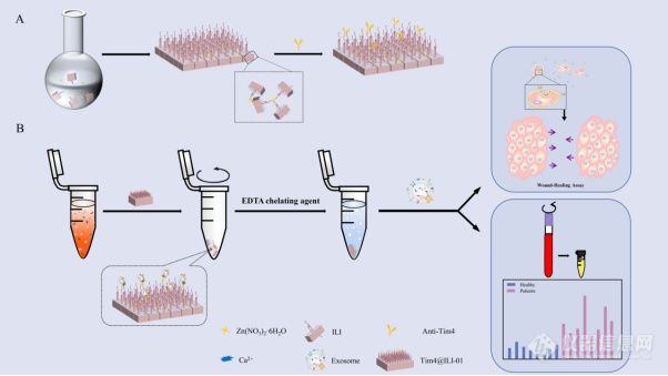
[align=center][font='times new roman'][size=21px][color=#000000]Tim4@ILI-01[/color][/size][/font][font='times new roman'][size=21px][color=#000000]免疫亲和材料用于外[/color][/size][/font][font='times new roman'][size=21px][color=#000000]泌[/color][/size][/font][font='times new roman'][size=21px][color=#000000]体的分离富集[/color][/size][/font][/align][align=left][font='times new roman'][size=20px][color=#000000]引言[/color][/size][/font][/align][font='times new roman'][size=16px]外[/size][/font][font='times new roman'][size=16px]泌[/size][/font][font='times new roman'][size=16px]体作为重要的信息载体,参与肿瘤的发生、进展、转移和化疗耐药等过程,近年来受到人们的广泛关注。外[/size][/font][font='times new roman'][size=16px]泌[/size][/font][font='times new roman'][size=16px]体携带大量的遗传物质,已成为液体活检最理想的分析目标。外[/size][/font][font='times new roman'][size=16px]泌[/size][/font][font='times new roman'][size=16px]体也是目前最有前途的非侵入性诊断和预后的生物标志物之一。为了进一步提高外[/size][/font][font='times new roman'][size=16px]泌[/size][/font][font='times new roman'][size=16px]体的临床应用价值,开发高效的分离技术具有重要的意义。目前,[/size][/font][font='times new roman'][size=16px]UC[/size][/font][font='times new roman'][size=16px]是外[/size][/font][font='times new roman'][size=16px]泌[/size][/font][font='times new roman'][size=16px]体分离的金标准。然而,这种方法需要繁琐的过程,并且具有耗时长([/size][/font][font='times new roman'][size=16px] 10 h[/size][/font][font='times new roman'][size=16px]),捕获效率低([/size][/font][font='times new roman'][size=16px]5[/size][/font][font='times new roman'][size=16px]%至[/size][/font][font='times new roman'][size=16px]25[/size][/font][font='times new roman'][size=16px]%)和重现性差的缺点。基于聚合物的外[/size][/font][font='times new roman'][size=16px]泌[/size][/font][font='times new roman'][size=16px]体分离试剂盒是通过添加聚合沉淀物以获得大量外[/size][/font][font='times new roman'][size=16px]泌[/size][/font][font='times new roman'][size=16px]体的方法。尽管该方法快速简便,但其他非外[/size][/font][font='times new roman'][size=16px]泌[/size][/font][font='times new roman'][size=16px]体污染物也可能共沉淀。因此,捕获的外[/size][/font][font='times new roman'][size=16px]泌[/size][/font][font='times new roman'][size=16px]体纯度相对较低。基于微流体的分离技术是利用外[/size][/font][font='times new roman'][size=16px]泌[/size][/font][font='times new roman'][size=16px]体的固有特征来捕获外[/size][/font][font='times new roman'][size=16px]泌[/size][/font][font='times new roman'][size=16px]体。但是,该技术不仅需要大量的临床样本,而且处理能力低也会影响后续的下游分析。最近,开发了一种基于抗体的亲和技术以提高外[/size][/font][font='times new roman'][size=16px]泌[/size][/font][font='times new roman'][size=16px]体的富集效率。例如,[/size][/font][font='times new roman'][size=16px]Boriachek[/size][/font][font='times new roman'][size=16px]等人设计了一种使用[/size][/font][font='times new roman'][size=16px]CD63[/size][/font][font='times new roman'][size=16px]抗体功能化的载金氧化铁纳米颗粒([/size][/font][font='times new roman'][size=16px]Au-NP Fe[/size][/font][font='times new roman'][size=16px]2[/size][/font][font='times new roman'][size=16px]O[/size][/font][font='times new roman'][size=16px]3[/size][/font][font='times new roman'][size=16px]NC[/size][/font][font='times new roman'][size=16px])用于分离和检测外[/size][/font][font='times new roman'][size=16px]泌[/size][/font][font='times new roman'][size=16px]体。将[/size][/font][font='times new roman'][size=16px]Au-NP Fe[/size][/font][font='times new roman'][size=16px]2[/size][/font][font='times new roman'][size=16px]O[/size][/font][font='times new roman'][size=16px]3[/size][/font][font='times new roman'][size=16px]NC[/size][/font][font='times new roman'][size=16px]材料分离得到的外[/size][/font][font='times new roman'][size=16px]泌[/size][/font][font='times new roman'][size=16px]体进一步识别胎盘碱性磷酸酶([/size][/font][font='times new roman'][size=16px]PLAP[/size][/font][font='times new roman'][size=16px])修饰的丝网印刷电极后,可以特异性检测出胎盘细胞分泌的外[/size][/font][font='times new roman'][size=16px]泌[/size][/font][font='times new roman'][size=16px]体。该方法可用于检测各种临床疾病的特定外[/size][/font][font='times new roman'][size=16px]泌[/size][/font][font='times new roman'][size=16px]体群体,尤其是妊娠并发症。然而,这些方法难以同时实现外[/size][/font][font='times new roman'][size=16px]泌[/size][/font][font='times new roman'][size=16px]体的高效捕获和无损释放,这极大地限制了外[/size][/font][font='times new roman'][size=16px]泌[/size][/font][font='times new roman'][size=16px]体生物学功能研究和在临床诊断中的应用。[/size][/font][font='times new roman'][size=16px]为了实现外[/size][/font][font='times new roman'][size=16px]泌[/size][/font][font='times new roman'][size=16px]体的临床价值,开发一种快速,有效且无损的外[/size][/font][font='times new roman'][size=16px]泌[/size][/font][font='times new roman'][size=16px]体分离方法具有重要的意义。金属有机框架材料作为一种特殊的多孔材料,因其具有高比表面积、良好的稳定性、较低的密度以及可调节的表面性能而引起人们的广泛关注。而基于抗体的免疫亲和捕获法具有灵敏度高、特异性好、且分离效率高等特点,在外[/size][/font][font='times new roman'][size=16px]泌[/size][/font][font='times new roman'][size=16px]体的分离富集方面具有很好的应用前景。将两者结合制备一种新的分离方法,实现外[/size][/font][font='times new roman'][size=16px]泌[/size][/font][font='times new roman'][size=16px]体的捕获和无损释放,为外[/size][/font][font='times new roman'][size=16px]泌[/size][/font][font='times new roman'][size=16px]体的下游临床诊断和检测提供强大的技术支持。[/size][/font][align=left][font='calibri'][size=29px]原理[/size][/font][/align][font='times new roman'][size=16px]本[/size][/font][font='times new roman'][size=16px]研究[/size][/font][font='times new roman'][size=16px]设计合成了一种[/size][/font][font='times new roman'][size=16px]Tim4@ILI-01[/size][/font][font='times new roman'][size=16px]免疫亲和材料用于分离富集外[/size][/font][font='times new roman'][size=16px]泌[/size][/font][font='times new roman'][size=16px]体。该分离方法具有处理时间短、非特异性粘附率低、捕获效率高以及无损释放外[/size][/font][font='times new roman'][size=16px]泌[/size][/font][font='times new roman'][size=16px]体等基本特征。如图所示,首先利用阳离子[/size][/font][font='times new roman'][size=16px]ILI[/size][/font][font='times new roman'][size=16px]作为有机配体,制备强亲水性的[/size][/font][font='times new roman'][size=16px]ILI-01 MOFs[/size][/font][font='times new roman'][size=16px]薄[/size][/font][font='times new roman'][size=16px]片作为捕获外[/size][/font][font='times new roman'][size=16px]泌[/size][/font][font='times new roman'][size=16px]体的基质材料。[/size][/font][font='times new roman'][size=16px]ILI[/size][/font][font='times new roman'][size=16px]-01 MOFs[/size][/font][font='times new roman'][size=16px]材料固有的亲水性能有效降低外[/size][/font][font='times new roman'][size=16px]泌[/size][/font][font='times new roman'][size=16px]体分离过程中的非特异性细胞粘附。此外,由于[/size][/font][font='times new roman'][size=16px]ILI[/size][/font][font='times new roman'][size=16px]-01 MOFs[/size][/font][font='times new roman'][size=16px]薄片上[/size][/font][font='times new roman'][size=16px]富含[/size][/font][font='times new roman'][size=16px]羧基,可以提高亲和力抗体的后续偶联效率,从而大大提高外[/size][/font][font='times new roman'][size=16px]泌[/size][/font][font='times new roman'][size=16px]体的捕获效率。在此,我们选择了针对外[/size][/font][font='times new roman'][size=16px]泌[/size][/font][font='times new roman'][size=16px]体膜上磷脂酰丝氨酸([/size][/font][font='times new roman'][size=16px]PS[/size][/font][font='times new roman'][size=16px])受体的新型[/size][/font][font='times new roman'][size=16px]Tim4[/size][/font][font='times new roman'][size=16px]抗体。已经证明[/size][/font][font='times new roman'][size=16px]Tim4[/size][/font][font='times new roman'][size=16px]蛋白分子与[/size][/font][font='times new roman'][size=16px]PS[/size][/font][font='times new roman'][size=16px]分子之间的特定相互作用可以有效地将外[/size][/font][font='times new roman'][size=16px]泌[/size][/font][font='times new roman'][size=16px]体从样品中分离出来,并且两者的结合存在[/size][/font][font='times new roman'][size=16px]Ca2+[/size][/font][font='times new roman'][size=16px]依赖特性,在中性条件下通过添加螯合剂可以轻易洗脱和回收外[/size][/font][font='times new roman'][size=16px]泌[/size][/font][font='times new roman'][size=16px]体。因此,开发的[/size][/font][font='times new roman'][size=16px]Tim4@ILI-01[/size][/font][font='times new roman'][size=16px]免疫亲和材料可以实现无损捕获和释放完整的外[/size][/font][font='times new roman'][size=16px]泌[/size][/font][font='times new roman'][size=16px]体,为后续临床诊断和治疗奠定了坚实的基础。[/size][/font][table][tr][td][align=center][img]https://ng1.17img.cn/bbsfiles/images/2021/08/202108012212485795_1809_5111497_3.png[/img][/align][/td][/tr][/table][align=center][font='times new roman']图[/font][font='times new roman']1 [/font][font='times new roman'] Tim4@ILI-01[/font][font='times new roman']免疫亲和薄片[/font][/align][font='times new roman'][size=18px][color=#000000]与常规外[/color][/size][/font][font='times new roman'][size=18px][color=#000000]泌[/color][/size][/font][font='times new roman'][size=18px][color=#000000]体分离方法的比较[/color][/size][/font][font='times new roman'][size=16px]进一步将所制备的[/size][/font][font='times new roman'][size=16px]Tim4@ILI-01[/size][/font][font='times new roman'][size=16px]免疫亲和材料与金标准“[/size][/font][font='times new roman'][size=16px]UC[/size][/font][font='times new roman'][size=16px]”进行比较。如[/size][/font][font='times new roman'][size=16px]图[/size][/font][font='times new roman'][size=16px]2[/size][/font][font='times new roman'][size=16px]所示,[/size][/font][font='times new roman'][size=16px]UC[/size][/font][font='times new roman'][size=16px]分离到的外[/size][/font][font='times new roman'][size=16px]泌[/size][/font][font='times new roman'][size=16px]体的大小分布明显[/size][/font][font='times new roman'][size=16px]更宽[/size][/font][font='times new roman'][size=16px],[/size][/font][font='times new roman'][size=16px]而[/size][/font][font='times new roman'][size=16px]Tim4@ILI-01[/size][/font][font='times new roman'][size=16px]免疫亲和材料捕获的外[/size][/font][font='times new roman'][size=16px]泌[/size][/font][font='times new roman'][size=16px]体显示出[/size][/font][font='times new roman'][size=16px]较[/size][/font][font='times new roman'][size=16px]窄的大小分布。此外,通过[/size][/font][font='times new roman'][size=16px]Tim4@ILI-01[/size][/font][font='times new roman'][size=16px]免疫亲和材料捕获的外[/size][/font][font='times new roman'][size=16px]泌[/size][/font][font='times new roman'][size=16px]体的[/size][/font][font='times new roman'][size=16px]众数[/size][/font][font='times new roman'][size=16px]和平均大小均明显小于[/size][/font][font='times new roman'][size=16px]UC[/size][/font][font='times new roman'][size=16px]捕获的外[/size][/font][font='times new roman'][size=16px]泌[/size][/font][font='times new roman'][size=16px]体。进一步比较[/size][/font][font='times new roman'][size=16px]了两种方法的捕获颗粒数和蛋白含量,[/size][/font][font='times new roman'][size=16px]Tim4@ILI-01[/size][/font][font='times new roman'][size=16px]免疫亲和材料富集法[/size][/font][font='times new roman'][size=16px]分离得到的[/size][/font][font='times new roman'][size=16px]颗粒数和蛋白含量分别比[/size][/font][font='times new roman'][size=16px]UC[/size][/font][font='times new roman'][size=16px]高[/size][/font][font='times new roman'][size=16px]5.2[/size][/font][font='times new roman'][size=16px]倍和[/size][/font][font='times new roman'][size=16px]2.4[/size][/font][font='times new roman'][size=16px]倍[/size][/font][font='times new roman'][size=16px]。[/size][/font][font='times new roman'][size=16px]之前的研究表明,[/size][/font][font='times new roman'][size=16px]颗粒[/size][/font][font='times new roman'][size=16px]-[/size][/font][font='times new roman'][size=16px]蛋白比与外[/size][/font][font='times new roman'][size=16px]泌[/size][/font][font='times new roman'][size=16px]体样品的纯度呈正相关[/size][/font][font='times new roman'][size=16px],[/size][/font][font='times new roman'][size=16px]而[/size][/font][font='times new roman'][size=16px]Tim4@ILI-01[/size][/font][font='times new roman'][size=16px]免疫亲和材料捕获的外[/size][/font][font='times new roman'][size=16px]泌[/size][/font][font='times new roman'][size=16px]体颗粒[/size][/font][font='times new roman'][size=16px]-[/size][/font][font='times new roman'][size=16px]蛋白[/size][/font][font='times new roman'][size=16px]比值[/size][/font][font='times new roman'][size=16px]比[/size][/font][font='times new roman'][size=16px]UC[/size][/font][font='times new roman'][size=16px]高[/size][/font][font='times new roman'][size=16px]6.7[/size][/font][font='times new roman'][size=16px]倍。因此,基于[/size][/font][font='times new roman'][size=16px]Tim4@ILI-01[/size][/font][font='times new roman'][size=16px]免疫亲和材料的富集法可以产生更多纯度更高的外[/size][/font][font='times new roman'][size=16px]泌[/size][/font][font='times new roman'][size=16px]体[/size][/font][font='times new roman'][size=16px],[/size][/font][font='times new roman'][size=16px]显示[/size][/font][font='times new roman'][size=16px]出明显[/size][/font][font='times new roman'][size=16px]的优势。[/size][/font][table][tr][td][align=center][img]https://ng1.17img.cn/bbsfiles/images/2021/08/202108012212488549_9401_5111497_3.jpeg[/img][/align][/td][/tr][/table][align=center][font='times new roman']图[/font][font='times new roman']2 [/font][font='times new roman']比较[/font][font='times new roman']Tim4@ILI-01[/font][font='times new roman']免疫亲和材料和[/font][font='times new roman']UC[/font][font='times new roman']分离的外[/font][font='times new roman']泌[/font][font='times new roman']体:([/font][font='times new roman']A[/font][font='times new roman'])[/font][font='times new roman']UC[/font][font='times new roman']分离的外[/font][font='times new roman']泌[/font][font='times new roman']体大小分布;([/font][font='times new roman']B[/font][font='times new roman'])众数和平均大小;([/font][font='times new roman']C[/font][font='times new roman'])颗粒计数和总蛋白量;([/font][font='times new roman']D[/font][font='times new roman'])颗粒数与总蛋白质量的比率[/font][/align][font='times new roman'][size=16px]进一步采用[/size][/font][font='times new roman'][size=16px]Western blotting[/size][/font][font='times new roman'][size=16px]对上述结果进行验证。如图[/size][/font][font='times new roman'][size=16px]3[/size][/font][font='times new roman'][size=16px]所示,[/size][/font][font='times new roman'][size=16px]通过[/size][/font][font='times new roman'][size=16px]Tim4@ILI-01[/size][/font][font='times new roman'][size=16px]免疫亲和材料[/size][/font][font='times new roman'][size=16px]富集的外[/size][/font][font='times new roman'][size=16px]泌[/size][/font][font='times new roman'][size=16px]体表面[/size][/font][font='times new roman'][size=16px]HSP70[/size][/font][font='times new roman'][size=16px]、[/size][/font][font='times new roman'][size=16px]CD63[/size][/font][font='times new roman'][size=16px]、[/size][/font][font='times new roman'][size=16px]TSG101[/size][/font][font='times new roman'][size=16px]、[/size][/font][font='times new roman'][size=16px]CD81[/size][/font][font='times new roman'][size=16px]的蛋白量分别比[/size][/font][font='times new roman'][size=16px]UC[/size][/font][font='times new roman'][size=16px]富集的[/size][/font][font='times new roman'][size=16px]高[/size][/font][font='times new roman'][size=16px]4.8[/size][/font][font='times new roman'][size=16px]倍、[/size][/font][font='times new roman'][size=16px]2.9[/size][/font][font='times new roman'][size=16px]倍、[/size][/font][font='times new roman'][size=16px]1.9[/size][/font][font='times new roman'][size=16px]倍、[/size][/font][font='times new roman'][size=16px]1.5[/size][/font][font='times new roman'][size=16px]倍[/size][/font][font='times new roman'][size=16px]。以上结果可表明[/size][/font][font='times new roman'][size=16px]所建立的[/size][/font][font='times new roman'][size=16px]基于[/size][/font][font='times new roman'][size=16px]Tim4@ILI-01[/size][/font][font='times new roman'][size=16px]免疫亲和[/size][/font][font='times new roman'][size=16px]材料[/size][/font][font='times new roman'][size=16px]富集法优于传统的[/size][/font][font='times new roman'][size=16px]UC[/size][/font][font='times new roman'][size=16px]法[/size][/font][font='times new roman'][size=16px]。[/size][/font][table][tr][td][align=center][img]https://ng1.17img.cn/bbsfiles/images/2021/08/202108012212491547_9181_5111497_3.jpeg[/img][/align][/td][/tr][/table][align=center][font='times new roman']图[/font][font='times new roman'] 3[/font][font='times new roman']比较[/font][font='times new roman']Tim4@ILI-01[/font][font='times new roman']免疫亲和材料富集法和[/font][font='times new roman']UC[/font][font='times new roman']分离的外[/font][font='times new roman']泌[/font][font='times new roman']体:([/font][font='times new roman']A[/font][font='times new roman'])外[/font][font='times new roman']泌[/font][font='times new roman']体标记蛋白的[/font][font='times new roman']Western blotting[/font][font='times new roman']结果;([/font][font='times new roman']B[/font][font='times new roman'])蛋白质含量的比较[/font][/align]


 我要推广仪器
 下载APP
下载APP