

方案详情文
智能文字提取功能测试中
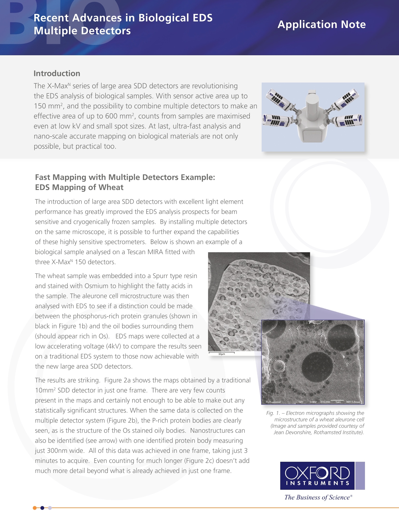
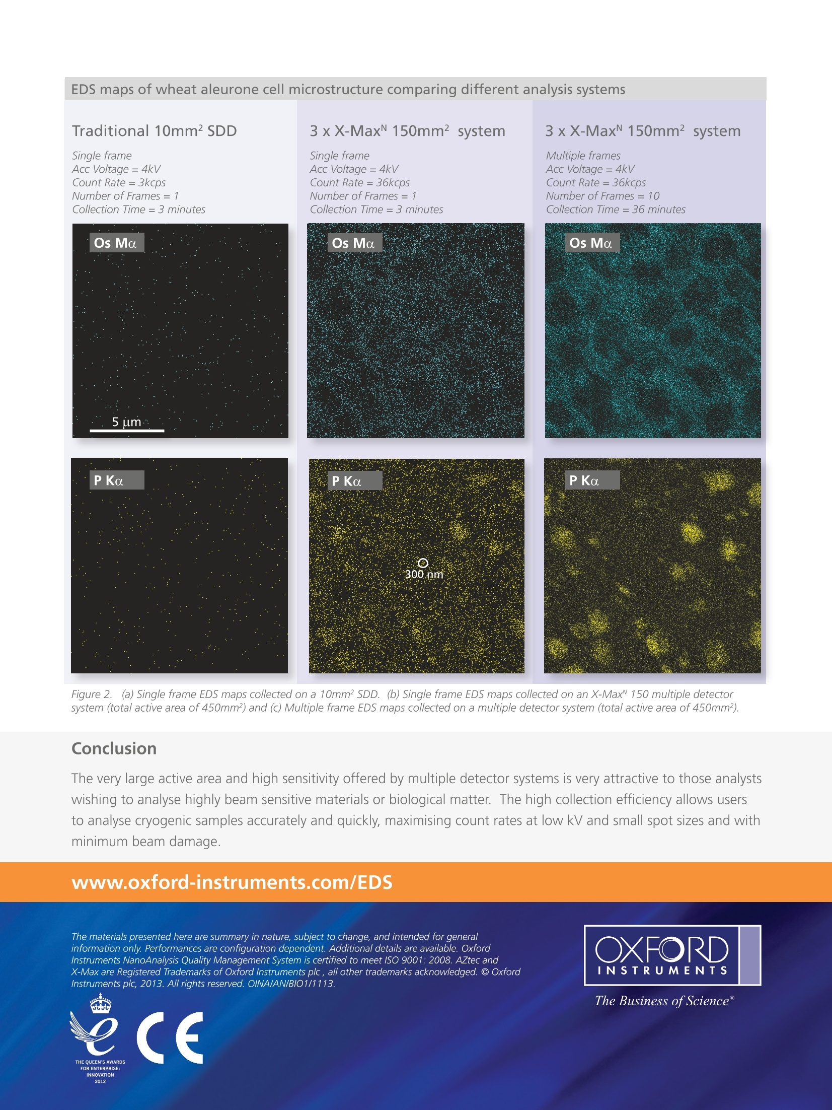
EDS maps of wheat aleurone cell microstructure comparing different analysis systems Introduction The X-MaxN series of large area SDD detectors are revolutionisingthe EDS analysis of biological samples. With sensor active area up to150 mm², and the possibility to combine multiple detectors to make aneffective area of up to 600 mm², counts from samples are maximisedeven at low kV and small spot sizes. At last, ultra-fast analysis andnano-scale accurate mapping on biological materials are not onlypossible, but practical too. Fast Mapping with Multiple Detectors Example:EDS Mapping of Wheat The wheat sample was embedded into a Spurr type resinand stained with Osmium to highlight the fatty acids inthe sample. The aleurone cell microstructure was thenanalysed with EDS to see if a distinction could be madebetween the phosphorus-rich protein granules (shown inblack in Figure 1b) and the oil bodies surrounding them(should appear rich in Os).EDS maps were collected at alow accelerating voltage (4kV) to compare the results seenon a traditional EDS system to those now achievable withthe new large area SDD detectors. The results are striking. Figure 2a shows the maps obtained by a traditional10mm² SDD detector in just one frame. There are very few countspresent in the maps and certainly not enough to be able to make out anystatistically significant structures. When the same data is collected on themultiple detector system (Figure 2b), the P-rich protein bodies are clearlyseen, as is the structure of the Os stained oily bodies. Nanostructures canalso be identified (see arrow) with one identified protein body measuringjust 300nm wide. All of this data was achieved in one frame, taking just 3minutes to acquire. Even counting for much longer (Figure2c) doesn't addmuch more detail beyond what is already achieved in just one frame. Fig. 1. - Electron micrographs showing themicrostructure ofa wheat aleurone cell(Image and samples provided courtesy ofJean Devonshire, Rothamsted Institute). Traditional 10mm² SDD 3xX-MaxN 150mm² system 3xX-Max 150mm² system Single frameAcc Voltage = 4kVCount Rate= 3kcpsNumber of Frames=1Collection Time=3 minutes Single frame Multiple frames Acc Voltage =4kV Acc Voltage=4kV Count Rate =36kcps Count Rate=36kcps Number of Frames=1 Number of Frames= 10 Collection Time =36 minutes Os Ma PKa PKa 300 nm Figure 2. (a) Single frame EDS maps collected on a 10mm² SDD. (b) Single frame EDS maps collected on an X-Max~ 150 multiple detectorsystem (total active area of 450mm²) and (c) Multiple frame EDS maps collected on a multiple detector system (total active area of 450mm²). Conclusion The very large active area and high sensitivity offered by multiple detector systems is very attractive to those analystswishing to analyse highly beam sensitive materials or biological matter. The high collection efficiency allows usersto analyse cryogenic samples accurately and quickly, maximising count rates at low kV and small spot sizes and withminimum beam damage. www.oxford-instruments.com/EDS The materials presented here are summary in nature, subject to change, and intended for general information only. Performances are configuration dependent. Additional details are available. Oxford ( Instruments NanoAnalysis Quality Management System is certified t o m e et I S O 9 0 01: 200 8. AZtec and X-Max are Registered Trademarks of Oxford Instruments p l c, a ll oth er trademar k s a ck n owledge d .@ Oxf ord Instruments plc, 2013. All rights reserved. OINA/AN/BIO1/1113. ) The Business ofScience" INNOVATION 2012L2 The X-MaxN series of large area SDD detectors are revolutionisingthe EDS analysis of biological samples. With sensor active area up to150 mm2, and the possibility to combine multiple detectors to make aneffective area of up to 600 mm2, counts from samples are maximisedeven at low kV and small spot sizes. At last, ultra-fast analysis andnano-scale accurate mapping on biological materials are not onlypossible, but practical too.
关闭-
1/2
-
2/2
产品配置单
牛津仪器科技(上海)有限公司为您提供《生物中微观结构检测方案(EBSD系统)》,该方案主要用于其他中微观结构检测,参考标准《暂无》,《生物中微观结构检测方案(EBSD系统)》用到的仪器有牛津仪器Ultim Extreme无窗超级能谱。
我要纠错
相关方案
 咨询
咨询