

方案详情文
智能文字提取功能测试中
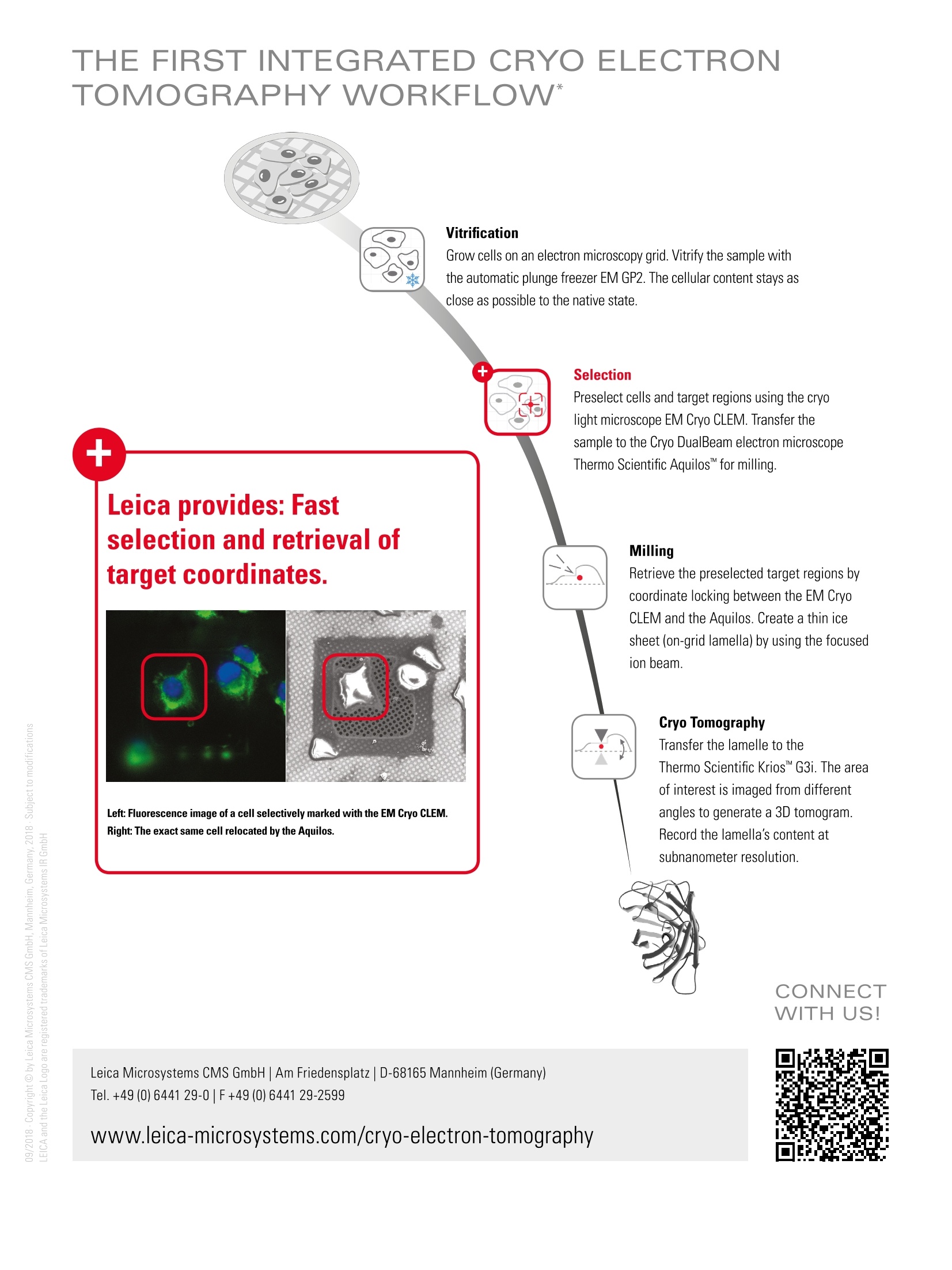
From Eye to Insight MICROSYSTEM S Segmentation of a cryo-electron tomogram. Proteasomes tethering to the nuclear pore complex (purple). Courtesy of Dr. Ben Engel, Dept. of Mol. Struct. Biology, MPI for Biochemistry, Martinsried, Germany Brain Research: Correlation of sample morphology and gene expression INVESTIGATION OF MOLECULES WITHIN THEIRSUBCELLULAR CONTEXT References ● Marx V., 2018, "Calling all cell biologists to try cryo-ET", Nature Methods 15:575-578. ● Vaites L.P., Harper,J.W.,2018,"Protein aggregates caught stalling", Nature 555,449-451. Oikonomou C.M., Jensen G.J., 2017,"Cellular electron cryotomography: towards structuralbiology in situ", Annual Review of Biochemistry 86: 873-896. Beck M., Baumeister W., 2016,"Cryo-Electron Tomography: can it reveal the molecularsociology of cells in atomic detail?", Trends in Cell Biology 26(11): 825-837. THE FIRST INTEGRATED CRYO ELECTRONTOMOGRAPHY WORKFLOW* Vitrification Grow cells on an electron microscopy grid. Vitrify the sample withthe automatic plunge freezer EM GP2. The cellular content stays asclose as possible to the native state. Leica provides: Fastselection and retrieval oftarget coordinates. Left: Fluorescence image of a cell selectively marked with the EM Cryo CLEM.Right: The exact same cell relocated by the Aquilos. Preselect cells and target regions using the cryolight microscope EM Cryo CLEM. Transfer thesample to the Cryo DualBeam electron microscopeThermo Scientific Aquilosfor milling. Milling Retrieve the preselected target regions by coordinate locking between the EM Cryo CLEM and the Aquilos. Create a thin ice sheet (on-grid lamella) by using the focused ion beam. Cryo Tomography Transfer the lamelle to the Thermo Scientific KriosG3i. The area of interest is imaged from different angles to generatea 3D tomogram. Record the lamella's content at subnanometer resolution Leica Microsystems CMS GmbH| Am Friedensplatz|D-68165 Mannheim (Germany)Tel.+49 (0)6441 29-0F+49(0) 6441 29-2599 www.leica-microsystems.com/cryo-electron-tomography
关闭-
1/2

-
2/2
产品配置单
徕卡显微系统(上海)贸易有限公司为您提供《活细胞中有丝分裂检测方案(显微图像分析)》,该方案主要用于其他中荧光成像检测,参考标准《暂无》,《活细胞中有丝分裂检测方案(显微图像分析)》用到的仪器有null。
我要纠错
相关方案
 咨询
咨询