方案详情文
智能文字提取功能测试中
ResearchGateSee discussions, stats, and author profiles for this publication at: https://www.researchgate.net/publication/331121770 Revised: 14 November 2018 Accepted: 16 January 2019Received: 14 February 2018DOI: 10.1002/jemt.23234WILEY MICROSCOPYRESEARCH&TECHNIQUE Rapid histologic diagnosis using quick fluorescence staining and tissueconfocal microscopy Article in Microscopy Research and Technique. February 2019 DOI: 10.1002/jemt.23234 CITATIONS0 READS40 14 authors,including: Hyunjin Kim Hee Jin Chang National Cancer Center Korea National Cancer Center Korea 34 PUBLICATIONS 671 CITATIONS 233 PUBLICATIONS 5,758 CITATIONS SEE PROFILE SEE PROFILE Yongdoo Choi So-Youn Jung National Cancer Center Korea National Cancer Center Korea 94 PUBLICATIONS 2,575 CITATIONS 109 PUBLICATIONS 1.302 CITATIONS SEE PROFILE SEEPROFILE RESEARCH ARTICLE Rapid histologic diagnosis using quick fluorescence stainingand tissue confocal microscopy Hongrae Kim1|Sunhye Leel|Hyunjin Kim²| Hee Jin Changg34|Yongdoo Choi?|Hong Man Yoon1.5 |Myeong-Cherl Kook4.5 So-Youn Jung°|Youngmi Kwon46Juehyung Kang’|Hongki Yoo’|Incheon Song°|Seungrag Lee’|Dae Kyung Sohn-1,3 iD Innovative Medical Engineering & Technology, National Cancer Center, Goyang, South Korea Biomarker Branch, National Cancer Center, Goyang, South KoreaCenter for Colorectal Cancer, National Cancer Center,Goyang, South Korea“Department of Pathology, National Cancer Center, Goyang, South KoreaCenter for Gastric Cancer, National Cancer Center, Goyang, South KoreaCenter for Breast Cancer, National Cancer Center, Goyang, South Korea'Department of Biomedical Engineering, Hanyang University, Seoul, South Korea°Nanoscope Systems, Inc., Daejeon, South Korea Osong Medical Innovation Foundation, Cheongju, Chungcheongbuk-do, South Korea Dae Kyung Sohn, Innovative Medical Engineering & Technology, National Cancer Center, 323 Ilsan-ro, Ilsandong-gu, Goyang, Gyeonggi-do 10408, South Korea. Email: gsgsbal@ncc.re.kr Review Editor: Paolo Bianchini Funding informationThe Ministry of Trade, Industry, and Energy,Grant/Award Number:10049773 Abstract With the development of advanced and minimally invasive surgical techniques, and in view ofthe functional and cosmetic aspects, the need for rapid and accurate diagnosis during surgery isincreasing. This study was conducted to develop a tissue diagnosis method using confocalmicroscopy after simple tissue staining that does not require freezing and slicing. At present,fluorescence staining with confocal microscopy is not generalized for real-time diagnosis duringsurgery. In this paper, we propose a fluorescence staining method using Hoechst 33342 andEosin that does not require tissue freezing and slicing. The proposed method can be used as partof a rapid tissue diagnosis method that is suitable for use in the operating room,although furtherresearch is required before it can be applied in clinical practice. KEYWORDS confocal microscopy, fluorescent image, minimally invasive surgery, quick staining method,tissue staining 1|1INTRODUCTION Histologic diagnosis requires the staining of nuclei and surroundingtissues, and the most commonly used stain is Hematoxylin andEosin (H&E). Hematoxylin stains the nuclei a dark blue color suchthat the internal structure can be observed. Eosin is used to stainthe surrounding tissue pink, orange, and red, in order to distinguishit from the nuclei and to facilitateobservation of the tissue struc-ture based on the shade and brightness of the connective tissuefibers and cytoplasms. In histologic diagnosis based on H&E staining, pathology slidesare commonly prepared and visually observed with an optical micro-scope. To create a tissue slide, the tissue is first fixed with paraffin,carbowax, celloidin, gelatin, etc., and then sliced thinly. The slide pro-duction process of a general tissue sample is as follows: Sample collection → Gross examination →Fixation - Washing→ Dehydration Cleaning → Impregnation or Infiltration →Embed-ding → Trimming → Microtome cutting → Stain →Mounting. As this process is lengthy, it cannot be used for tissue diagnosisduring surgery. Instead, when rapid tissue diagnosis is required, an optimal cutting temperature (OCT) compound is used to quick-freezethe tissue before it can be thinly sliced (Bancroft & Gamble, 2008). Incancer surgery, access to sentinel lymph node biopsy results is essentialin order to determine the required extent of the surgery. In skin cancersurgery, such as Mohs surgery, several frozen section biopsies arerequired during surgery in order to properly consider the cosmetic andfunctional aspects. This type of frozen section tissue diagnosis requiresapproximately 15-40 min (Kim, Hubbard, McManus, Mason Jr., &Pruitt Jr., 1985; Rajadhyaksha, Menaker, Flotte, Dwyer, & Gonzalez,2001; Udelsman, Westra, Donovan, Sohn, & Cameron, 2001); however,because of its impact, significant effort has been focused on reducingthis time. Intraoperative diagnoses using confocal microscopes wererecently introduced, and are now being employed extensively in surger-ies requiring intraoperative diagnosis, such as Mohs surgery (Bennassar,Vilata, Puig, & Malvehy, 2014; Gareau et al., 2008). This study was conducted to confirm the possibility of tissue diag-nosis via confocal microscopy after simple tissue staining withoutfreezing and slicing, for rapid and easy tissue diagnosis during surgery. 2ihNMATERIALSAND METHODS In order to verify the rapid method of tissue diagnosis, we first selectedthe staining reagents from among various fluorescent reagents andestablished the staining method. Then, animal experiments were con-ducted to examine the performance of the selected staining method,and clinical trials performed to determine the possibility of distinguish-ing between normal and cancerous tissue. 2.1 | Tissue staining for confocal microscopy In confocal microscopy, fluorescent staining dye is used to measurethe fluorescence signal of a specific wavelength emitted by an excita-tion light source of a specific wavelength. Dyeing reagents of differentwavelengths, such as H&E of different colors, are selected to stain thenuclei and surrounding tissues. Tables 1 and 2 list commercially avail-able fluorescent dyes (Bruchez Jr., Moronne, Gin, Weiss, & Alivisatos,1998; Chattopadhyay et al., 2006; Dewan,Ahmad, & Swamy, 2014;Hotz, 2005; Lajunen & Kubin, 1986;Veta et al., 2013). In this study, Eosin Y dye (Ex 488 nm/Em 499-633 nm) was selectedOstain theepproteins,and Hoechst333422 (Ex 405 nm /Em410-1,313 nm), which is a fluorescent dye with a different fluorescencewavelength than that of Eosin Y, was selected to stain the nuclei. A porta-ble confocal microscope (K1-Fluo_RT, Nanoscope Systems, Korea) wasused to capture confocal images using the corresponding fluorescencesignals. To minimize the time required for tissue diagnosis, we directlystained the tissues in 2-mm thicknesses without first making tissue slices.We implemented the following optimized quick Hoechst tissue stainingmethod, which required a total estimated time of less than 2 min, throughrepeated dyeing experiments (data not shown). Tissue washing in 95% ethanol for 10 s →Tissue staining for30 s with Eosin →Washing in 95% ethanol for 20 s → Washing in100% ethanol for 5 s → Washing with distilled water (D/W) for 5 sbefore undergoing Hoechst nuclear staining →Nuclear staining for30 s with Hoechst Washing in 100% ethanol for 3 s. TABLE 1 Fluorescent dyes used to stain the proteins in thecytoplasms Absorption Emission Fluorochrome maximum peak maximum peak Cascade blue 380,401 419 Cascade yellow 399 549 Pacific blue 405 456 Fluorescein (fitc) 495 520 R-phycoerythrin (R-PE) 495,584 576 PE-Texas red 495,584 620 PE-cyanine5 495,584 670 Peridinin-chlorophylI 490 677 Eosin Y 546 561 Allophycocyanin 650 660 2.2 Animal experiments for evaluating confocalmicroscopic images Animal experiments were performed using nude mice to assess thequalities of the fluorescence staining and fluorescence imaging. Thisstudy was reviewed and approved by the Institutional Animal Careand Use Committee (IACUC) of the National Cancer Center ResearchInstitute (NCC-15-229B). In order to compare the results of the conventional H&E stainingmethod with those of fluorescent staining using Hoechst 33342 andEosin, two histopathological slides of the same cross section were pre-pared using conventional frozen section methods involving OCT com-pound. The first slide was stained using H&E stain and observed underan optical microscope. The second slice was stained using Hoechst33342 and Eosin, and fluorescence images were then obtained using aconfocal microscope (LSM780, Zeiss, Germany). Tumor tissues wereobtained after subcutaneously injecting tumor cells into a nude mouse. A tissue confocal microscope (K1-Fluo_RT) was used to observethe tissue itself following fluorescence staining without first making aslide of the tissue slice. The images were obtained using the followingobjective lens: magnification: 20x, numerical aperture (NA): 0.75,working distance: 0.65 mm. Fast axis scanning was conducted using aresonant scanner at 8 kHz, whereas the slow axis was scanned using amotorized stage moving at 4.88 mm/s. As the image was constructedat 1024 per fast axis line, this gave a pixel dwell time of 0.61 ns. The TABLE 2Fluorescent dyes used to stain the nuclei Absorption Emission Fluorochrome maximum peak maximum peak DAPI 372 476 Hoechst33342 395 450 Hoechst33258 352 455 Vybrant Dyecycle violet 369 437 Propidium iodide 342,535 617 Ethidium bromide 320,518 605 Arcridine orange 503 530 (DNA), 640 (RNA) 7-Sminoactinomycin-D 546 647 Thiazole orange 512 533 Vybrant Dyecle green 506 534 Vybrant Dyecle orange 519 563 measured illumination power on the sample was 5 mW. The tissue wasdivided into two sections, and half was fluorescently stained to obtain afluorescence image of the section. The remaining tissue was stainedwith H&E staining using a frozen section method and OCT compound,and then observed with an optical microscope. After fluorescence stain-ing, the tissue samples were placed in a confocal dish, as shown inFigure 1, before being observed with a tissue confocal microscope. Abright-field microscope was used to acquire images of the pre-sectioned4 um-thick H&E slides whereas a confocal microscope was used toacquire images of the unsectioned tissue. In order to calibrate the non-horizontal part of the sample during tissue scanning, the depth from thetissue cross-section was scanned using the alternative method shown inFigure 2. The image was then reconstructed based on the sharpnessand contrast of the scanned image, to obtain an optimal scan image.Using image processing techniques, all of the fluorescence imagesobtained using the tissue confocal microscope were made to have acolor similar to those stained by H&E staining (Yoon et al.,2016). 2.3Clinical experiments to assess differentialdiagnoses between tumors and normal tissues To assess the possibility of differential diagnoses between tumors andnormal tissues with a tissue confocal microscope, we conducted clinicalexperiments according to the principles of the Declaration of Helsinki,and all patients provided written informed consent. The protocolwas approved by the Institutional Review Board of our institution(NCC2016-0087). In three cases of colorectal cancer, three cases of breast cancer,and three cases of gastric cancer,normal and cancerous tissues wereobtained during surgery and used in the experiments. Each tissue was first cut to a size of about 3×3 mm². Then, thecenter of the tissue was cut and divided into two equally sized sections.One side was prepared as a pathological slide with H&E staining for theoptical microscope, and the other side was fluorescently stained in atissue state for the tissue confocal microscope (K1-Fluo_RT). All fluo-rescence images were modified with an image processing technique toexhibit colors similar to those resulting from H&E staining. In each case,the same surfaces from both images were compared. 3 RESULTS With our quick Hoechst tissue staining method, we successfullyobtained tissue images after approximately 2 min of tissue harvesting.An additional 5 min was required to obtain whole fluorescence imagesof a ~3×3 mm² tissue section with the confocal microscope. Wefound that the images obtained from the animal and clinical experi-ments could be used for rapid tissue diagnosis without the need fortissue slicing 3.1 | Animal experiments To confirm the use of Hoechst 33342 and Eosin staining in tumortissue, two histologic slides of tumor tissues were prepared andcompared. In the H&E stained slide, only one image with stainednuclei and cytoplasms could be obtained using the optical microscope.In contrast, the confocal microscope acquired images showing onlynuclear or only cytoplasmic staining, or both nuclear and cytoplasmicstaining simultaneously. When the acquired images were compared, itwas observed that the nuclei were not uniform in size and had unevenshapes. These characteristics were observed in the tumors in bothslides. This indicates that the characteristics of the tumor could beconfirmed accurately in the fluorescence image stained by Hoechst33342 and Eosin. The images obtained by H&E staining and by fluo-rescence staining using Hoechst 33342 and Eosin are shown inFigure 3. The images in Figure 3 were not significantly different in terms oftheir visual acuity, even though one was obtained using a confocalmicroscope (LSM 780) from a slide of tissue slices,and the other wasobtained using a tissue confocal microscope (K1-Fluo_RT) after fluo-rescence staining without first making a tissue slide. 3.22| Clinical experiments Both tumorous and normal tissues were obtained in three cases ofbreast cancer, three cases of colorectal cancer, and three cases ofgastric cancer. The tissue was divided into halves, one of which wasfrozen and then used to prepare slides, while the other half was FIGURE 1 Tissues in confocal dish: (1) top and (2) scanning surfaces of sample [Color figure can be viewed at wileyonlinelibrary.com] FIGURE 2 Scanned images by depth of human colon tumor tissue:(1-4) scans of sample at a depth of 8.4 um;(5) image reconstructed byselecting a cleanly scanned image from each frame [Color figure can be viewed at wileyonlinelibrary.com] directly stained by Hoechst 33342 and Eosin for use in the tissue con-focal microscope. The resulting images are shown in Figure 4. After histological staining, the images obtained with the confocalmicroscope could be used to identify the characteristics of the tumor,but it was found that the staining was not uniform in some tissues.This is thought to be because the fluorescence reagent was unable touniformly penetrate the tissue deeply, and the tissue could not be uni-formly pressed onto the microscope lens for clean scanning. In addi-tion, it was confirmed that the fluorescence dyeing did not becomeconstant when the time from collection to dyeing of the tissue wasextended. 4 DISCUSSION In this paper, we proposed a method of performing fluorescence stain-ing using Hoechst 33342 and Eosin without tissue freezing and slicingfor rapid and easy tissue diagnosis during surgery. The purpose of thismethod is to facilitate rapid diagnosis during surgery via confocal microscopy. Rapid tissue biopsy assays such as the technique presentedin this paper can be very useful for repeated frozen diagnostic methodssuch as Mohs surgery, or minimally invasive surgical methods such assentinel lymph node biopsy. In addition, various immuno-stainingmethods can be applied through a confocal microscope. Therefore, it isexpected that this approach will also be applicable to the fast immuno-chemical staining method, which confirms gene mutations in tissue. With the development of advanced and minimally invasive surgicaltechniques, and considering functional and cosmetic aspects, the needfor rapid and accurate diagnosis during surgery is increasing. A fluores-cence staining method with confocal microscopy has not been general-ized for use in real-time diagnosis during surgery. Fluorescence stainingwith confocal microscopy has the following advantages. First, it can beused to simultaneously identify various structures in tissues by employ-ing fluorescent materials of various colors. Secondly, antibodies orsimilar compounds can be bound to a fluorescent dye reagent toconfirm various genetic or protein characteristics expressed in tissues.Thirdly, digital images can be acquired with a confocal microscope,which enables the use of a diverse range of accurate processing and FIGURE 3 Histologic images of tumor (subcutaneous): (1) optical microscopic image after staining using H&E, (2) LSM780 confocal microscopicimage after staining using Hoechst 33342 & Eosin,and (3) K1-Fluo_RT confocal microscopic image after staining using Hoechst 33342 & Eosin[Color figure can be viewed at wileyonlinelibrary.com] FIGURE 4 (1) Histologic image of colonic normal mucosa from optical microscope after H&E staining. (2) Pseudo-color image of the same colonicnormal mucosa from the K1-Fluo_RT confocal microscope after Hoechst 33342 & Eosin staining. (3) Histologic image of gastric cancer from theoptical microscope after H&E staining. (4) Pseudo-color image of the same gastric tumor from the K1-Fluo_RT confocal microscope after Hoechst33342 & Eosin staining. (5) Histologic image of breast cancer from the optical microscope after H&E staining. (6) Pseudo-color image of the samebreast cancer from the K1-Fluo_RT confocal microscope after Hoechst 33342 & Eosin staining [Color figure can be viewed atwileyonlinelibrary.com] analysis techniques. Finally, a confocal microscope can acquire cross-sectional images of both the surface and inner layers of the tissue with-out requiring thin tissue sections or slides. In our study, tissue images were obtained after about 2 min oftissue harvesting, and an additional 5 min were required to obtainwhole fluorescence images of a ~3×3 mm² tissue section with aconfocal microscope. Because no pathological slides are needed forfrozen sections, this method can significantly reduce the timerequired for diagnosis. In addition, only a few simple fluorescencedyeing reagents and a confocal microscope are required for use inthe operating room. As the images obtained from a confocal micro-scope are not familiar to many clinicians, we also proposed amethod to convert these images into familiar H&E stained images.Only a small number of cases performed in this study could notestablish a standard to distinguish cancer from normal tissue,although it was interpreted by pathologist. However, because theresultant image is already in digital format, it can be utilized effi-ciently for image processing and quantitative analysis with even alarge number of cases. Therefore, once artificial intelligence tech-niques become commonplace, they will facilitate rapid tissue diag-nosis in the operating room. The results of our experiments highlight several problems thatremain unresolved. The first is that fluorescence staining is notuniform as time passes after sample collection. This is due to tissuedegradation caused by omitting the cell and tissue fixing step inorder to facilitate rapid diagnosis. As time is of the essence in this application, this degradation may not be a concern. However, inorder to standardize and generalize the tissue fluorescence stainingprocess, this problem requires resolution. Secondly,it is difficult tomaintain good-quality tissue after fluorescence staining and observa-tion using the confocal microscope. This is because confocal micro-scopes use lasers for fluorescence measurements, which can resultin water evaporation and tissue degradation. In fact, this problemmay be related to the abovementioned tissue fixation problem. Evenif a digital image is stored by a confocal microscope, the difficulty (orimpossibility) of re-measuring or re-observing the same tissue after asingle measurement may cause additional problems. In addition, theadvantage of the confocal microscope is that it has the potential toidentify cancer cells in the deeper parts of the tissue, not on the sur-face. However, when the tissue was measured using the stainingmethod and equipment developed in this study, it was confirmed thatthe brightest fluorescence image was observed at a depth of 10-15 umfrom the tissue surface. This may lead to the inconvenience ofobtaining a tissue section repeatedly or cutting the obtained tissueto obtain an image of the tissue section in order to confirm whethercancer cells are present at the interface during surgery. Althoughthere is a limit to the ease of use of the proposed method in theoperating room owing to the clinical application of the equipmentdeveloped at the current level, this limitation may be removed in thefuture. Some limitations have already been solved by the develop-ment of dyeing methods that show deeper penetration and thedevelopment of instruments including laser power. In conclusion, we have developed a tissue diagnosis method usingconfocal microscopy that facilitates rapid tissue diagnosis in the oper-ating room. Further research is required before this technology can beapplied in clinical practice. ACKNOWLEDGMENTS This research was supported by the Ministry of Trade, Industry,andEnergy (10049773) and by the Chungcheongbuk-do Value CreationProgram through the Osong Medical Innovation Foundation of Korea,funded by the Chungcheongbuk-do. This study was reviewed andapproved by the Institutional Animal Care and Use Committee(IACUC) of the National Cancer Center Research Institute. We wouldlike to thank Editage (www.editage.co.kr) for English language editing. ORCID Dae Kyung Sohnn https://orcid.org/0000-0003-3296-6646 REFERENCES ( Bancroft, J . D ., & Gamble, M. (2008). The o ry and p ractice of histological t echniques. China: Elsevier H ealth Sciences. ) ( Bennassar, A., V ilata, A., Puig, S. , & Malvehy, J. (2014). Ex vivo f luores-cence confocal microscopy for fast e v aluation o f tumour ma r gins dur- ing Mohs surgery. B ritish Journal of Dermatology,170(2), 360-365. ) ( Bruchez, M., Jr., Moronne, M., Gin, P., Weiss, S., & Alivisatos, A . P. (1998). Semiconductor nanocrystals as fl u orescent bi o logical labels. Science, 281(5385),2013-2016. ) Chattopadhyay, P. K., Price, D. A., Harper, T. F., Betts, M. R., Yu, J.,Gostick, E., ... Roederer,M.(2006).Quantum dot semiconductor nano-crystals for immunophenotyping by polychromatic flow cytometry.Nature Medicine, 12(8),972-977. Dewan, M. A., Ahmad, M. O., & Swamy, M. N. (2014). A method for auto-matic segmentation of nuclei in phase-contrast images based on intensity, convexity and texture. IEEE Transactions on Biomedical Cir-cuits and Systems,8(5),716-728. Gareau, D. S., Li, Y., Huang, B., Eastman, Z., Nehal, K. S., &Rajadhyaksha, M.(2008). Confocal mosaicing microscopy in Mohs skinexcisions: Feasibility of rapid surgical pathology. Journal of BiomedicalOptics, 13(5), 054001. Hotz, C. Z. (2005). Applications of quantum dots in biology: An overview.Methods in Molecular Biology, 303, 1-17. Kim, S. H., Hubbard, G. B., McManus, W. F., Mason, A. D., Jr., &Pruitt, B. A., Jr. (1985). Frozen section technique to evaluate early burnwound biopsy: A comparison with the rapid section technique. Journalof Trauma,25(12),1134-1137. ( Lajunen, L. H . , & Kubin, A . (1986). Determination of trace amounts of m ol y b-denum in p l ant tissue by solvent extraction-atomic-absorption and direct-current plasma emission spectrometry. Talanta, 33(3), 265-270. ) ( Rajadhyaksha, M ., Menaker, G. , F l o tte, T . , D wyer, P. J., & G o nzalez, S. ( 2001). Confocal examination of nonmelanoma cancers in t h ick skin exci- sions to p otentially guide Mohs micrographic surgery without frozen his-topathology. Journal of Investigati v e Dermatolog y ,117(5), 1 137-1143. ) ( Udelsman, R., W estra, W . H. , Donovan, P. I ., S o hn, T. A., & Cameron, J. L. ( 2001). R andomized pr o spective evaluation of frozen-section ana l ysis forfollicular neoplasms of the thyroid. Annals of Surgery,233(5), 716-722. ) ( Veta, M., van D iest, P. J. , Kornegoor, R., Huisman, A., Viergever, M . A., &Pluim, J. P. (2013). Automatic n uclei segmentation i n H & E stained breast cancer h istopathology images. PLoS One, 8(7), e70221. ) ( Y oon, W. B., K i m, H., Kim, K. G. , Choi, Y. , Chang, H. J., & Sohn, D. K.(2016). Methods of hematoxylin and Eosin ima g e information acquisi-tion and optimization i n confocal microscopy. He a lthcare Inf o rmaticsResearch, 22(3),238-242. ) All content following this page was uploaded by Hyunjin Kim on June The user has requested enhancement of the downloaded file. Microsc Res Tech. wileyonlinelibrary.com/journal/jemt◎ Wiley Periodicals, Inc. 随着先进和微创外科技术的发展,从功能及美容角度,对于在外科手术期间快速准确诊断成为了一项困扰科研人员的额难题。一般情况下制备组织切片样品需要以下的步骤:Gross examinationFixationWashingDehydrationCleaningImpregnation or InfiltrationEmbedding-TrimmingMicrotome cuttingStainMounting.由于该过程很长,因此不能用于手术期间的组织实时诊断。2019年由韩国高阳市国家癌症中心,汉阳大学生物医学工程系,韩国大田Nanoscope System,Inc ,韩国忠清北道Osong医疗创新基金会等联合研究开发一种使用共聚焦显微镜检查的简单组织染色后不需要冷冻和切片的组织诊断方法。 该方法可作为适合在手术室使用的快速诊断一部分组织的方法。当需要快速诊断组织时,可以使用最佳切割温度(OCT)化合物在将组织切成薄片之前快速冷冻(Bancroft&Gamble,2008年)。在癌症手术中,获得前哨淋巴结活检结果对于确定所需的手术范围至关重要。在诸如Mohs手术的皮肤癌手术中,为了正确考虑外观和功能方面,在手术过程中需要进行几次冷冻切片活检。这种类型的冷冻切片组织诊断需要大约15-40分钟。但是,由于其影响,人们已集中精力减少这种时间。最近引入了使用共聚焦显微镜进行术中诊断的方法,使用快速的 Hoechst 组织染色方法,我们在大约 2 分钟的组织采集后可成功获得组织图像。我们发现,从动物和临床实验获得的图像可用于快速组织诊断,而无需组织切片。 在这项研究中,我们研究了选择曙红Y染料(Ex 488 nm / Em 499–633 nm)对蛋白质染色,选择Hoechst 33342(Ex 405 nm / Em 410–1313 nm)染色,该荧光染料具有不同的荧光波长选择比曙红Y染色的细胞核染色。使用便携式共焦显微镜(K1-Fluo_RT,Nanoscope Systems,韩国)使用相应的荧光信号捕获共焦图像。虽然,荧光共聚焦显微镜在手术期间的实时诊断还未普及,但是K1-Fluo荧光共聚焦显微镜进行荧光染色有着以下的优势。首先,通过采用各种荧光材料进行染色,它可以用来同时识别组织中的各种结构。 其次,使用荧光染料试剂绑定到抗体或类似化合物中,可以确认组织中各种遗传或蛋白质的表达特征。第三,通过K1-Fluo荧光共聚焦显微镜可以获得数字荧光图像,从而进行各种精确的数据处理和分析。最后,利用K1-Fluo的Z-STACK功能 可以获取需要的薄组织的三维立体图像,从而可以分析细胞组织的各种三维立体数据。目前,韩国各生命工程学院,医药研究所已经成为K1-Fluo 的忠实客户,正如之前所说的,未来K1-Fluo 有望成为微创外科手术行业的一项设备。
关闭-
1/7

-
2/7

还剩5页未读,是否继续阅读?
继续免费阅读全文产品配置单
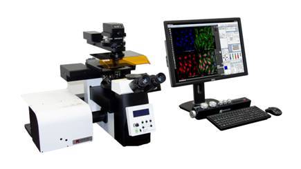
Nanoscope Systems,Inc.为您提供《细胞中生物检测方案 》,该方案主要用于细胞中生物检测,参考标准《暂无》,《细胞中生物检测方案 》用到的仪器有 K1-Fluo ABM 激光荧光共聚焦显微镜。
我要纠错
推荐专场
相关方案


 咨询
咨询


