方案详情文
智能文字提取功能测试中
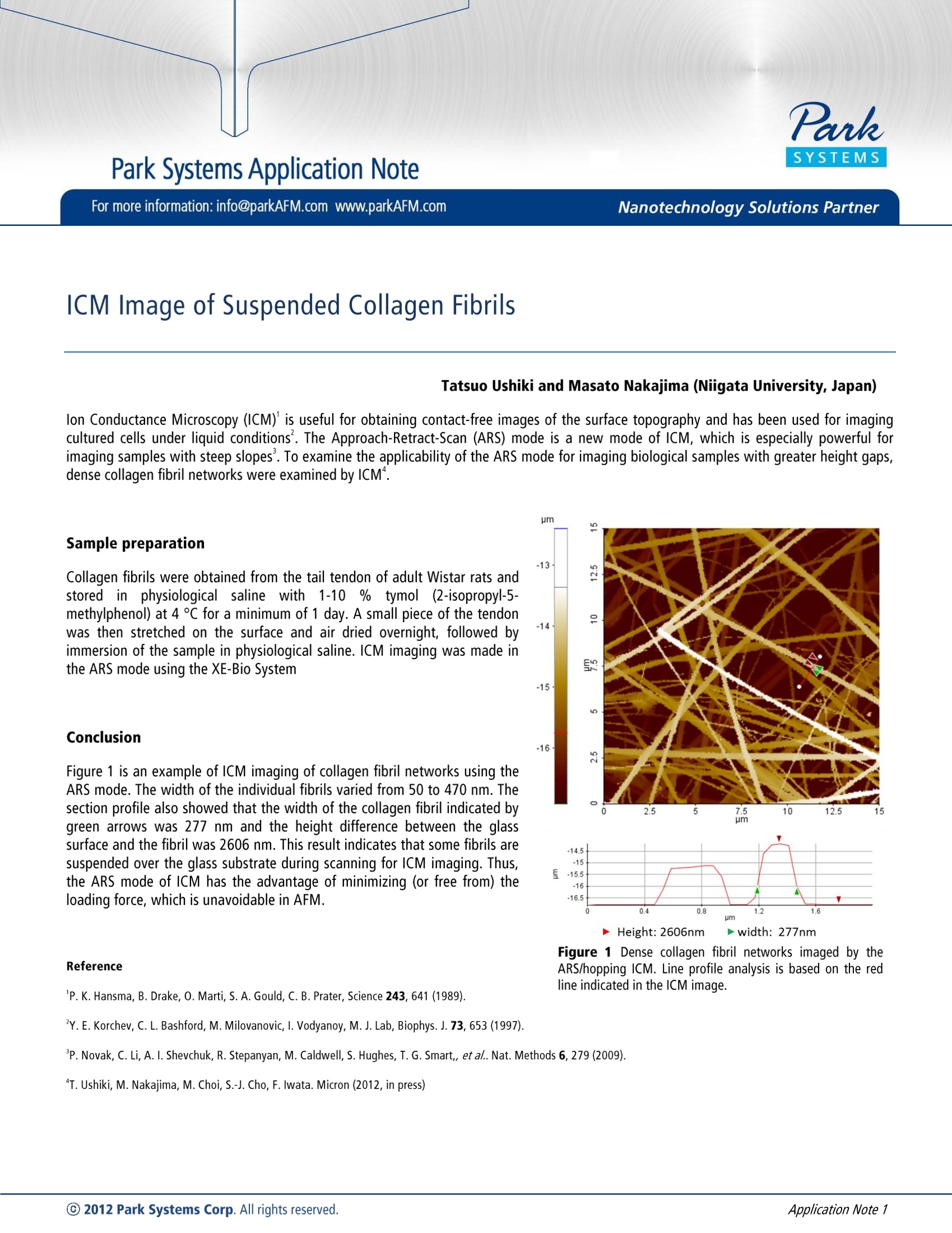
Park Systems Application NoteC) 2012 Park Systems Corp. All rights reserved.Application Note 1 For more information: info@parkAFM.com www.parkAFM.com Nanotechnology Solutions Partner ICM Image of Suspended Collagen Fibrils Tatsuo Ushiki and Masato Nakajima (Niigata University, Japan) lon Conductance Microscopy (ICM) is useful for obtaining contact-free images of the surface topography and has been used for imagingcultured cells under liquid conditions. The Approach-Retract-Scan (ARS) mode is a new mode of ICM, which is especially powerful forimaging samples with steep slopes . To examine the applicability of the ARS mode for imaging biological samples with greater height gaps,dense collagen fibril networks were examined by ICM. Sample preparation Collagen fibrils were obtained from the tail tendon of adult Wistar rats andstored in physiological saline with1-10% tymol (2-isopropyl-5-methylphenol) at 4 ℃ for a minimum of 1 day. A small piece of the tendonwas then stretched on the surface and air dried overnight, followed byimmersion of the sample in physiological saline. ICM imaging was made inthe ARS mode using the XE-Bio System Conclusion Figure 1 is an example of ICM imaging of collagen fibril networks using theARS mode. The width of the individual fibrils varied from 50 to 470 nm. Thesection profile also showed that the width of the collagen fibril indicated bygreen arrows was 277 nm and the height difference between the glasssurface and the fibril was 2606 nm. This result indicates that some fibrils aresuspended over the glass substrate during scanning for ICM imaging. Thus,the ARS mode of ICM has the advantage of minimizing (or free from) theloading force, which is unavoidable in AFM. Reference pm -14.5 -15 E -15.5 -16 -16.5 0.4 0.8 1.2 1.6 pm Height: 2606nm D width: 277nm Figure 1 Dense collagen fibril networks imaged by theARS/hopping ICM. Line profile analysis is based on the redline indicated in the ICM image. P. Novak, C. Li, A. I. Shevchuk, R. Stepanyan, M. Caldwell, S. Hughes, T. G. Smart,, et al.. Nat. Methods 6, 279 (2009). "T.Ushiki, M. Nakajima, M. Choi, S.-J. Cho, F. Iwata. Micron(2012, in press)
关闭-
1/1
产品配置单
Park帕克原子力显微镜为您提供《ICM image of suspended Collagen Fibrils》,该方案主要用于其他中null检测,参考标准《暂无》,《ICM image of suspended Collagen Fibrils》用到的仪器有null。
我要纠错
相关方案
 咨询
咨询