

方案详情文
智能文字提取功能测试中
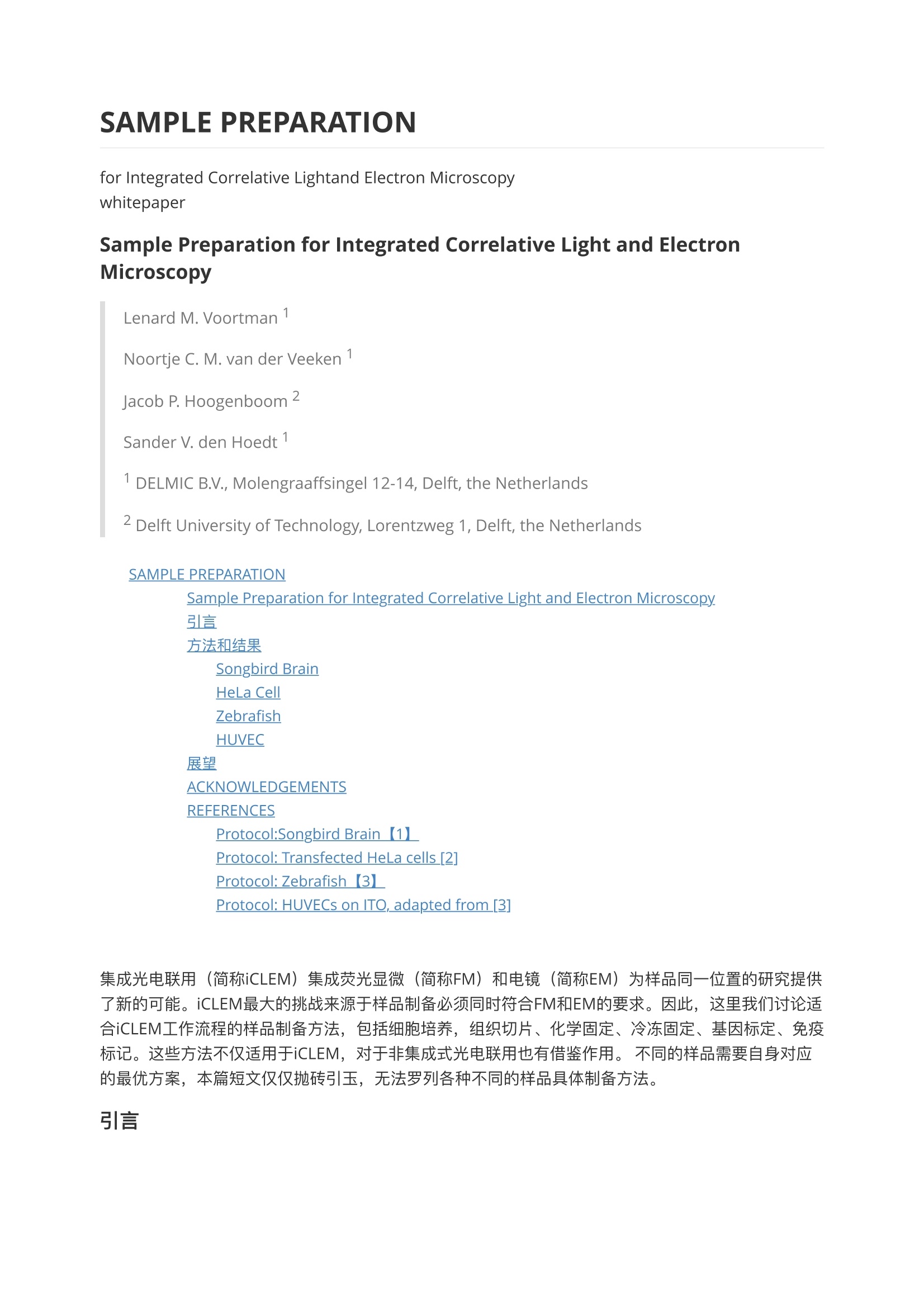
SAMPLE PREPARATION for Integrated Correlative Lightand Electron Microscopy whitepaper Sample Preparation for Integrated Correlative Light and ElectronMicroscopy Lenard M. Voortman ' Noortje C. M. van der Veeken Jacob P. Hoogenboom Sander V. den Hoedt ' ' DELMIC B.V., Molengraaffsingel 12-14, Delft, the Netherlands Delft University of Technology, Lorentzweg 1, Delft, the Netherlands SAMPLE PREPARATION Sample Preparation for Integrated Correlative Light and Electron Microscopy引言 方法和结果 Songbird Brain HeLa Cell Zebrafish HUVEC 展望 ACKNOWLEDGEMENTS REFERENCES Protocol:Songbird Brain 【1L Protocol: Transfected HeLa cells [2] Protocol: Zebrafish【3】 Protocol: HUVECs on ITO, adapted from [3] 集成光电联用(简称iCLEM) 集成荧光显微(1(简称FM))和电镜((简称EM)为样品同一位置的研究提供了新的可能。iCLEM最大的挑战来源于样品制备必须同时符合FM和EM的要求。因此,这里我们讨论适合iCLEM工作流程的样品制备方法,包细细胞培养,组织切片、化学固定、冷冻固定、基因标定、免疫标记。这些方法不仅适用于iCLEM,对于非集成式光电联用也有借鉴作用。,不同的样品需要自身对应的最优方案,本篇短文仅仅抛砖引玉,无法罗列各种不同的样品具体制备方法。 引言 过去的近10年,光电联用作为一种非常有用的科研工具越来越受到关注。主要原因在于CLEM同时结合了FM和EM各自的优点:FM能有效获取组织、细胞和细胞器内特定组件的官能信息;EM的高分辨能力可以获得清晰的超微结构信息。相互搭配互补,FM为EM观察的特定感兴趣的区域 (region ofinterest, 简称ROI)提供靶向。 尽管好处很多, CLEM还是遭遇很多的挑战和困难,高成本,费时并且要求很高的实验技巧。传统的非集成式关联,需要在完全不同的显微镜单独拍摄,并且FM和EM要求的样品制备方法也完全不兼容。由于FM和EM在原理上根本性区别,因此从FM到EM转移时还需要额外的样品处理步骤。这样的流程造成样品形变,引起关联的不准确。另外,要在EM下重新准确导航定位到FM采集的兴趣区域 (ROI)也是一项巨大挑战,尤其对大尺寸样品的定位更加困难。 iCLEM(集成式光电关联)有效的解决了以上痛点。将FM和EM有机集成在一起,根本性消除了样品在不同显微镜间转移带来的挑战。因为FM和EM观察的同同区域,因此无需对ROI的导航定位。另外由于样品是一次制样,无需额外的中间处理环节,一致性强,无形变无收缩,观察结果更可信。 对于iCLEM的样品制备方法也是一个全新的研究领域,到目前为止仅有少量的相关文章发表。对于非集成式光电联用的样品制备方法相对较多[1-3]。iCLEM样品制备的主要困难在于,EM一般要求样品采用重金属染色提高衬度,而这些重金属会脆灭荧光。另外, EM要求真空环境,因此样品需要干燥,干燥的环境影响荧光信息[4]。最新发表的文章显示SEM的真空度也一定程度影响荧光[5]。随着 iCLEM这样强大的科研工具的更多应用,我们预计越来越多的样品制备方法的研究文章将发表。 这里我们展示用于iCLEM的一些样品制备方法,特别选取具有代表性的几种制备方法供大家参考。 请注意,本篇文章的目的不是iCLEM样品制备方法的详细指南,另外我们也不讨论生物相关或应用。还有,我们仅仅是抛砖引玉,介绍iCLEM样品制备的可能性。我们主要展示iCLEM样品制备没有根本性的障碍。同时也可以看到iCLEM可以非常高效,简单,准确地获得图片的叠加。 方法和结果 来自荷兰delmic公司的SECOM平台,世界首创集成光电联用iCLEM。样品安装方式也是非常独特和方便,见图1示意。 Fig. 1. Illustration of how the specimen is ploced inside theSECOM platform. Thee-beam scars from obove whereas the lightmicroscope objective i situated below the cover glass To preventcharging of the sample due to thee-beam, the cover glass is coatedwith indium tin oxide (ITO) and cannected to the sample carrierusing carbon tope 样品直接放置或培养在ITO玻璃((玻璃表面镀一层氧化钢锡)上上。ITO对于可见光是完全透明,同时具有导电性, 可以导走SEM观察时在样品上积累的电荷[6]。 每种制备方法对应的结果展示在图2,简要描述见本文末尾。如果对于细节感兴趣,我们建议您完整阅读相应文章。 Songbird Brain 神经突出的连接方式对于理解神经的工作机理至关重要。结合EM我们可以观察到神经囊和突出密度,同时FM荧光标记可以标定特定的神经种类[7]。特别指出,尽管本次研究中的EM染色脆灭了原始的荧光标记,,但使用荧光抗体在切片上还可以重新标定,这说明制样方法可以很好的保护抗原性实现片上免疫标记。尽管这个protocol是特定适用于神经样品,相信类似方法可以拓展到其他领域的样品制备。 HeLa Cell 本次研究的目的是获得细胞膜内 lipid diacylglycerol的分布[5]。因此protocol设计成保持GFP和mCherry荧光活性,同时染色树脂包埋切片获得EM需要的衬度。关于使用的包埋剂的详细描述,请参考文章[5]。文章中作者还发现一个有趣的现象,快速冷冻替代方法【8】可能对于保持iCLEM需要的GFP的荧光活性至关重要。 Zebrafish 由于重金属染色对于荧光活性的影响,我们尝试使用无重金属染色剂染色。很明显,.,荧光活性得到更好的保存。当然负面影响就是EM衬度明显下降。无论如何,这样的方法得到的衬度基本能满足EM的观察。当然由于衬度的下降,细胞膜很难观察清楚。后续试验采用了低剂量的饿酸,以此达到某种平衡。 在FM下, GFP和E2-Crimson信号非常好。特别注意,,E图2中红色结构实际上是色素细胞而非特定标记。 HUVEC 人脐静脉内皮细胞Human umbilical vein endothelial cells (简称HUVEC)包含棒状结构称之为Weibel-Palade体的存储颗粒,其中含有Von Willebrand因子 (VWF)。这些细胞器在血液凝结过程发挥着重要作用。 我们使用样品快速制备,在一天内完成固定、免疫标定、脱水和光电关联观察。因此此管没有使用额外的EM染色,但是整个细胞在低电压下依然呈现良好的衬度,非常清晰[9]。荧光信息也保持异常完美。甚至在脱水保存在冰箱一个月后,样品中的荧光活性依然能获得较好的荧光像。 Fig. 2. Correlative light and electron micrographs using the SECOM platform(DELMIC B.V.,Delft) installed on a Quanta 250 FEG (FEI Company, Eindhoven). 1st row: fluorescenceimage. 2nd row: scanning electron micrograph. 3rd row: overlay of FM and EM. Columns 1to 4: projection neurons in songbird brain, HeLa cell expressing GFP-C1, Zebrafish and human umbilical veinendothelial cells labeled for Von Willebrand factor. Columns 1, 3 and 4: EM imaging usingseconday electron detector and FM imaging with Nikon Plan Apo 60x /0.95 lens, multicolorLED light engine, Clara CCD camera (Andor Technology, Belfast). Column 2: EM imagingusing the vCD backscatter detector and FM imaging with Nikon Plan Apo 100x /1.40 oilimmersion lens using vacuum compatible immersion oil, laser light source, Zyla sCMOScamera (Andor Technology, Belfast). 展望 本篇展示了几种用于iCLEM样品的制备方法,每种方法不限于文中展示的样品,根据需要可以拓展到更多种类的样品制备。。神经研究的示例,显示树脂包埋切片后,在片上进行免疫标记可以获得非常好的关联图像。HeLa细胞示例,表明采用冷冻替代可能是一种非常好的保持GFP荧光活性的方法。 在Zebrafish的样品制备过程中,还有很多可以提升的空间。无论如何,我们将它和HUVECS的样品制备protocols展示出来,即使没有额外的衬度增强, EM还是足够获得超微结构细节。 尽管荧光保持能力由于iCLEM的集成会受到限制,我们还是能找到EM与FM之间的平衡。对于不同应用,这种平衡需要研究者进行精细的调整。 集成光电联用最大的好处是,去除了从FM转移到EM的样品中间制备环节,最大程度的获得高准确度的关联,在大小不同视场间无缝转换。iCLEM提供了无缝的拍摄流程,既快又准的解决方案。 ACKNOWLEDGEMENTS Songbird brain samples were kindly provided by Dr Thomas Templier and Prof Dr Richard H RHahnloser, University of Zurich and ETH Zurich. Sample preparation and imaging of transfectedHeLa cells was performed by Dr Christopherj Peddie and Dr Lucy M Collinson, Cancer ResearchUK London Research. Zebrafish samples were provided by Rohola Hosseini and Gerda Lamers,Leiden University. We gratefully acknowledge the help of MarjonJ. Mourik, Leiden UniversityMedical Center, with the sample preparation of HUVECs. REFERENCES ( [1] Robert Kirmse and Eric H ummel. Co r relative Microscopy Protocols. Carl Zeiss Microscopy GmbH, June 2013. W eb. 28 April 2014 ) ( [2] Published Sample Preparation Protocols. FEl Company. Web.28 April 2 0 14 ) ( [3] Thomas Muller-Reichert and Paul Verkade, eds. Correlative Light and Electron Microscopy.Vol. 111.Academic P r ess, 2012. ) ( [4] Matthia A. Karreman, et al. "Optimizing immuno-labeling for correlative fluorescence and electron microscopy on a single specimen."journal of s t ructural biology 180.2 (2012):382-386. ) [5] ChristopherJ. Peddie, et al. "Correlative and integrated light and electron microscopy of in-resin GFP fluorescence, used to localise diacylglycerol in mammalian cells."Ultramicroscopy(2014). 6] Helma Pluk, et al. "Advantages of indium-tin oxide coated glass slides in correlative scanningelectron microscopy applications of uncoated cultured cells."journal of microscopy 233.3 (2009):353-363. [7] Daniele Oberti, Moritz A. Kirschmann, and Richard HR Hahnloser."Correlative microscopy ofdensely labeled projection neurons using neural tracers." Frontiers in neuroanatomy 4 (2010).[8] Kent L. McDonald and Richard I. Webb."Freeze substitution in 3 hours or less."journal ofmicroscopy 243.3 (2011):227-233. [9] Nalan Liv. "Protocol for Simultaneous Correlative Light Electron Microscopy with HighRegistration Accuracy" PhD Thesis (2014). Protocol:Songbird Brain 【1】 Fixation Perfuse 20 pl heparin and 5 ml 0.9% NaCl Fix 20 minn2% PFA and0.075% GA in phosphate buffer (0.1 M, pH 7.4) Remove brain Post-fix1 h 2% PFA and: 0.075% GA in phosphate buffer (0.1 M, pH7.4)Cut 60-um-thick sagittal vibratome sections Localize the area of interest using a widefield fluorescence micro-scope Electron microscopy staining Wash cacodylate buffer (0.1 M, pH 7.4) post-fix 40 min 1.5% potassium ferrocyanide and 1% OsO4 in cacodylate buffer (0.1M, pH 7.4) 1h 1%OsO4 in cacodylate buffer(0.1 M, pH 7.4) 1h 1% uranyl acetate in distilled water Dehydrate Flat-embed Durcupan ACM resin, cure for 48 h at52°C Sectioning Localize the area of interest using a light microscope Resection and attach to a blank resin block Serial section, 60-90 nm Collect sections on ITO coated coverslips Immunofluorescence staining Etch 10 minn1% periodic acid Wash 15 times ddH20 Wash 2 times 10 min Tris and PBS (TPBS, pH 7.4) Pre-block 30 min5% goat serum inTPBS Block 10 min 1% goat serum in TPBS Primary 1.5 h 1:50 Rabbit anti Alexa488 and 1:50 Rab- bit anti Texas Red Wash 4 times 10 min Tris-HCl buffer (0.05 M, pH 7.5) Secondarv 1.5h 1:50 Alexa 546 anti-rabbit Wash 15 times ddH20 Protocol: Transfected HeLa cells [2] Cell culture Maintain cells in DMEM supplemented with 10% foetal bovine serum Transfect cells with GFP-C1 and mCherry-H2B constructs Fixation was performed 18-24 h after transfection High pressure freezing Spin cells in an Eppendorf tube to form a pellet Resuspend in equal volume of media and 10% BSA, maintain at 37°C Spin down a volume ofcells in a blocked 200 ul pipette Cut away the end of the tip and pipet into membrane carriersLoad into high pressure freezer Store carriers containing frozen cells under liquid nitrogen Quick freeze substitution Modified version of the method described in [8] Substitution media 5% H20 and 0.1% or 0.2% UA in in acetone Transfer to moulds filled with100% acetone and incubate for 15 min Wash 3 times 15 min 100% acetone Infiltration 3h 220%,40%, 60%, 80%,100%HM20 Incubate overnight100% HM20 4 changes offresh resin polymerize48h 360 nm UV light Sectioning Trim blocks from the moulds and store at room temperature in the dark Protocol: Zebrafish 【3】 Labels GFP and E2-Crimson Fixation Fixation 2 h PHEM fixativeRinse 3 times 10 min PHEM Dehydration Dehydration and resin Infiltration on a rotator Dehydrate 15 min 50%, 15 min 70%, 15 min 80%, 15min 90%, 15 min 100% Ethanol Embedding 1h or overnight 1:1 LR-white in Ethanol 2 times 1h LR-white Polymerization 24h UV Chamber in cold room Protocol: HUVECs on ITO, adapted from [3] Cell culture** Clean slides wash in ethanol, dry, glow discharge (to make surface hydrophilic topromote cell adhesion)Sterilize 15 min UVCoat 1% gelatin in PBS (0.1 M, pH 7.4)Trypsinize cells and seed directly onto ITO slides Grow cells up to desired confluency, 37°C, 5% CO2 Fixation Fix30 min 2,5% PFA and 0,25% GAin phosphate buffer Immunofluorescence staining Wash 2 times Tris-HCI buffer (0.05 M, pH 7.4) Block 20 minn0.1% Triton X-100 and 5% Normal Goat Serum (NGS) in PBS Primary 1 h 1:500 Rabbit anti Human VWF in PBS-5% NGS Wash 2 times 5 min PBS Block 5min PBS-5% NGS Secondarv 30 min 1:100 Goat anti-Rabbit Alexa 568 in PBS-5% NGS Wash 2 times 5 min PBS Stain 20 min 1:20 Phalloidin Alexa 488 in PBS Wash 3 times 5 min PBS Dehydration 2 quick washes;770% Ethanol Dehydrate5 min 70%, 5 min 80%, 5 min 90%, 5 min 100%, 15 min 100%, 15 min 100% Etha-nol Air dry slides 关于Delmic B.V. DELMIC B.V. 是位于荷兰delft市的一家专注于电子显微领域的高科技创新电镜附件公司,提供电镜集成解决方案。。目前主要产品,SECOM平台集成荧光显微与电子显微镜系统实现光电联合应用以及SPARC平台高性能角分辨阴极荧光成像系统。 DELMIC技术起源于代尔夫理工的 Charged Particle Optics课题组,第一个产品即是光电联用SECOM平台。SECOM集成在SEM上实现光镜电镜联用,快速荧光成像并以优于50nm的对位精度与电镜像的完美自动叠加。2011年末,公司获得荷兰国家原子分子研究所 AMOLF Polman团队创新研发的angle-resolved阴极荧光成像 (ARC)技术授权。该技术全球创新首创,Polman团队因此获得2014年MRS创新大奖。Delmic继续研发ARC技术并推出商用化产品SPARC平台,实现了全球唯一具有角分辨解析功能的高性能纳米级荧光成像系统。这些系统无缝集成在SEM上,广泛应用于从纳米光电子到生命科学各个科研领域,帮助科学家开辟新的研究途径。 DELMIC致力于持续创新,为科学家提供高性能的极致创新集成解决方案。 关于苏州德尔微仪器有限公司 苏州德尔微仪器有限公司是位于苏州生物纳米园的创新服务公司,提供电镜实验室的高端仪器和设备。与全球创新科学实验仪器公司紧密合作,致力于创造和引进独特的实验方法和表征手段,为中国电镜在纳米科技,先进材料和生命科学等领域的突破提供最先进的工具。 作为荷兰Delmic B.V.公司中国代理商,德尔微仪器提供集成光电用用和高性能角分辨荧光成像平台及相关服务。基于我们的使命,德尔微与电镜主机、电镜附件、样品制备备商商泛合作,致力于创新样品制备工艺和装备、极致探测手段和表征方法、定制化工程服务和解决方案。 助力科学,探索致发现! 电镜实验室专业工程团队。 EM Labs'Engineering Team 地址:苏州市工业园区星湖街218号生物纳米园A7楼203室,215123
关闭-
1/11
-
2/11
还剩9页未读,是否继续阅读?
继续免费阅读全文产品配置单
苏州德尓微仪器有限公司为您提供《细胞中超结构信息检测方案(电镜部件)》,该方案主要用于其他中超结构信息检测,参考标准《暂无》,《细胞中超结构信息检测方案(电镜部件)》用到的仪器有SECOM-集成式光电联用平台。
我要纠错
相关方案
 咨询
咨询