

方案详情文
智能文字提取功能测试中
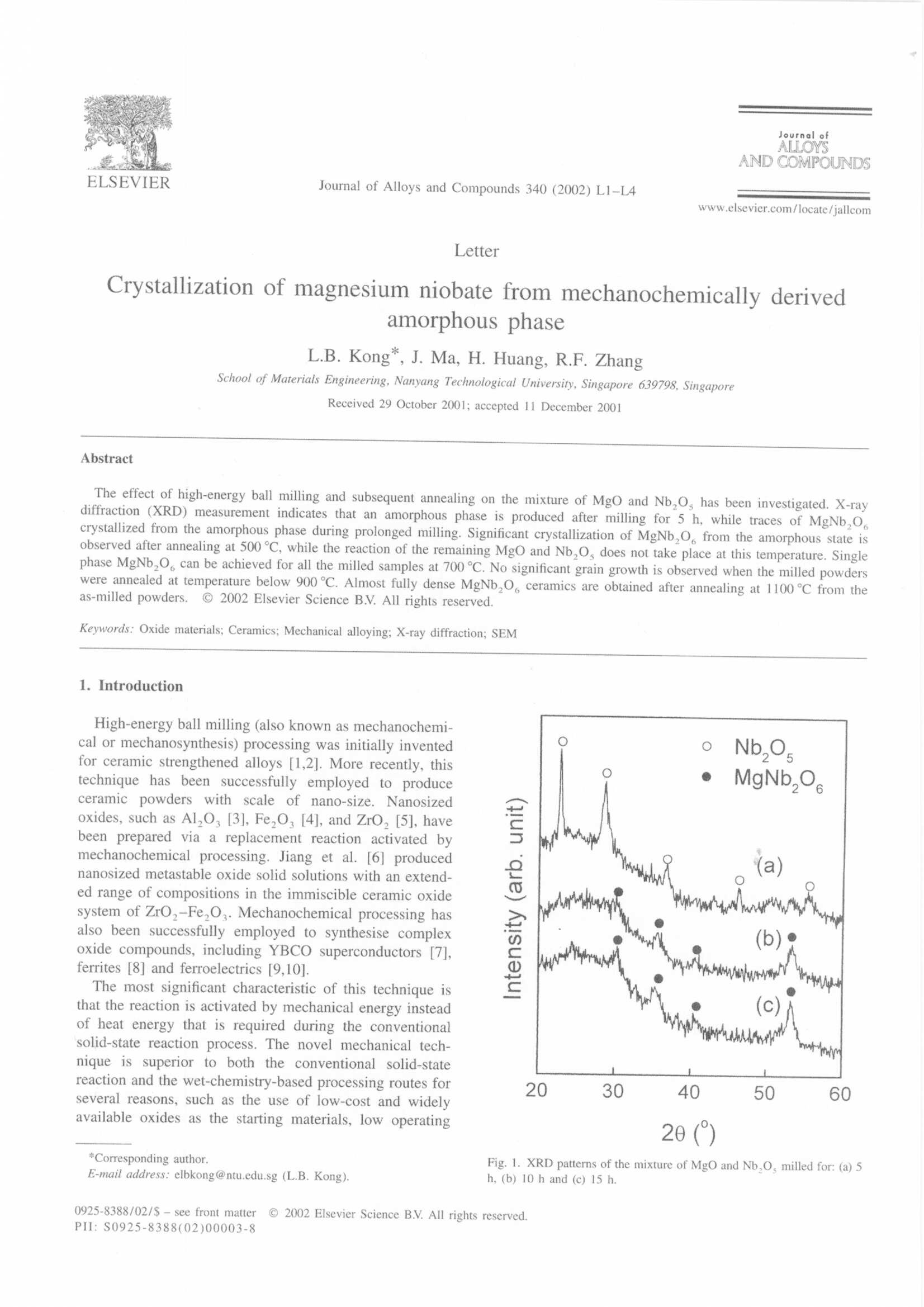
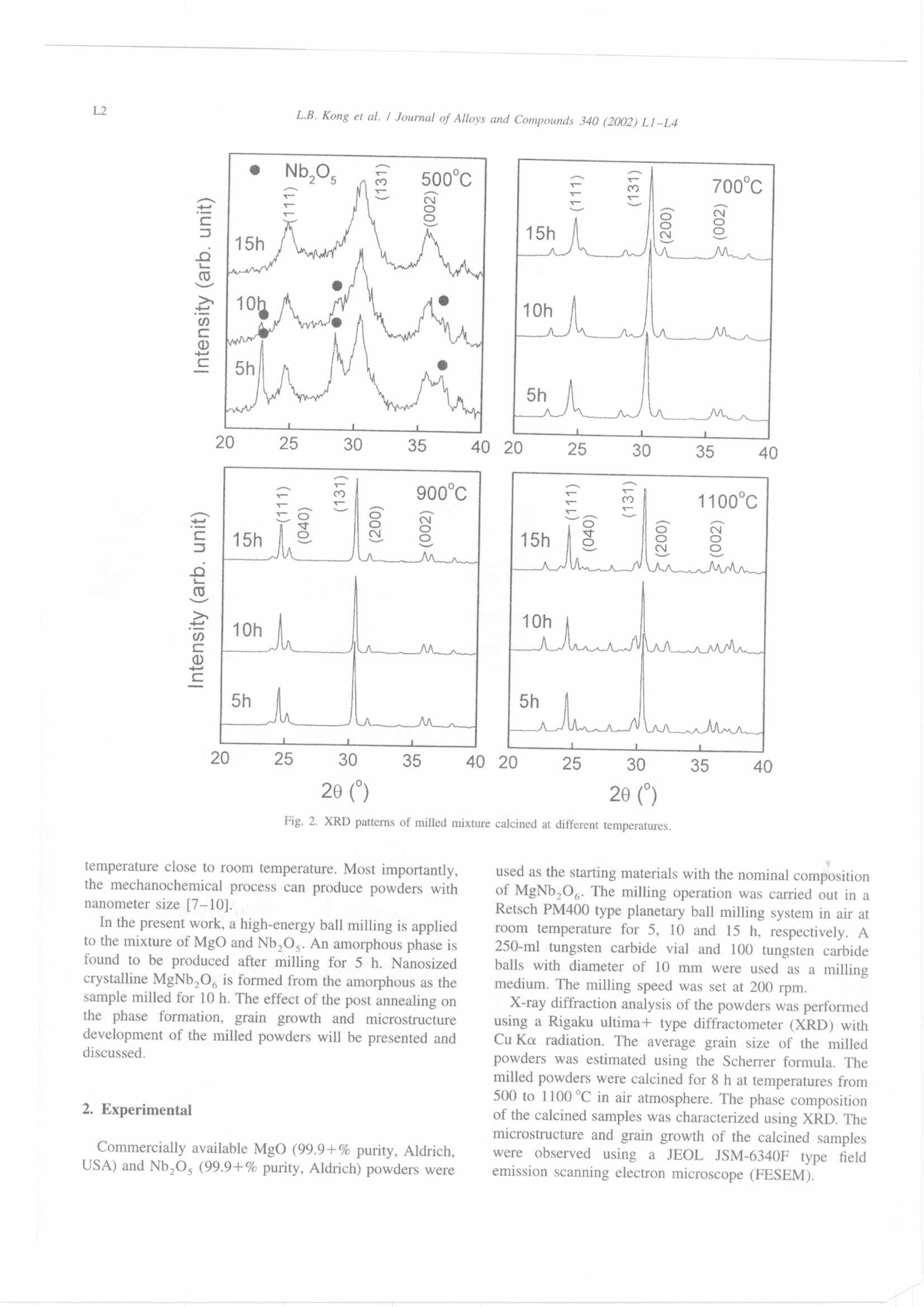
Journal ofALLOYSAND COMPOUNDSJournal of Alloys and Compounds 340 (2002) L1-L4www.elsevier.com/locate/jallcom L2L.B. Kong et al./ Journal of Alloys and Compounds 340 (2002) L1-L4 C 2002 Elsevier Science B.V. All rights reserved.PII: S0925-8388(02)00003-8 Letter Crystallization of magnesium niobate from mechanochemically derivedamorphous phase L.B. Kong*, J. Ma, H. Huang, R.F. Zhang School of Materials Engineering, Nanyang Technological University, Singapore 639798, SingaporeReceived 29 October 2001; accepted 11 December 2001 Abstract : effect of high-energy ball milling and subsequent annealing on the mixture of MgO and Nb,O, has been investigated. X-raydiffraction (XRD) measurement indicates that an amorphous phase is produced after milling for 5 h, while traces of MgNb,O,crystallized from the amorphous phase during prolonged milling. Significant crystallization of MgNb,O from the amorphous state isobserved after annealing at 500℃, while the reaction of the remaining MgO and Nb,O, does not take place at this temperature. Singlephase MgNb,O, can be achieved for all the milled samples at 700 ℃. No significant grain growth is observed when the milled powderswere annealed at temperature below 900C. Almost fully dense MgNb,O ceramics are obtained after annealing at 1100℃ from theas-milled powders. C 2002 Elsevier Science B.V. All rights reserved. Keywords: Oxide materials; Ceramics; Mechanical alloying; X-ray diffraction; SEM High-energy ball milling (also known as mechanochemi-cal or mechanosynthesis) processing was initially inventedfor ceramic strengthened alloys [1,2]. More recently, thistechnique has been successfully employed to produceceramicpowdersSVwithscale: of nano-size. Nanosizedoxides, such as Al,O,[3], Fe,O, [4], and ZrO, [5], havebeen prepared via a replacement reaction activated bymechanochemical processing. Jiang et al. [6] producednanosized metastable oxide solid solutions with an extend-ed range of compositions in the immiscible ceramic oxidesystem of ZrO,-Fe,O,. Mechanochemical processing hasalso been successfully employed to synthesise complexoxide compounds, including YBCO superconductors [7],ferrites [8] and ferroelectrics [9,10]. The most significant characteristic of this technique isthat the reaction is activated by mechanical energy insteadof heat energy that is required during the conventionalsolid-state reaction process. The novel mechanical tech-nique is superior to both the conventional solid-statereaction and the wet-chemistry-based processing routes forseveral reasons, such as the use of low-cost and widelyavailable oxides as the starting materials, low operating 20() 20() 20() Fig. 2. XRD patterns of milled mixture calcined at different temperatures. temperature close to room temperature. Most importantly,the mechanochemical process can produce powders withnanometer size [7-10]. In the present work, a high-energy ball milling is appliedto the mixture of MgO and Nb,O,. An amorphous phase isfound to be produced after milling for 5 h. Nanosizedcrystalline MgNb,O, is formed from the amorphous as thesample milled for 10 h. The effect of the post annealing onthe phase formation, grain growth andi microstructuredevelopment of the milled powders will be presented anddiscussed. 2. Experimental Commercially available MgO (99.9+% purity, Aldrich,USA) and Nb,O, (99.9+% purity, Aldrich) powders were used as the starting materials with the nominal compositionof MgNb,O. The milling operation was carried out in aRetsch PM400 type planetary ball milling system in air atroom temperature for 5, 10 and 15 h, respectively. A250-ml tungsten carbide vial and 100 tungsten carbideballs with diameter of 10 mm were used as a millingmedium. The milling speed was set at 200 rpm. X-ray diffraction analysis of the powders was performedusing a Rigaku ultima+ type diffractometer (XRD) withCu Ko radiation. The average grain size of the milledpowders was estimated using the Scherrer formula. Themilled powders were calcined for 8 h at temperatures from500 to 1100℃ in air atmosphere. The phase compositionof the calcined samples was characterized using XRD. Themicrostructure and grain growth of the calcined sampleswere observed using a JEOL JSM-6340F type ffieldemission scanning electron microscope (FESEM). Fig. 1 shows the XRD patterns of the mixture of MgOand Nb,Os milled for different time durations. For thesample milled for 5 h, the main diffraction peaks areattributed to Nb,O,. No MgO diffraction peak is observedin the XRD pattern probably because it is too weakcompared with that of Nb,O.. It is also hard to determinethe diffraction of MgNb,O。 crystallines. However, thehump existing between 20°and 40° in the XRD pattern(Fig. la) indicate that amorphous phase is formed as aresult of the high-energy ball milling after milling for 5 h.As the millingitime duration increases to 10 h. thediffraction peaks of Nb,Os are totally disappearing fromthe XRD pattern, while a trace of MgNb,O, can bedistinguished as shown in Fig. 1b. At the same time, theamorphous hump becomes more obvious than in Fig. la.Further increase in milling time duration leads to increasedcrystallization of MgNb,O, which is evidenced by itsincreased diffraction peaks. The above observation indicates that the formation ofMgNb,O, via the high-energy ball milling process in thepresent work could follow a sequence of refinement ofOxides, amorphization of the oxides and crystallization ofMgNb,o。 from the amorphous matrix. This is differentfrom the formation of other compounds such as leadtitanate (PbTiO,3on PT),lead zirconate titanate(PbZro.48Tio.52Os or PZT), leadmagnesium niobate(PbMgi/3Nb230 cor PMN) and bismuth titanate(Bi Ti,O), all of which were directly obtained via thehigh-energy ball milling without the present of amorphous The effect of calcination temperature on the crystalliza-tion of MgNb,O, from the milled powders were studiedby calcining the samples at different temperatures. XRDpatterns of the calcined powders are shown in Fig. 2. Aftercalcining at 500℃ for 8 lh, crystalline MgNb,O。 isobserved in all the three samples. For the 5-h milledsample, diffraction peaks of Nb,O, are still appearing inthe XRD pattern, which is similar with that observed in theas-milled powder (Fig. la). A slight trace of Nb,O.diffraction is also detected in the sample milled for 10 h.No Nb,O, is observed in the 10-h as-milled samples asseen in Fig. 1b, which may be due to the fact diffractionpeaks of Nb,O, is too weak to be detected by XRDmeasurement. This observation implies that annealing at500 C for 8 h is enough for MgNb,O, to crystallize fromthe amorphous produced as a result of the high-energy ballmilling, but not sufficient to promote the reaction of theremained Nb,Os and MgO even though they were sig-nificantly refined by the mechanochemical treatment. How-ever, the broadened diffraction peaks indicate that thecrystallized MgNb,0,possesses a small grain size andpoor crystallinity. This result also strongly confirms thepresence of the amorphous state produced by the millingprocess. As the annealed temperature increases to 700℃, all thethree samples demonstrate a single phase of MgNb,0Thelattice constantt of theoorthorhombicgstructuredMgNb,O。 is estimated from the XRD patterns to bea=5.699(2), b=14.192(4) and c=5.033(3), which is ingood agreement with the literature data (JCPDS No. 33875). The absence of Nb,O, and the increased andsharpened diffraction peaks of MgNb,O, indicate that thereaction of the remained MgO and Nb,O, is complete andthe crystallization of MgNb,O, from the amorphous is Fig. 4. SEM photographs of the milled mixture calcined at 1100 ℃ for 8h: (a) 5 h, (b) 10 h and (c) 15 h. greatly enhanced at this temperature. This temperature issignificantly lower than required by the conventional solid-state reaction process. Further increase in calcinationtemperature leads only to increase in diffraction peaks ofMgNb,O, indicating the grain growth MgNb,O, as aresult of high-temperature annealing. The grain growth behavior and microstructural develop- ment of the milled powders were examined by scanningelectron microscopy. SEM photographs of the calcinedpowders are shown in Figs. 3 and 4. The samples calcinedat 900℃ for 8 h exhibit porous structure with an averagegrain size of less than 1 pm, while the 1100℃ calcinedsamples demonstrate nearly fully dense structure. Rod-likegrains with about 10 pm length and 1-2 pm thickness areobserved for all the three samples. For the samplesannealed at low temperatures (500 and 700℃), SEMresults are not shown here because of their poor qualities.However, the grain size of the low-temperature calcinedsamples is almost the same as that of the as-milledpowders. 4. Conclusions High-energy ball milling process has been applied to themixture of MgO and Nb,O,. Amorphization of the mixtureas a result of the high-energy ball milling is observed aftermilling for 5 h. Small fraction of MgNb,O, is formed inthe sample milled for 10 h. Further crystallization ofMgNb,O, from the amorphous state can be achieved byannealing the samples at 500℃. However, the reaction ofthe remained MgO and Nb,O, cannot take place at thistemperature. Both the crystallization andreaction areenhanced as the calcination temperature increases to700℃. No significant grain growth is observed when themilled powders were annealed at temperature below900 ℃. Only porous samples can be obtained at 900℃C,while 1100℃ annealing leadss to almost fully denseMgNb,O,ceramics. References ( [1 ] P . S . G i l man , J . S . B e nj a mi n, Ann u. Rev . Mat er . Sci. 13 (1983)279-300. ) ( [2] N.J. W e lh am, P . E . Wi lli s, T. K e rr , J . Am. Ceram. Soc . 83 (1) (2000) 33-4 0 . ) ( [3] J . D i ng, T . T s uzu ki , P. G . McCormi c k, J. Am. Ceram . S oc. 79 (11)(1996) 295 6 -29 5 8 . ) [4] J. Ding. T. Tsuzuki, P.G. McCormick, Nanostruct. Mater. 8 (6)(1997) 739-747. [5] S.C. Dodd, K. Raviprasad, P.G. McCormick, Scripta Mater. 44(2001) 689-694. [6] J.Z. Jiang, F.W. Poulsen, S. Morup.J. Mater. Res. 9(3) (1994)535-540. [7] M. Simoneau, G. L'Esperance, M.L. Trudeau, R. Schulz, J. Mater.Res. 9 (3)(1994) 535-540. [8] G. Nicoara, D. Fratiloiu, M. Nogues, J.L. Dormann, F. Vasiliu,Mater. Sci. Forum 235-238 (1) (1997) 145-150. [9] J. Xue, D. Wan, S.E. Lee, J. Wang, J. Am. Ceram. Soc. 82 (7)(1999) 1687-1692.
关闭-
1/4
-
2/4
还剩2页未读,是否继续阅读?
继续免费阅读全文产品配置单
弗尔德(上海)仪器设备有限公司为您提供《铌酸镁晶体中机械化学研磨方法检测方案(研磨机)》,该方案主要用于其他中机械化学研磨方法检测,参考标准《暂无》,《铌酸镁晶体中机械化学研磨方法检测方案(研磨机)》用到的仪器有德国莱驰行星式球磨仪/机Retsch PM400。
我要纠错
推荐专场
研磨机、研磨仪、粉碎机、球磨机
更多


相关方案
 咨询
咨询