

方案详情文
智能文字提取功能测试中
M1105Figure 1: Comparison of SVP Load Flow Imaging ofSubvisible Protein Particles: Moving Towards Best Practices Abstract Purpose: Flow imaging uses camera-based techniques to image particles in the 1 to 100pm sizerange. As protein particles in this size range have the potential to cause an immunogenic response,these methods have become the most widely used technique to detect and quantify subvisibleparticles (SVPs) in injectable protein products. Methods: A number of different protein formulations were evaluated for SVP levels using bothMFI and FlowCAM" instruments. While high counts of SVPs occur in some samples, many of theparticles do not appear to be protein in nature.Approaches are described to minimize the impact ofnon-protein SVPs on the determination of protein particulates. Results: A wide range of non-protein particulates have been observed, including air bubbles,silicone oil droplets, fibers, and inorganic crystals. In addition, optical artifacts can occur in highconcentration formulations and particles can settle due to stratification. As a result, samplingtechniques and analysis parameters must be developed for each specific protein product. Ourexperience indicates that both sample handling and post-processing algorithms can reduce therelative content of foreign particles, making the quantitation of protein particulates more accurateand precise. Conclusion: The importance ofexamining the actual images and taking prudent steps to minimizethe appearance of non-protein SVPs is critical for obtaining reliable data on micron-sized proteinparticulates. These observations and recommendations are intended to encourage discussion aboutthe best methods to use when employing camera-based techniques to detect and quantify proteinparticulates. Purpose Several different therapeutic proteins were evaluated for levels of subvisible particles (SVPs) usingeither MFI"" or FlowCAM° instruments. In some cases, both instruments were used. The purposewas not to compare the two different platforms, but to develop the best practices for flow imaginganalysis. In doing so, a number of critical aspects were identified as requiring further investigation.Many of these issues are tied to image quality. Hopefully, this work serves as the basis for anongoing discussion within the biopharmaceutical industry on the best methods for data collectionand analysis when evaluating protein products for SVPs by flow imaging. Methods & Results The following general sections look at several topics of interest when using flow imaging tocharacterize SVPs in protein formulations: MFI/FlowCAM Comparison ofSVP load: To date, there has not been a published study comparing the subvisible particle (SVP) load for thesame samples using these two flow imaging platforms. Below are shown the particle distributionsgPfor four samples of the same lyophilized peptide stored for twelve weeks after reconstitution. Whilethe numbers do vary, there is fairly good agreement between the methods, at least in terms of totalparticle counts per unit volume. Figure 1 (continued): Comparison of SVP Load Comparison oflmage Quality: While particle counts might be similar, there are differences in image quality that are important toconsider. In this study, the same sample of an aqueous solution containing crystals was examinedusing both MFI and FlowCAM. The FlowCAM can be fitted with two different objectives (4xand 10x). The differences in image quality are striking. Better image quality means more reliablequantitation of particle properties like size, translucency and aspect ratio. Sort Edge Gradient Classified: 0 2-5um Figure 2: FlowCAM Images for SVPs in 2pm-5pm Range (above):4x Magnification (left) and 10x Magnification (right) Par 0-5.00w Figure 3: MFI Images for SVPs in 2pm-5pm Range (above): 5x Magnification Formulation 2 Sample Handling: Stratification: In high concentration liquidformulations, it has often been observed thatthe particle counts per mL are greater near thebottom of the samples than near the top. (Seeexample on right) Air Bubbles/Filtration: Many of the smaller spherical particles in a sample are likely airbubbles (although surfactants and oil droplets could be present as well). Filtration of the samplecould alleviate much of this artifact. However, Figure 4: Stratification Found in High Concentration Samples no study has been published on the effects of sample filtration or degassing on flow imaging. Image Evaluation: Careful examination of the images is required for two reasons. First, artifacts can occur that canskew proper quantitation of the number of SVPs. Second, many of the particles observed by flowimaging are not protein-based. Image Analysis Software/Image Classification: Given that many of the particles observed by flow imaging are likely not protein in nature,especially for smaller particles, it is critical to have a method that removes these non-proteinparticles from consideration in an automated and unbiased fashion. The first detailed attempt at developing such a template to ‘filter' protein from non-protein particlesused data from MFI (Strehl et al., Pharm. Res. 2012,29: 594-602). The best template employedfour successive filters: aspect ratio, circularity, maximum object intensity and standard deviation ofthe object intensity. FlowCAM Software allows for templates to be created that can filter or classify particles based onindividual characteristics or a series of properties that may be specific to a particle type (see Table1 below). These classification templates allow one to segregate particles based on a composite ofproperties in an automated, unbiased fashion. Those images that display qualities associated withnon-protein particles such as droplets or bubbles fall into individual classes, while the remainingparticles that do not sort to specific criteria will be held in a general class for all remaining particles. Area(ABD) Circularity (Hu) Diameter (ESD) Geodesic Aspect Ratio Perimeter Transparency Area (Filled) Aspect Ratio Compactness Convex Perimetelier Elongation Fiber Curl Geodesic Length Geodesic Thickness Roughness Sigma Intensity Volume(ABD) Volume (ESD) Circle Fit Convexity Fiber Straightness Intensit Sum Intensit Width Circularity Diameter (ABD) Filter Score Length Symmetry Using these templates, one can automatically filter any set of particle images collected with theFlowCAM instrument running the latest version of the software. An example of a ClassificationTemplate is as follows: 1. First Class -Removes any particles less than 3 um in size. There is insufficient resolution togather reliable estimates of image properties for smaller size particles.2. Second Class -Remove droplet particles using a specifically designed filter. iSS-3. Third Class - Capture all remaining particles in the 3 to 100 um size range which shouldprimarily consist of an enriched protein particle population. 4. Additional Processing- If these particles can be reassigned in an unbiased fashion, thenadditional processing should be done. Otherwise, they need to be considered close enough inproperties to protein particulates that they cannot be excluded from consideration. Conclusions It appears that methods may need to be developed for each protein system, or at least for classesof protein formulations. Systematic studies are needed to evaluate the impact of sample handling,viscosity, refractive index, etc. In addition, it appears that it should be possible to enrich the particlepopulation in terms of protein-containing particulates by using templates to remove artifacts andforeign materials in an automated and unbiased fashion. However, no such studies currently appearin the literature. 目的:流动成像使用基于相机技术对1至100μm尺寸范围内的颗粒进行成像。 由于该范围内的蛋白质颗粒具有引起免疫原性反应的潜力,因此这些方法已成为检测和定量注射蛋白质产品中的亚可见颗粒(SVP)的最广泛使用的技术。方法:使用MFI™和FlowCAM®仪器评估许多不同的蛋白质配方的SVP水平。 虽然在一些样品中发生了大量的SVP,但许多颗粒本质上看起来不是蛋白质。 描述了使非蛋白质SVP对蛋白质颗粒测定的影响最小化的方法。
关闭-
1/1

产品配置单
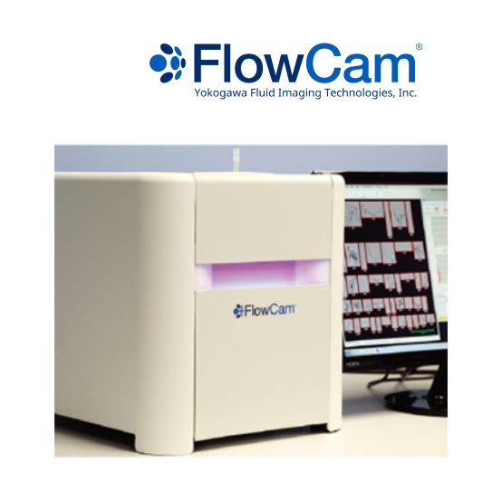
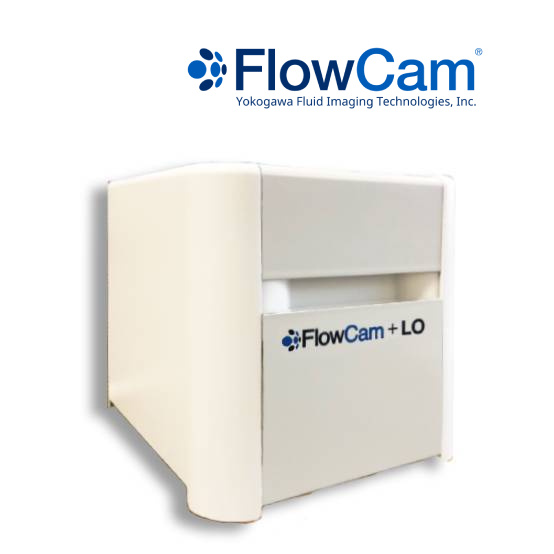
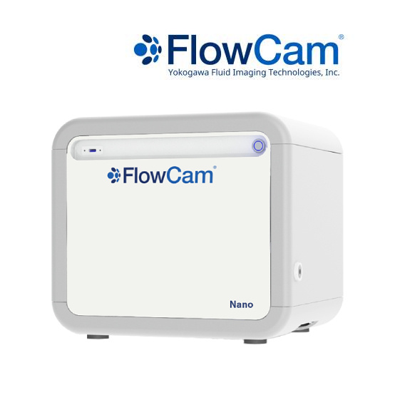
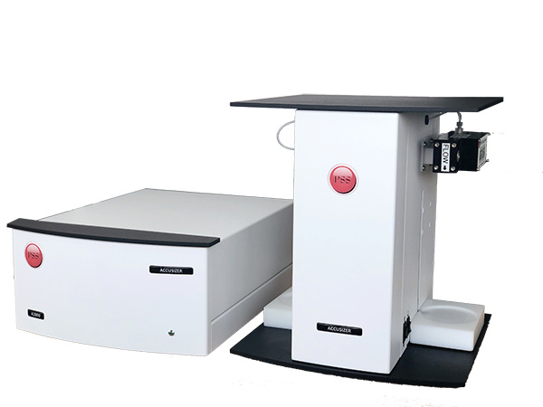
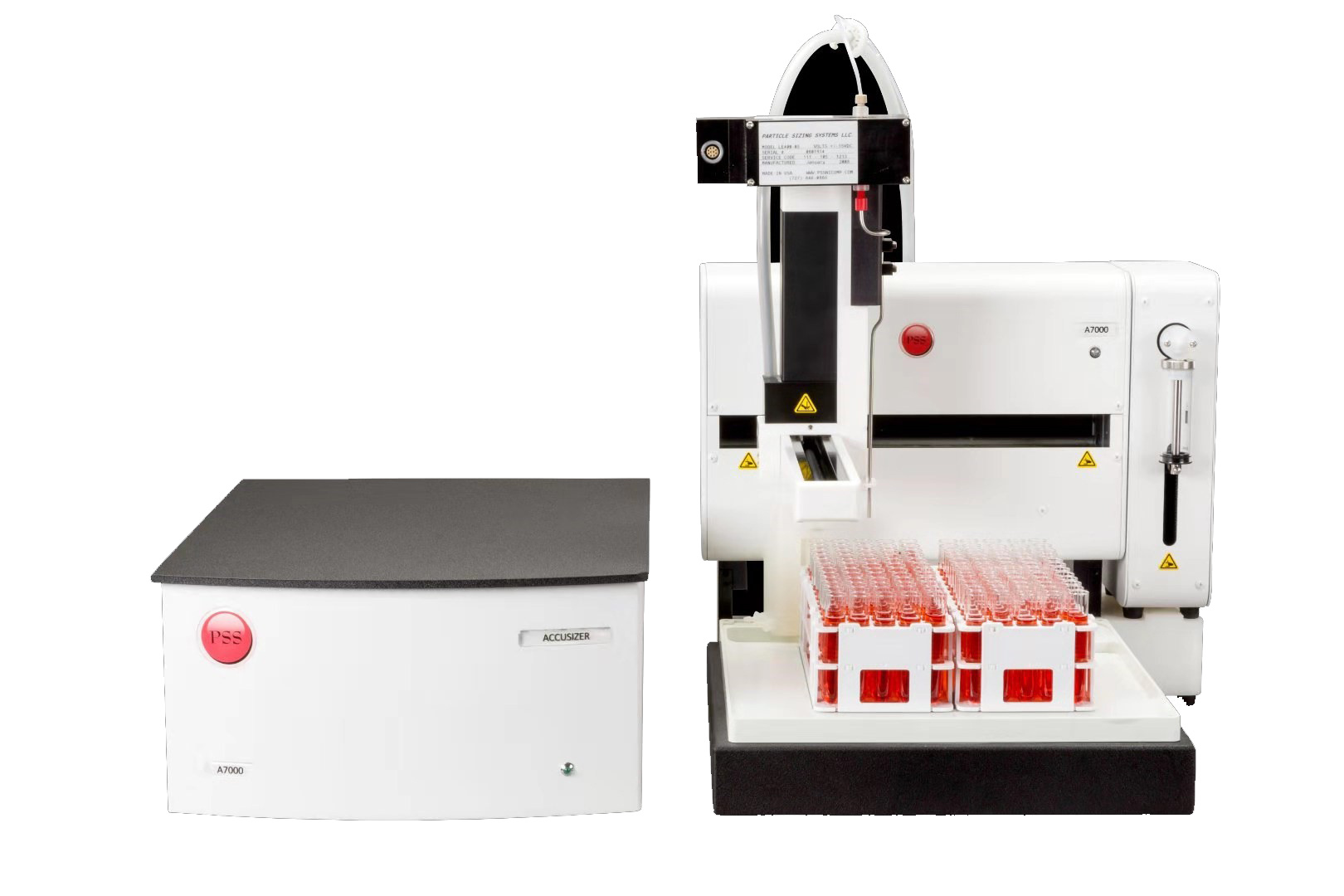
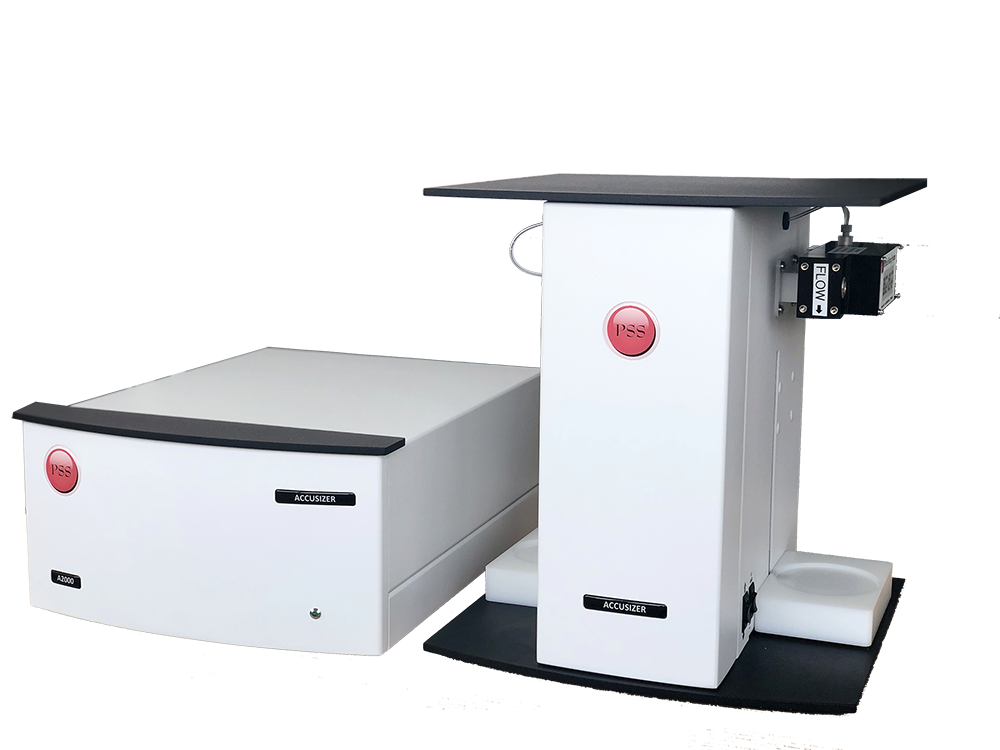
大昌华嘉科学仪器为您提供《亚可见蛋白颗粒中流动成像检测方案(图像粒度粒形)》,该方案主要用于其他中流动成像检测,参考标准《暂无》,《亚可见蛋白颗粒中流动成像检测方案(图像粒度粒形)》用到的仪器有流式颗粒成像分析系统FlowCam®8100、颗粒成像法+光阻法分析系统 FlowCam® + LO、纳米流式颗粒成像分析系统 FlowCam® Nano。
我要纠错
推荐专场
图像粒度粒形分析仪
更多

相关方案
 咨询
咨询