

方案详情文
智能文字提取功能测试中
analyticaistryArticlepubs.acs.org/acCite This: Anal. Chem. XXXX, XXX, XXX-XXX ArticleAnalytical Chemistry Noninvasive and Accurate Detection of Hereditary Hearing LossMutations with Buccal Swab Based on Droplet Digital PCR Fang Wang, Lingxiang Zhu, Baoxia Liu, Xiurui Zhu, Nan Wang Tao Deng," Dongyang Kang,lunmin Pan,DwWenjun Yang,Huafang Gao,* and Yong Guo*8D 'Human Genetic Resource Center, National Research Institute for Health and Family Planning, 12 Da Huisi Raod, Beijing 100081,People’s Republic of China Chinese Academy of Medical Sciences, Graduate School of Peking Union Medical College, 9 Dongdan Three Road, Beijing100730, People’s Republic of China SDepartment of Biomedical Engineering, School of Medicine, Collaborative Innovation Center for Diagnosis and Treatment ofInfectious Diseases, Tsinghua University, 30 Shuangqing Road, Beijing 100084, People’s Republic of China "Beijing CapitalBio Medical Laboratory, 88 Kechuang Six Street, Beijing 101111, People’s Republic of China -Department of Otorhinolaryngology Head Neck Surgery, Chinese PLA General Hospital, 28 Fuxing Road, Beijing 100853, P. R.China "MOE Key Laboratory of Protein Sciences, School of Life Sciences, Tsinghua University, 30 Shuangqing Road, Beijing 100084,People’s Republic of China TargetingOne Corporation, 268 Chengfu Road, Beijing 100190, People’s Republic of China Supporting Information ABSTRACT: Hereditary hearing loss is a common clinicalneurosensory disorder in humans and has a high demand forgenetic screening. Current screening techniques using peripheralblood or dried blood spots (DBSs) are invasive. Therefore, thisstudy aims to develop a noninvasive and accurate detectionmethod for eight hotspot deafness-associated mutations based onbuccal swab and droplet digital PCR (ddPCR). First, this methodwas evaluated for analytic performance including specificity,detection limit, dynamic range using plasmid DNA. The specificitywas 100% and the detection limit was 5 copies. The dynamic rangeof this ddPCR-based method was from 10 to 10°copies/uL. Next, the method was found to accurately quantify mitochondrial gene heteroplasmy rate as low as 1% for both m.1494C>T andm.1555A> G sites. Then, we demonstrated that buccal swab was a reliable sample. DNA can be extracted and accuratelyquantified after a buccal swab had been stored for 90 days at either room temperature or -20°C. Finally, clinical samples (23DBSs and 42 buccal swabs) were tested to further evaluate the accuracy and clinical applicability of this method. All clinicalsamples were accurately quantified and genotyped. This noninvasive and accurate method is highly promising as a geneticscreening method for deafness-associated mutations due to its high sensitivity and accuracy. earing loss is one oH:f the most common neurosensorydisorders in humans. It can be caused by both geneticand environmental factors, among which 50-60% is caused bygenetic factors. The incidence of congenital hearing loss isapproximately 1-3/per 1000.4 In China, nearly 30000 infantsare born with congenital sensorineural hearing loss each year.The high incidence of hearing loss is due to the 4-5% carrierrate of deafness-associated mutations in Chinese popula-tions. To date, more than 100 genes associated with hearingloss have been iidentified. Despite the high geneticheterogeneity, a large proportion (70-80%)) of deafnesscases in China are caused by several hotspot mutationsina small number of genes including GJB2, SLC26A4, andMTRNR1.10,11 Therefore, it is feasible to carry out screeningfor deafness-associated mutations. Such genetic screenings have high diagnostic value because patients carrying mutationsof congenital deafness can be identified earlier and receivelanguage rehabilitation training at the proper time, which has aprofound effect on language development, communication,mental health, and career planning. In addition, peoplecarrying mitochondrial gene mutations, once identified, canavoid using aminoglycoside drugs. Currently, various techniques including microarra.14,15y,mass spectrometry (MS),16,17 anadn dr next generation sequencing(NGS)18,19 1have been applied to screen deafness-associated ( Received: March 11, 2018 ) ( Accepted: : July 1 0, 2018 ) ( Published: July 10, 2018 ) mutations in China. The CapitalBio Deafness Gene MutationDetection Array (CapitalBio Corporation, Beijing, China) ismicroarray-based and screens 9 hotspot mutations through 2-3 drops of heel blood from newborns. This array has been usedin more than 20 provinces and cities in China and nearly 3million people have been screened. Among them, nearly6000 children and 60000 family members carrying mitochon-drial gene mutations were identified so they can avoid the riskof deafness caused by the misuse of aminoglycoside drugs. Qiet al.tested 20 hotspot mutations with 9317 neonates’ driedblood spots via matrix-assisted laser desorption-ionizationtime-of-flight MS. The sensitivity and specificity of the methodwere both 99.9%. Wei et al. identified 5 novel variants from104 deafness genes and three micro-RNA regions usingperipheral blood samples from 23 unrelated Chinese familyprobands by targeted genomic capture and massively parallelsequencing. The above techniques have played an importantrole in deafness-associated mutation screening, but the DNAsamples were mainly isolated from the peripheral blood ordried blood spots (DBSs), which were obtained throughinvasive sampling ,memthods. Noninvasive detection methods fordeafness-associated mutation screening are in urgent need buthave rarely been reported. Buccal swab, an ideal substitute for the blood,21,22ischaracterized by an easy and noninvasive sampling procedure,convenient transportation, and good performance in geneticresearch. The collection of buccal swab does not requireprofessional personnel and any participants can collect thebuccal swabs by themselves through simple training, withoutthe pain of a needle prick. Apart from being noninvasive,buccal swab can be stored at room temperature andtransported by mail.23,24 In addition, buccal swab has beenapplied to many genetic research fields and showed satisfactoryperformance.25,26 Adriaanse et al.25 demonstrated that buccalswab is suitable for HLA typing at both outpatient clinics andas self-administrated sampling performed at home. However,the DNA extracted from buccal swabs of different participantsvaried in yield, integrity, and quality, which may affect theaccuracy and reproducibility. We hypothesized that buccalswab may be a reliable and suitable source of DNA for dropletdigital PCR (ddPCR)-based methods. ddPCR isa new technology that enables absolutequantification of nucleic acids with high analytical sensitivityand precision. ddPCR splits PCR reagents into tens ofthousands of nanoliter or picoliter partitions by a microfludicchip, so that each droplet contains 0 or 1 DNA template. AfterPCR amplification and fluorescence detection, the targetnucleic acids are calculated from the number of positive andnegative droplets by Poisson statistics.28 Compared to qPCR,ddPCR enables the absolute quantification of nucleic acidswithout the need of a standard curve.Moreover, each dropletis an independent and closed reaction environment, which canreduce the possibility of interdroplet contamination and theeffect of PCR inhibitors. ddPCR has been widely applied tomolecular diagnostic tests, including measurement of copynumber variations,single nucleotide variant analysis, cancerbiomarker discovery,and infectious pathogenic microorgan-ism identification. Huang et al.3 tested 131 HCC patientsusing ddPCR, and the HBV copy numbers were successfullydetected in all clinical samples. Here, based on buccal swab and ddPCR, a noninvasive andaccurate method for detecting eight deafness-associatedhotspot mutations(GJB2:: c.3SdelG, c.176_191del16, c.235delC, c.299_300delAT; MTRNR1: m.1494C > T,m.1555A >G; SLC26A4: c.2168A> G, c.IVS7-2A > G)was developed and systemically investigated. First,iweevaluated the specificity, detection limit, and dynamic rangeusing plasmid DNA. Second, we tested this method inquantifing the heteroplasmy rates for m.1494C >T andm.1555A> G sites. Heteroplasmy rate is one of the factors thatare associated with the complicated clinical phenotypes ofmitochondrial-associated disease.4 Few reports have ad-dressed the detection of heteroplasmy rate, especially lowlevel heteroplasmy rate, for aminoglycoside-induced hearingloss. Third, we examined the reliability of buccal swabs underdifferent preservation conditions. Finally, 23 DBSs and 42buccal swabs were tested to validate the accuracy and clinicalapplicability of this method. EXPERIMENTAL SECTION Clinical Samples. Ethics approval was granted by EthicsCommittee of the National Research Institute for Health andFamily Planning (Beijing, China). Buccal swabs were collectedaccording to the "Protocol of buccal swab collection” (seeSupporting Information). Samples of 10 volunteers from ourlab and 32 patients carrying deafness-associated mutations(homozygotes, heterozygotes, or compound heterozygotes)from Chinese PLA General Hospital (Beijing, China) werecollected. Genomic DNA was extracted from buccal swabusing Hi-Swab DNA Kit (Tiangen, Beijing, China). DBSs(diameter 6 mm) were provided by Beijing CapitalBio MedicalLaboratory (Beijing, China) and stored at room temperature.Genomic DNA was extracted from DBSs using TIANampBlood Spots DNA IKit(Tiangen). Genomic DNA wasquantified using Qubit 3.0 Fluorometer (Thermo FishierScientific, MA). The estimated copy number was calculated bythe following eq 1, in which 1 ng human genomic DNA isconsidered to have 300 copies, E is the estimated copynumber/uL (in copies/uL), Y is the total yield of DNA (inng), and V is the total elution volume (in pL). Plasmid Preparation. To validate the feasibility of thismethod, plasmids containing wildtype and mutant DNAs ofthe eight detection sites were constructed (see SupportingInformatioaran). The plasmids were constructed with pGM-Tvector and linearized by ScaI-HF (New England Biolabs, MA). Primers and Probes. To design primers and probes, weselected sequences of 100-130 bp long of the eight detectionsites. Primers and probes were designed and evaluated withOligo 7 and Primer Express 3.0 software (Table S-2). Primersand probes were synthesized and purified by TSINGKEBiological Technology Corporation (Beijing, China) andInvitrogen (Shanghai, China). Probes were fluorescentlylabeled (wildtype probes:5'-FAM/3'-MGB; mutant probes:S'-VIC/3'-MGB). Workflow of ddPCR. We carried out ddPCR with theQX200 Droplet Digital PCR system (Bio-Rad, CA) accordingto the manufacturer’s instructions. After ddPCR mixturepreparation and thermal cycling, the plate was loaded on aQX200 Droplet Reader for fluorescence detection. Finally, datawere analyzed using Quantalife Software 3.0 (see SupportingInformation). Specificity of ddPCR. To validate the specificity of thismethod, ddPCR amplification experiments using the wildtype group (WT), mutant group (Mut), heterozygous group (Het),and negative control group(NC) were performed for eachdetection site. The specificity was verified by analyzing theddPCR results with the expected ones. Approximately 6000copies of wild and mutant plasmid DNA were added to WTand Mut, respectively. Het contained 6000 copies each of wildand mutant plasmid DNA, and sterile water was added to NC.The ddPCR experiment was conducted in triplicate. Dynamic Range and Detection Limit. To examine thedynamic range and detection limit of the method, serialdilutions of plasmid DNA were subjected to ddPCR. PlasmidDNA was quantified using Qubit 3.0 and estimated copynumber was calculated. Then, we diluted the plasmid DNA inthe following concentrations (copies/pL): 10, 10, 10, 10,50, 10, 5, 1, and NC. The ddPCR experiments for eachdetection site were carried out in three groups (WT, Mut, andHet), and each group was conducted in triplicate. Heteroplasmy rate. To evaluate the quantificationaccuracy of the method for mitochondrial gene heteroplasmyrate, standard samples of m.1494C > T and m.1555A> G siteswith different mutation loads were prepared and quantified byddPCR. First, the wildtype and mutant plasmid DNA werediluted to the same concentration (10000 copies/puL). Next,plasmid DNA was mixed at different volume ratios andstandard samples were constructed: 0, 1, 2, S, 10, 30, 50, 70,90, 95, 98, 99, and 100%. The ddPCR experiment wasconducted in triplicate. Heteroplasmy rate was calculated bythe following eq 2, in which W is the wildtype copy number, Mis the mutant copy number Mutation Detection and Quantification Using DBSs.To verify the sensitivity and accuracy of the ddPCR-basedmethod, we quantified DNAs isolated from different sizedDBSs using Qubit 3.0 to determine the optimal size and thentested 23 previously genotyped DBSs of the optimal size usingddPCR. DBSs including six wildtype, one c.23SdelC(homozygote), and one c.299_300delAT (homozygote) werecut into 1/2, 1/4, and 1/8 of the original size. The results ofddPCR were compared with the microarray and Sangersequencing provided by Beijing CapitalBio Medical Labo-ratory. Validation of the preservation condition of buccalswab. To validate the reliability of buccal swabs, samplesstored under different conditions (temperature and time) werequantified using Qubit 3.0 and ddPCR. First, buccal swabsfrom 10 volunteers in our lab were collected. The samples from5 of the volunteers were stored at room temperature, and theremaining samples were stored at room temperature for 7 daysand then transferred to -20 °C. Then, three buccal swabs ofeach volunteer were quantified after storage of differentperiods, including 0 (6 h), 7, 30, and 90 days. The optimalpreservation conditions were determined according to the totalyield of DNA, which was quantified using Qubit 3.0 andddPCR. Moreover, to validate whether a long period of storagecauses damages to the genomic DNA, DNA integrity, and theamount of the targeted genomic DNA of three volunteers’buccal swabs preserved for different time and at differenttemperatures were tested by Agilent 2100 (Agilent, CA) andddPCR. Mutation Detection and Quantification Using BuccalSwabs. To verify the accuracy and clinical applicability, buccal swabs of 32 patients from Chinese PLA General Hospital andthe 10 volunteers were collected and stored at roomtemperature for about 30 days. Genomic DNA was extractedfrom buccal swabs and quantified using Qubit 3.0 and ddPCR.The ddPCR results of the 10 volunteers and 32 patients wereverified by Sanger sequencing. Statistical Analysis. Some statistica terms were analyzedincluding dynamic range, detection limit, statistical significanceand relative uncertainty. The dynamic range was defined asdescribed a range in which the copy number quantified byddPCR scales linearly with the estimated copy number byQubit 3.0 (R²>0.98) with an acceptable level of precision(CV≤ 25%).The detection limit was defined as describedthe lowest concentration in a sample that can be reliablydetected in all replicates, but not necessarily quantified.Statistical significance was regarded as significant when p <0.05 by Pair-Sample t-Test through SPSS 22.0 (IBM, NY). Therelative uncertainty under 95% confidence (U9sr) was definedas eq 3, in which s is the experimental standard deviation and Xis the mean value. The relative error was defined as eq 4, in which X was thecopy number measured by ddPCR, and X was the estimated. copy number by Qubit 3.0. RESULTS AND DISCUSSION Assay Design. As shown in Figure 1, the workflow ofddPCR-based mutation detection mainly consisted of fivesteps: sample preparation, preparation of reaction mixture, Figure 1. Illustration of the ddPCR-based assay. The workflow of thisassay consists of five main steps: sample preparation, preparation ofreaction mixture, droplet generation, PCR amplification, andfluorescence detection and data analysis. In sample preparation,DNA was isolated from plasmids, DBSs, or buccal swabs. Theextracted DNA was used as templates for ddPCR experiments. Afterfluorescence detection, data were analyzed through statisticalsoftware. Figure 2. Specificity of ddPCR-based assay. (A) 1D droplet plots ofFAM and VIC amplitude for eight detection sites (from 1 to 8:c.35delG, c.176_191del16, c.23SdelC, c.299_300delAT, m.1494C >T, m.1555A> G, c.2168A> G, and c.IVS7-2A> G). Four groups ofexperiments (from left to right: WT, Mut, Het, NC) were performedfor each detection site. (B) Mean copy numbers in triplicatesquantified by ddPCR of c.35delG and c.235delC sites. Error bars:standard deviation of the three replicates of each group. Figure 3. Dynamic range of the ddPCR-based assay. 1D droplet spotsof FAM and VIC for c.235delC WT (A), Mut (B), and Het (C) wereshown on the left. The estimated copy numbers (copies/uL) were10, 10, 10’, 10, 50, 10, 5, 1, and NC. Linear fitting lines ofc.235delC WT (A), Mut (B), Het (C) were shown on the right. Errorbars represent the standard deviation of the three replicates at eachtarget concentration. Figure 4. Linear fitting lines plotted against different heteroplasmyrates for m.1494C> T (A) and m.1555A> G sites (B). Error barsdenote the standard deviation of the three replicates at eachheteroplasmy rate. The scatter plot in the top left corner representsdetails of heteroplasmy rate from 0% to 12% and from 88% to 100%. Figure 5. Quantification results of DBSs by the Qubit 3.0 and ddPCR.(A) Total yield of DNA from eight DBS samples of different sizes:from left to right is 1/2, 1/4, and 1/8 of the original size. (B) 2Ddroplet plots of H610462 and H5C2386 for heteroplasmy ratequantified by ddPCR. (C) 2D droplet plots of K772670 and K771955for heteroplasmy rate quantified by ddPCR. droplet generation, PCR amplification, and fluorescencedetection and data analysis. For hotspot mutations andmitochondrialgene heteroplasmy rate can be detectedsimultaneously. Specificity of ddPCR. To validate the specificity of thismethod, four groups of ddPCR amplification experimentsincluding WT, Mut, Het, and NC were performed in triplicatesfor each detection site. As shown in Figures 2A and S1A,B, thespecificity of ddPCR experiments was 100% as expected. ForNC, all experiments performed in this study showed nopositive signal in either the FAM or the VIC channel, B 700 Figure 6. Quantification results of buccal swabs under differentstorage conditions by the Qubit 3.0 and ddPCR. Total yield of DNAof buccal swabs stored at -20C (A) and at room temperature (B). indicating that there was no contamination in the ddPCRsystem. For WT, positive signal was detected in the FAMchannel and no signal was detected in the VIC channel. ForMut, positive signal was detected in the VIC channel and nosignal was detected in the FAM channel. For Het, positivesignals were detected in both the FAM and VIC channels.From the 1D droplet plot, we could see two positive dropletbands in the Het because some of the wildtype and mutantplasmids were dispersed into the same droplet and were bothamplified. As shown in Figures 2B and S-1C and Table S-3, thecopy numbers of WT and Mut templates quantified by ddPCRwere in accordance with the estimated copy numbers in allthree settings (WT, Mut, and Het). Moreover, the ratio ofwildtype to mutant plasmid in Het was close to 1:1, indicatingthat there was no interference between them during PCRamplification. The number of accepted droplets per reactionwas 15123±1396. As shown in Table S-3, the Ugsr of differentdetection sites ranged from 0.65% to 8.27%. Dynamic Range and Detection Limit of ddPCR. Toexamine the dynamic range of the method, serial dilutions ofplasmid DNA were quantified by ddPCR and the results ofthree replicates were analyzed. The number of accepteddroplets per reaction was 15866±1611. As shown in Figures 3and S-2, the ddPCR signals were linear over the range from 10to 10° copies/uL with R²≥0.9976 for all eight sites. As shownin Table S-4, CV of eight detection sites were below 25% whenthe concentrations above 10 copies/uL. Therefore, the lineardynamic range of this ddPCR-based assay was ranging from 10to 10 copies/uL. As shown in Table S-4, the detection limitthat can be reliably detected in all replicates was found to be Scopies for all 8 detection sites. In addition, the U9sr of mostdetection sites was less than 10% when the concentrationabove 10 copies/uL. Heteroplasmy Rate Quantification. To estimate theaccuracy of the ddPCR-based method for mitochondrial geneheteroplasmy rate measurement, standard samples withdifferent mutation loads of m.1494C>T and m.1555A> Gsites were constructed and quantified by ddPCR. As shown inFigure 4 and Table S-S, the ddPCR signal was linear over amutation load ranging from 0% to 100% with R²>0.9995 forboth sites. The results showed that this method couldaccurately quantify as low as a 1% mutant heteroplasmy ratefrom wildtype plasmid DNA, and a 1% wildtype heteroplasmyrate from mutant plasmid DNA. The number of accepteddroplets per reaction was 15949 ±1492. As shown in Table S-5, the Uosof different standard samples was less than 10%when the heteroplasmy rate above 10%. Moreover, thismethod detected low heteroplasmy rates in four DBSs andthree buccal swabs that werepreviously identified ashomoplasmic by Sanger sequencing, iindicating that theddPCR-based method was more sensitive than Sangersequencing. Therefore, this method has a good sensitivityand specificity for mitochondrial gene heteroplasmy ratedetection. Heteroplasmy rate is one of the most important character-istics of mitochondrial DNA mutations. Evaluating mitochon-drial heteroplasmy level is important for assessing mitochon-drial dysfunction and severity of mitochondrial diseases.Heteroplasmy level may b+e one of the factors that contributeto the complex clinical phenotypes of mitochondrial-associateddiseases.4 Currently, there is no "golden standard" for thedetection of mitochondrial gene heteroplasmy rate. Fewreports have addressed the detection of heteroplasmy rate,especially low-level heteroplasmy rate, for aminoglycoside-induced hearing loss. This ddPCR-based quantificationmethod provided a reliable approach for the analysis ofcomplicated mitochondrial heteroplasmy for hearing loss dueto tthe advantage of its high12accuracy and absolutequantification. Performance of ddPCR for DBSs. To verify thesensitivity of this method for DBSs, six wildtype, onec.23SdelC homozygote, and one c.299_300delAT homozygoteDBSs (diameter 6 mm) were cut into 1/2, 1/4, 1/8 of theoriginal size and quantified. As shown in the Figure SA andTable S-6, as the size of DBSs decreased, the total yield ofDNA decreased accordingly. However, as shown in Table S-7,the DNA from 1/8 size DBSs was enough for a successfulddPCR reaction. Therefore, the ddPCR-based methodsignificantly reduced the usage of DBSs and a single DBScan potentially be used for multiple genetic tests. In addition, we tested 23 previously genotyped DBSs(diameter 6 mm) including three wildtype, 11 heterozygous,and nine mutant DBSs to further verify the accuracy andspecificity of this method. All DBS samples were cut into 1/8size and quantified using Qubit 3.0 and ddPCR. As shown inTable S-7, the detection rate of all 23 DBSs was 100% and theconcordance rate was 100% by comparing the results ofddPCR with microarray and Sanger sequencing. The numberof accepted droplets per reaction was 13941±1691. The totalyield of DNA extracted from 1/8 DBS varied among differentDBSs and ranged from 0.19 to 1.73 ng/uL. In addition, ddPCRalso detected low heteroplasmy rates in two m.1494C > TDBSs (H610462 and HSC2386) and two m.1555A>G DBSs(K772670 and K771955), which were erroneously identified ashomoplasmy by microarray and Sanger sequencing. As shownin Figure SB,C and Table S-7, the heteroplasmy rates of the Table 1. Information of 42 Buccal Swabs and Quantification Results of ddPCR above four DBS were 97.3, 96.3, 98.1, and 98.7%, respectively.The 1D droplet plots of the 23 DBSs are shown in Figure S-3. Storage of Buccal Swabs. To validate the storagereliability of buccal swabs, samples of 10 volunteers underdifferent conditions were quantified using Qubit 3.0 andddPCR. As shown in Figure 6, regardless of the storagetemperature (room temperature or -20 °C), there were nosignificant differences in total DNA yield between 7- and 90-day storages (p=0.282 for room temperature, and p = 0.311for -20 C). The total DNA yield of BS002, BS0O7, andBSO08 appeared higher in samples from a longer storage time,which may be due to sample fluctuation caused by differentrubbing strength during sample collection. Moreover, as shownin Figure S-4 and Table S-9,there was no significant differencein total DNA yield between 0- and 7-day storages (p=0.712). In addition, as shown in Table S-8, the copy numbers ofgenomic DNA from buccal swabs after have been stored 90days could reliably quantified by ddPCR. The DNA frombuccal swabs stored for 90 days was still sufficient for thisddPCR method. Several papers reported that the DNA isolated from buccalcells was often degraded, leading to poor performance ingenetic testingT.herefore, buccal swabs stored1 underdifferent conditions were tested in this study. Because of thetransportation period of regular mail is usually 7 days in China,we tested buccal swabs after they had been stored for 7, 30,and 90 days. As shown in Figure S-5, the length of genomicDNA fragments was approximately 700 bp to 10 kb. In thisstudy, the amplicon size of eight detection sites was 100-130bp. As shown in Table S-10, the quantification results of Qubit 3.0 and ddPCR showed the same trend over time, and thedetection rates for deafness-associated mutations were 100%Although the total yield of genomic DNA from buccal swabdecreased after 90-day storage, it did not affect the detection ofthe mutations. Buccal swab is a simple, rapid, and noninvasive samplingmethod, which is more acceptable than invasive methods,particularly for children. After sampling, buccal swabs can betransported by mail,23,24 which is easily carried out among alarge population." In our future studies, it will be interesting totest longer storage periods (6 months or 1 year) of buccalswabs under different temperatures. In addition, the copynumbers quantified by ddPCR were in accordance with theestimated copy numbers. The results indicated that exogenousDNA such as oral bacterial genomic DNA did not affect theaccuracy or reproducibility of the ddPCR. Overall, whetherstored at room temperature or -20 C, buccal swab was areliable sample source for this method, even after 90 days ofstorage. Performance of ddPCR for Clinical Buccal Swabs. Tovalidate the accuracy and clinical applicability of this methodfor buccal swabs, samples from 10 volunteers in our lab and 32patients (who had been previously genotyped) from ChinesePLA General Hospital were tested. As shown in Table 1, the 42participants consisted of 12 males and 30 females ranging inage from 1 month to 41 years with an average of 23.5 years.There were 10 wildtypes, 30 heterozygotes, one c.23SdelChomozygote, and one c.299 300delAT and c.IVS7-2A> Gcompound heterozygote, covering six detection sites(c.176_191del16, c.23SdelC, c.299_300delAT, m.1555A>G, c.2168A>G, and c.IVS7-2A>G). The total yield of DNAextracted from single buccal swabs ranged from 0.44 to 60.00ng/uL. The copy numbers quantified by ddPCR were inaccordance with the estimated copy numbers calculated fromthe total yield. The number of accepted droplets per reactionwas 14286 ±1756. All samples were successfully characterizedby this method. The results of ddPCR were 100% inaccordance with Sanger sequencing. The 1D droplet plotsare shown in Figure S-6 and the results of Sanger sequencingare shown in Figures S-7 and S-8. Currently, various techniques have been applied to deafness-associated mutation screening, and most studies were invasivedetection methods using blood samples. These techniquesneed a certain amount of template DNA, and buccal swabscannot be used because of its low yield and purity. In thisstudy, we developed a ddPCR-based method, which greatlyreduced the amount of template DNA needed due to its highsensitivity. Although the total DNA yield was different betweendifferent buccal swabs, this method successfully analyzed all 42samples. Therefore, this method is highly sensitive and can beused with buccal swabs. In this study, we selected eight hotspot deafness-associatedmutations, which cover approximately 70-80% hereditaryhearing loss cases,28,9 to examine the specificity and accuracyof the ddPCR-based method. Although only eight detectionsites were tested in this study, the results showed that it issuitable to use the ddPCR method with buccal swabs formutation detection. Importantly, m.1494C>T and m.155SA>G mitochondrial gene mutations were included in our paneland the two mutations have values in medication guidancesuch as choosing aminoglycoside drugs. m.1494C > T andm.1555A > G mutations have been associated with amino-glycoside-induced nonsyndromic hearing loss.38 The ddPCR- based assay is easy to perform and can be readily expandedwithout complex optimization for ddPCR amplification,therefore it is necessary to further increase the number ofmutations that contribute to hearing loss in future research. Infuture studies, multiple fluorescent barcodes can be added formultiplex PCR amplification, which could enhance detectionthroughput and decrease detection cost. Noninvasive prenataltesting (NIPT) is still a great challenge for hearing lossdiagnosis. Several reports on NIPT of hearing loss showed thatthe specificity and detection rate of NIPT was not satisfied.39,40Therefore, it would be interesting to use ddPCR for thedevelopment of a method for NIPT including testing cell-freefetal DNA (cffDNA) from maternal plasma and testing fetalcells from cervix cells of pregnant women. CONCLUSIONS In this study, we developed a noninvasive and accurate methodto detect hotspot mutations and quantified mitochondrial geneheteroplasmy rate simultaneously based on buccal swabs andddPCR. The specificity, detection limit, dynamic range, andreproducibility were evaluated. Furthermore, 23 DBSs and 42buccal swabs were analyzed to examine the clinical applicabilityof this method. This method had a good performance forplasmid DNA, DBSs, and buccal swabs in terms of sensitivity,specificity, and accuracy. Therefore, this approach is expectedto become a competitive and promising genetic screeningmethod for deafness-associated mutations. ASSOCIATED CONTENT S Supporting Information The Supporting Information is available free of charge on theACS PublicationswebsiteatDOI:10.1021/acs.anal-chem.8b01096. Detailed methods and results; figures showing specific-ity, dynamic range of ddPCR-based assay, 1D dropletplots for DBSs and buccal swabs, and Sanger sequencingresults of buccal swabs; tables presenting information onprimers and probes, quantification results of thespecificity analysis, dynamic range of the eight detectionsites, quantification results of DBSs and buccal swabs,and heteroplasmy rate (PDF). AUTHOR INFORMATION Corresponding Authors *E-mail: yongguo@tsinghua.edu.cn. Tel: +86-10-6278 3960. *E-mail: gaohuafang@nrifp.org.cn. Tel.:+86-10-6215 4091. ORCIDO Junmin Pan: 0000-0003-1242-3791 Yong Guo:0000-0003-3175-3008 Notes The authors declare no competing financial interest. ACKNOWLEDGMENTS We acknowledge Beijing CapitalBio Medical Laboratory forgenerously supplying the DBS samples and Chinese PLAGeneral Hospital for support in the collection of buccal swabsamples. The research was supported by Chinese NationalNatural Science Foundation (Nos. 81772288 and 81572083),the Central Public Interest Scientific Institution Basal ResearchFund (No. 2016GJZ01), and the Fund from TargetingOneCorporation (No. 20162000009). ( (1) Morton, C. C.;N a nce, W. E. N. Engl. J.Med. 2 0 06, 354, 2151- 2164. ) ( (2) Yuan, Y . Y .; You, Y . W .; Huang, D. L. ; Cui, J. H . ; Wang, Y. ; Wang, Q; Y u , F . ; Kang, D . Y . ; Yuan, H. J; H a n, D. Y.; D a i, P. J.Transl. Med. 2 009, 7, 79. ) ( (3) Mehl, A . L.; Thomson, V . P e diatrics 2002, 1 09, e7. ) ( (4) Firkser, L. L.; Sun, S. Pediatrics 1997, 99, e4. ) ( ( 5) D ai, P.; Liu, X.; Yu, F . ; Zhu, Q. W. ; Yuan, Y. Y.; Y ang , S. Z.; S un, Q.; Yuan, H. J.; Yang, W. Y.; Huang, D . L . ; Han, D. Y. C h in.J. Otol.2006,4,1-5. ) ( ( 6) D ai, P .; Yu, F .; Han, B .; Liu, X. Z.; Wang, G. J .; Li, Q; Yuan, Y . ) Y.; Liu, X.; Huang, D. L.; Kang, D. Y.; Zhang, X.;Yuan, H.J.; Yao, K.; ( H ao, J. S .; He,J.; H e, Y.; Wang, Y. Q.; Ye , Q. ; Yu , Y. J . ; L i n, H . Y.;et al. J . Transl. Med. 2009, 7, 26. ) ( (7) h ttp://hereditaryhearingloss.org ( accessed Jun 21, 2018). ) ( (8) Wei,Q . J.; W ang, S . ; Yao,J.;Lu, Y. J.; Chen, Z. B.; Xing, G. Q; Cao, X . J. T ransl. Med. 2013, 1 1 , 1 6 3. ) ( (9) H u,X. Y. ; Liang, F. H. ; Zh a o, M.; Gong, A.; B e rry, E. R.; S h i, Y .;Wang, Y. X.; Chen, Y.; L i u, A.; Qu, C . Y. Int. J. Pediatr. Otorhi. 2012, 76, 1 474-1480. ) ( ( 10) Wang,Q. J .; Zh a o, Y. L.; Ra o , S. Q; Guo , Y. F . ; H e, Y.; L an , L.; Yang, W. Y . ; Zheng, Q. Y.; Ru b en, R. J.; Han, D. Y.; Shen, Y. I n t. J.Pediatr.Otorhi . 2011, 7S, 535-542. ) ( (11) Ouyang,X. M.; Yan, D.; Yuan, H. J . ; Pu, D.; Du, L. L.; Han, D. Y.; Liu, X. Z. J. J . Hum. Genet. 2009, S4, 131-140. ) ( (12) Y oshinaga-Itano, C. Ment. Re t ard. De v . Disabil. Res. Rev. 2003, 9, 252-266. ) ( (13)Lancet, T . L ancet 2016, 387, 2351-2351. ) ( ( 14) Wu, H. ; Fe n g, Y.; Jiang, L.; Pa n , Q.; Liu , Y. Y.; L iu , C.; He, C.F.; Chen, H. S.; Liu, X. M.; Hu, C .; Hu, Y. Q .; Mei, L. Y . P LoS O n e 2016, 11, e0151909. ) ( . (15)Li, C. X.; Pan, Q; Guo, Y. G.; Li, Y . ; Gao, H. F.; Zhang, D.; Hu, H.; X ing, W . L . ; M i tchelson, K. ; Xi a , K. ; Da i , P. ; Ch e ng, J. Hum. Mutat. 2008,29,306-314. ) ( (16) Peng, Q . ; H u ang, S.; Li a ng, Y.; Ma, K.; Li, S. P.; Ya n g, L.; Li , W. R.; Ma, Q.; Liu, Q; Zhong, B . M .; L u, X . M . Genet. T est. Mol.Biomarkers 2016, 20, 603-608. ) ( . (17 ) Chun, J. Y.; Shin, S. K.; Min, K. T . ; Cho, W.; Kim,J.; Kim, S O.; Hong, S. P. J . Mol.Diagn . 2014, 16, S73-S83. ) ( (18) W ei, Q.J; Zhu, H. M.; Qia n , X. L.; Chen, Z. B.; Yao,J.; Lu, Y. J.; Cao, X.; Xing, G. Q . J. Transl. Med. 2014, 12, 311. ) ( (19) E liot S hearer, A .; D eluca, A. P.; H ildebrand, M. S. ; Tay-lor, KR.; Gurrola, J.; Scherer, S .; Scheetz, T. E.; Smith, R. J. H. Proc. Natl. Acad. S ci. U. S. A. 2010, 107, 21104-211 0 9. ) ( (20) h ttp://cn.capitalbio.com / gxba/xwzx/gsxw/2017ndsjd/2 6 665.sh t ml ( accessed Jun 30, 2018). ) ( ( 21 ) Said, M.; Cappiello, C.; Dev a ney, J. M.; P odini, D.; Beres, A. L.; Vukmanovic, S . ; R a is-Bahrami, K.; L uban, N . C.; Sandler, A . D.; Tatari-Calderone, Z . S ci. Rep. 2014, 4, 4286. ) ( (22) Thomson, D. M.; Brown, N. N. ; Clague, A. E. Clin. Chim. Acta 1992, 207,169-174. ) ( ( 23) F r eeman, B.; Powell, J.; B all, D.; Hill , L.; Craig, I.; Plom i n, R. Behav. Genet. 1997, 27,251-257. ) ( (24) F reeman, B.; Smith, N.; Curtis, C.; Huckett, L.; Mi l l, J.; Craig,I. W. Behav. Genet. 2003, 33 , 67-72. ) ( (25) A driaanse, M. P.; V reugdenhil, A . C.; V astmans, V . ;Groeneveld, L . ; M olenbroeck, S.; Sc h ott, D. A.; Vo o rter, C. E.; ) Tilanus, M. G. J. Gastroenterol. Hepatol. 2016, 31,1711-1716. ( (26) M inucci, A.; C anu, G.; Concolino, P . ; Guarino, D . ; Boc-cia, S.; F icarra, S.; Zuppi, C.; Giardina, B.; Capoluongo, E. C l in. C h im. Acta 2014,431,125-130. ) ( (27) H indson, B. J.; N e ss, K. D. ; Ma s quelier, D. A .; Belgrader, P.; Heredia, N. J . ; Makarewicz, A. J. ; Br i ght, I . J.; Lucero, M. Y.; Hiddessen, A. L.; Legler, T. C.; Kitano, T. K.; Hodel, M. R.; Petersen, J . F.; Wyatt, P . W .; Steenblock, E . R . ; Shah, P. H. ; Bo u sse, L. J .;Troup, C. B.; Mellen, J. C.; Wittmann, D. K.; et al. A nal. C hem. 2 0 11, 83, 8604-8610. ) ( (28) Pinheiro, L. B.; Coleman, V. A.; Hindson, C. M. ; Herrmann,J;Hindson, B . J.; Bhat, S.; Emslie, K. R. Anal. Chem. 2012,8 4 , 1 0 03- 1011. ) ( ( 29) Hughesman, C. B.; Lu, X. J. D.; Liu, K. Y . P.; Z hu, Y.; T owle, R. M.; H aynes, C.; P oh, C. F. S ci. Rep.2017, 7 , 1 1855. ) ( (30) Decraene, C. ; Si l veira, A. B.; Bidard, F. C. ; Va l lee, A. ; Mi c hel,M.; M elaabi, S .; Vincent-Salomon, A.; S aliou, A.; Houy, A.; Milder, M .; L antz, O.; Ychou, M . ; Denis, M. G. ; Pi e rga, J. Y.;Stern, M . H.; P roudhon, C. C lin. Chem. 2 018, 6 4, 317-328. ) ( (31) K im, M. K.; Woo, S. M.; Park, B.; Yoon, K. A.; Kim, Y.H.; Joo, J.; L ee, W. J.; H an, S . S.; P ark, S.J.; K ong, S. Y. Clin. Chem. 2018, 64,726-734. ) ( (32) Koepfli, C.; Nguitragool, W.; Hofmann, N. E.; Robinson, L.J; Ome-Kaius, M.; Sa t tabongkot, J.; Felger, I.; Mueller, I. Sci. Rep. 2016, 6,39183. ) ( ( 33) H u ang, J. T . ; L i u, Y. J.; Wang, J.; Xu, Z. G . ; Y a ng, Y . ; Shen, F.;Liu, X. H.; Zhou, X .; L iu, S. M. C lin. Chem. 2 015, 61,290-296. ) ( (34) Ballana, E.; Govea, N.;Ci d , R. ; Ga r cia, C. ; Arribas, C.; Ro s ell, J.; Estivil l , X . Hum . Mutat. 2008, 29, 248-257. ) ( (35) European N etwork of GMO Laboratories (ENGL). Definition ofminimum performance requirements for analytical methods o f GMO testing; JRC95544; Joint Research Center: Ispra (VA), Italy, Oct 2015. a han? ( 36) Livy, A.; L ye, S.; Jagdish, C. K.;Hanis, N . ; S h armila, V.; Ler, L.W.; P ramod, B. Indian J. Clin. B iochem. 2 012, 2 7 , 28-33. ) ( (37) Abouta, S. ; King, I. B.; A b out a, J. S . ; Thornquist, M. D.; Bigler, J ; P atterson, R. E.; K ristal, A. R.; Shattuck, A. L .; Potter, J. D.; White, E. Cancer Epidemiol. Biomakers Prev. 2002, 11, 1130-1133. ) ( (38) Ballana, E.; M orales, E . ; Rabionet, R.; Montserrat, B.; Ve n tayol, M .; B r avo, O.; G as parini, P. ; Es t ivill, X. B i ochem. Bi o phys. R es . Commun. 2006, 341,950-957. ) ( ( 39) Han, M. Y.; Li, Z. F . ; W ang, W. L.; Huang, S. S.; Lu , Y. P. ; Gao, Z. Y .; Wang, L . X . ; K a ng, D. Y. ; Li, L . W . ; Liu, Y. Q.; Xu, M. N.; Cram, D. S.; D ai, P. Genet. Med. 2017, 1 9,1309-1316. ) ( (40) C hang, M. Y.; Kim, A. R.; Kim, M. Y.; Kim, S . ; Y oon,J.; Han,J. J.; Ahn, S.; Kang, C .; C hoi, B. Y. Sci. Rep. 2016, 6, 3 715. ) AO XXXX American Chemical SocietyDOI: acs.analchem.bnal. Chem.XXXX, XXX, XXX-XXX BDOI:acs.analchem.bnal. Chem. XXXX, XXX, XXX-XXX dPCR用于遗传性耳聋检测,Anal. Chem., 2018近日,清华大学医学院生物医学工程系郭永实验室合作在《Analytical Chemistry》发表题为”Noninvasive and Accurate Detection of Hereditary Hearing Loss Mutations with Buccal Swab Based on Droplet Digital PCR”的研究论文,该研究以数字PCR技术为基础,以口腔拭子为检材,建立了一种精准可靠、灵敏度高、适合在人群中开展耳聋基因突变无创检测的新方法。耳聋是一种常见的遗传病,遗传异质性高、人群携带率高,诊断价值大,普筛需求大。中国遗传性耳聋的突变基因携带率约4-5%,发病率约为1-3/1000,每年大约新增6万聋儿。目前耳聋突变检测方法有基因芯片、质谱、基因测序等技术,检测样本来源主要是成人静脉血、新生儿足跟血,需要针刺取样,属于侵入性取样方式。基于上述技术开发的遗传性耳聋无创检测方法较少报道。 数字PCR作为一种新兴并快速发展的检测技术,具有单分子检测灵敏度和绝对定量的能力,在生物、医学、农业和检验检疫领域引起广泛关注。本研究基于数字PCR的高灵敏度和绝对定量的优势,以口腔拭子为检测样本,建立了一种耳聋无创检测新方法。该研究对耳聋8个常见突变位点检测的特异性、动态范围、最低检测限等重要参数进行了系统验证,通过无创取样的口腔拭子对方法的准确性和临床适用性进行了研究,见图1。研究显示,该方法特异性为100%,最低检测限为5拷贝,在10-105拷贝范围内定量的准确性和重复性较好,并实现了对药物性耳聋低至1%的突变异质率精准定量检测,所有临床样本检测结果与Sanger测序一致率为100%。图1.耳聋基因突变无创检测方法示意图该研究对口腔拭子在不同保存条件下的有效期进行了研究,结果显示口腔拭子无论是在常温还是-20℃保存,经过长达90天的保存仍能被数字PCR技术准确检出,这为口腔拭子在常温运输和保存后进行检测提供了科学基础。基于数字PCR对口腔拭子样本进行检测,样本易得,灵敏度高,准确可靠,具有良好的临床适用性和规模化筛查应用潜力。已毕业博士生王芳为本文第一作者,郭永研究员对该研究进行了合作指导,这项研究得到了解放军总医院、博奥生物集团有限公司和北京新羿生物科技有限公司的大力支持。本课题得到国家自然科学基金委、中央公益性科研机构基金、企业横向合作课题等的经费资助。来源:清华新闻网
关闭-
1/8

-
2/8

还剩6页未读,是否继续阅读?
继续免费阅读全文产品配置单
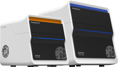
新羿制造科技(北京)有限公司为您提供《口腔拭子中dPCR用于遗传性耳聋检测检测方案(PCR)》,该方案主要用于鼻/咽拭子中遗传检测,参考标准《暂无》,《口腔拭子中dPCR用于遗传性耳聋检测检测方案(PCR)》用到的仪器有新羿TD-1 微滴式数字PCR系统、ddPCR 通用扩增试剂、微液滴生成油及微液滴检测油。
我要纠错
相关方案
 咨询
咨询