方案详情文
智能文字提取功能测试中
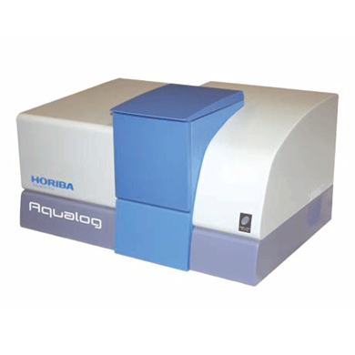
Water-Quality Measurements with theHORIBA Jobin Yvon AquaLog-R Adam M. Gilmore, Ph.D. HORIBA Scientific 3880 Park Avenue Edison, NJ 08822 TEL:732-494-8660×135 Email: adam.gilmore@horiba.com Abstract Water-quality is a significant environmental concern. Our AquaLogspectrofluorometer andanalysis software facilitate identification and quantification of natural and man-made sources ofcolored dissolved organic matter (CDOM) for water quality. Introduction Due to the increasing shortages of fresh, unpolluted drinking and irrigation water resources andpollution of the oceans globally, the analysis of colored dissolved organic matter (CDOM) is akey research area of international significance. Analysis of the often dilute and complex mixturesof CDOM components in water is made much easier using fluorescence technology, because itcan yield parts-per-billion (ppb) sensitivity for many organic compounds and it is relativelyinexpensive, simple to use, and nondestructive in nature compared to other techniques currentlyemployed. Fluorescent CDOM can comprise both natural and man-made components. The naturalcomponents primarily consist of decomposition products of plant and animal materials, includinghumic and fulvic acids, proteins, and aromatic amino-acid constituents. Man-made fluorescentcomponents can include petroleum products, fertilizers, pesticides, herbicides, sewage,pharmaceuticals, and-of increasing concern—toxic nanomaterials. CDOM influences naturalbodies of water in several ways. Two of the primary effects are on the absorbance and lightpenetration-depth and on the oxygen demand (both biological and chemical), both of which arevital to sustaining life in water. CDOM is directly related to the oxygen demand, and henceviability, of natural water bodies because it consumes oxygen upon photodegradation, especiallyunder the influence of UV light exposure. The most common CDOM fluorescence-measurement technique is known as the excitation-emission map or EEM,2. An EEM is acquired by scanning the excitation (absorbance)spectrum of the sample while simultaneously acquiring the fluorescence emission spectrum ateach excitation wavelength coordinate. The EEM is important for CDOM analysis because, incontrast to a typical two-dimensional absorbance scan of a water sample, the EEM exhibits athird dimension to provide both the absorbance and emission spectra of all fluorescentcomponents. Thus an EEM strongly increases selectivity and potentially facilitates not onlyqualitative but quantitative analysis of all the samples’components. One challenge in using fluorescence to analyze these samples is that, in most CDOM samples,the fluorescence spectral components can exhibit significant overlapping features. Currentmethodologies to separate, identify and quantify the components have focused on conventionalnon-linear least-squares spectral deconvolution as well as a variety of multivariate techniques. The latter multivariate methods include, among others, Partial Least Squares (PLS), PrincipalComponent Analysis (PCA), and the recently popularized Parallel Factor Analysis or PARAFACmethod, which we focus on in this review. The key issues surrounding analysis of the EEMs with PARAFAC are the importance ofreproducible and accurate spectral and instrumental correction of the datal1.2h6.719] A majorrequired spectral-correction method discussed below involves the coordinated evaluation of theabsorbance spectrum of the sample in order to correct for the optical processes known as“inner-filter effects”(IFEs)[10]-[13]. IFEs manifest in concentrated CDOM samples as both absorbanceof the excitation beam (primary inner filter effect), and also sample fluoresence signals(secondary inner-filter effect), where the absorbance spectra and emission spectral regions ofcomponents overlap. IFE correction is significant in light of its role in referencing spectrallibraries. In most CDOM analyses, component identification is based on a public or privately-generatedspectral library. Obviously, both intra- and interlaboratory comparisons must rely on traceableand reproducible spectral-correction procedures for the identifications to be validated. This isof great concern given the diversity of instruments and conditions under which EEMs arereported in the literature for CDOM. Hence the final section of this article outlines our view ofthe future of fluorescent CDOM measurement for both research and analytical applications withregard to several prominent international efforts to standardize the EEM methodology. A Focus on HORIBA's Fluorescence Instrumentation for Water Quality:The AquaLog@ When laboratory budgets and portability considerations are important, the bench-top AquaLog°is suitable for many CDOM laboratories because it exhibits the world’s highest water-sensitivityrating for a benchtop fluorometer; it employs a thermoelectrically-cooled charge-coupled device(CCD) array detector for ultimate speed and sensitivity, is portable (can be operated directly on a ship), is fully corrected for the excitation source and instrument response, and also is equippedwith the transmission detector needed for IFE correction. Fig. 1. The optical layout for a bench-top AquaLog@ configured for rapidly measuringexcitation-emission maps ofcolored dissolved organic matter. Numerical labels designate majorcomponents of the unit, described in the paragraph below. The AquaLog (Fig. 1) is optimally configured for CDOM analysis using a xenon-arc lightsource to excite many UV-absorbing CDOM components of interest (1). The configurationshown also makes measurements of turbid and solid samples easier by virtue of an aberration-corrected double-grating excitation monochromator to remove stray light (2). Central to theAquaLog@ is the sample compartment that is optically configured for simultaneous absorbanceand fluorescence (90 degrees) data acquisition (3). The AquaLog@ is equipped with a referencedetector (4a) to monitor and ratiometrically correct both the excitation source’s spectrum for the emission detector and the absorbance detector’s signals. A transmission detector (4b) is attachedto the AquaLog@'s sample compartment to record the sample's transmission/absorbancespectrum under the same spectral-bandpass and -resolution conditions as the fluorescence EEMdata, which is required for accurate IFE corrections. Most important for the CDOM application,however, is that the AquaLog@ is configured with an aberration-corrected 140 mm focal-lengthspectrograph with a thermoelectrically-cooled CCD detector (4c), which enables EEM collectionat rates faster than one per minute. The CCD provides an unrivaled multichannel signal-to-noiseadvantage compared to scanning monochromator single-channel-detector-based systems. Speedis important because many CDOM studies involve hundreds of EEM samples. The system’scontrol electronics are contained in the base (5). An Overview of the Method of Excitation-Emission Mapping withAbsorbance (IFE) Analysis of CDOM A typical EEM (Fig. 2A) for a CDOM sample involves scanning the excitation wavelength from240-500 nm and acquiring the emission spectra from 250-600 nm. Spectral-bandpass and-resolution conditions are now generally accepted to be 5 nm by the research communityl.Because the excitation axis is scanned and the excitation source exhibits wavelength-dependentintensity features, the emission detector signal must be divided by the respective excitation-source intensity monitored using the reference (R) detector at each excitation coordinate.Additionally both the R detector and emission (S) detector signals require correction for theinstrument’s spectral responsivity, which conventionally includes subtracting the dark-currentsignal from each detector in addition to multiplying the R and S signals by their respectiveexcitation (Xcorrect) and emission (Mcorrect) spectral correction factors. Hence, the final EEMsignal is recorded in our AquaLog@ software using the general formula SR where S.=(S-dark) xMcorrect and Re=(R-dark) × Xcorrect. In conjunction to the EEM fluorescence signal,the sample’s absorbance spectral data (Figure 2B) can be collected in parallel by measuring thetransmission (4) detector signal formula I=A/Re, where Ac=(A-dark) and Re=(R -dark) ×Xcorrect. To measure the absorbance spectrum of the sample, one must also measure the I=(A/Re) of a blank or reference sample to calculate Abs=log (I/I) as a function of wavelength.The conventional reference or blank sample is usually highly purified water (18.2 MQ,<2 ppbtotal organic carbon). Note that the blank sample can serve several additional purposes asdescribed below relative to correcting and processing the EEM data for qualitative andquantitative spectral analysis. Fig. 2. An EEM of the Pony Lake FulvicAcid standard sample from theInternational Humic Substance Society(A). This map,and all maps shownbelow, were collected using 5 nm opticalbandpass at 0.1 second integration timeper emission spectrum. Panel B showsthe absorbance spectrum of the sampleshown in (A) measured under the samebandpass and integration timeconditions. EEM Data Processing: Instrumental, Spectral, and IFE Correction withAquaLog@ Software Figure 3A shows for a blank sample of ultra-pure water that the current practice for EEMsinvolves measuring the excitation and emission scan ranges, which includes their overlapregions. These overlap regions manifest in intense signals from the monochromated excitationsource in the emission detector’s response; these lines are caused by both the first- (and second-)order Rayleigh-scattering features consistent with the well-known grating equation. Figure 3Aalso shows another spectral feature, associated with the ultra-pure water sample, known as thewater Raman scattering line. The Raman scattering line is related to the Rayleigh scattering lineby a constant energy shift of 3382 cm. Most CDOM component libraries contain spectra forwhich the artifactual Rayleigh and water-Raman spectral features have been removed, and henceEEM data must be processed to remove these features systematically.Figure 3B shows the raw EEM for a CDOM sample, namely an aliquot of a standard sample known as the Pony LakeFulvic Acid (PLFA) sample sourced from the International Humic Substance Society (IHSS).Here the main contours of the CDOM components are observed, along with the Rayleigh andRaman line scattering features. The AquaLogsoftware package can remove both artifacts.Figure 3C shows the result of subtracting the blank EEM data in Fig. 3A from the CDOMsample data in Fig. 3B,which effectively removes the Raman scatter line. Figure 3D shows theresults of applying the Rayleigh-masking algorithm, which nullifies the signal intensities for boththe first- and second-order Rayleigh lines. As mentioned above, the water Raman signal of ultra-pure water is often used to normalize the signal intensities of the CDOM sample EEMs; similarlymany researchers choose to normalize the CDOM EEMs based on a unit of fluorescence fromT1116],[12]Tquinine sulfate (QSU) 11.[6][12]. Importantly, the AquaLog"EEM-processing software performsboth the water Raman and QSU normalization. Fig. 3. The fundamental instrument-correction operations forprocessing an EEM, including blank subtraction and Rayleigh-line nullification. The EEM of an ultra-pure water sampleserving as the blank or reference sample showing thecharacteristic first- and second-order Rayleigh scattering lines(white) and water Raman scattering line in yellow (A). Anuncorrected EEM of the Pony Lake Fulvic Acid standardsample (B). Panel C shows the result of subtracting the EEMdata from the blank sample (A) from the sample in (B). PanelD shows the results ofthe algorithm that nullifies the first- andsecond-order Rayleigh lines from the map shown in (C). following simple equation applied to each excitation-emission wavelength coordinate of theEEM 111]. where Fideal is the ideal fluorescence-signal spectrum expected in the absence of IFE, Fobs is theobserved fluorescence signal, and AbSEx and AbsEm are the measured absorbance values at therespective excitation and emission wavelength-coordinates of the EEM. A number of advancedalgorithms described in the literature can also account for variations of the optical geometricalparameters of the cuvette path-length, beam- or slit-width, and positioning/shifting of the cuvette10]-[1relative to the excitation and emission beam paths [10]-[13] Figures 4A and B show a comparison of EEMs collected using both a concentrated (Abs254nm ~0.8) PLFA sample to one diluted with ultra-pure water (Abs254 nm ~0.2) in Figures 4C and D.The absorbance value at 254 nm is an industry standard for evaluating the total CDOMconcentration [8]. The EEMs on the left show the uncorrected (Fobs) while the EEMs on the rightshow the IFE corrected (Fideal) for the corresponding samples. It is clear that for the uncorrected,higher-absorbing sample in Figure 4A that the spectral contours >300 nm on the excitation axisare stronger compared to the contours below 300 nm. This is because the IFE effects arestrongest in the UV regions (below 300 nm) where the overlap of component excitation andemission spectra is largest. The IFE correction (Fig. 4B) clearly reconstitutes the same contoursseen with the dilute sample (Fig.4C) and the IFE correction had little effect on the dilute sampleEEM in Figure 4D. Fig. 4. A comparison ofthe influence ofthe inner-filter-correction algorithm onEEMs ofconcentrated (top row) anddilute (bottom row) samples ofthe PonyLake Fulvic Acid standard sample. Theuncorrected EEMs are shown in panels Aand C; the corrected maps are shown inB and D, respectively. Likewise, Figure 5 shows in more detail the influence of the IFE corrections by comparing theabsorbance spectra and the integrated excitation spectra of the PLFA EEM-data samples from adilution series. Figure 5A shows the absorbance spectra for the PLFA dilution series which areparticularly featureless with a quasi-exponential decrease from the UV to visible regions. Figure5B shows the plot of the absorbance values at 254 nm from Figure 5A versus the dilution factorused in the experiment. The data in Figure 5B exhibit a highly significant linear trend up to thepeak absorbance near Abs~0.8. Figure 5C shows that the Fobs for the integrated excitationspectra indicates there is a strong reduction in the intensity below 400 nm. Figure 5D presents nocorrelation between the absorbance values at 254 nm (x-axis) and the excitation intensity valuesat 254 nm (y-axis) beyond Abs~ 0.2 due to the increasing IFE. However, as shown in Figure 5E,when the IFE is corrected as described above, the excitation spectral intensity is recovered,closely paralleling the absorbance spectra (Fig.5A) for the all samples. The highly significantlinear relationship between the excitation intensity at 254 nm and the absorbance at 254 nmshown in Figure 5F confirms the validity of the IFE correction and its value for concentratedCDOM analysis, allowing comparison to dilute sample spectra. Fig.5. Comparison of the concentration-dependence ofthe absorbance spectra (A and B) andthe excitation spectra ofthe Pony Lake Fulvic Acid sample before (C and D) and after (E and F)inner-filter correction. The samples were diluted from full strength to 100-fold as indicated inthe legends. Plots of the absorbance versus wavelength are in A; B shows the linear relationship ofthe absorbance at 254 nm and the dilution factor. Plots of the excitation spectra representingthe integral ofthe excitation axis are shown in C versus wavelength. Panel D shows plots oftheabsorbance value at 254 nm against the Nexcitation intensity at 254 nm; the red arrow illustratesthe nonlinear region when Abs > 0.2. Panel E shows plots of the excitation spectra from (C)after inner-filter correction, while (D) shows the plot of the linear relationship between theexcitation intensity at 254 nm and the absorbance at 254 nm. Spectral Analysis and Component Identification: Conventional NonlinearLeast-Squares and Multivariate Approaches As required by the CDOM research community, the concerted application of the instrumentalspectral corrections, Rayleigh-line masking, water-Raman subtraction, Raman or QSUnormalization and IFE correction are readily enabled by the EEM-processing tools in ourAquaLogsoftware. As mentioned above, the purpose of the spectral corrections and EEM-processing is to make the identification and quantification easier of the CDOM components thatare usually based on a reference-component library. Here we focus attention on a popular andpromising library-based multivariate technique for CDOM analysis, namely, PARAFAC, whichhas been documented extensively by researchers including many using HORIBA’s fluorescenceinstruments [1[3]-5]Importantly the AquaLog@ software offers direct access to a MatLabconsole for purposes of processing data using the PARAFAC tools in N-way Toolbox, a public-domain package especially developed for CDOM analysis [4. The modeling advantages ofPARAFAC center on its ability to simultaneously evaluate the EEM data as a matrix, and toenvelop multiple (often hundreds) of EEMs simultaneously for increased statistical significance13H-5]. PARAFAC has been successful at identifying a wide range of CDOM componentsincluding humic and fulvic acids, tryptophan- and tyrosine-like substances, quinones, severalpolycyclic aromatic hydrocarbons, and distinguishing microbial, marine and terrestrial CDOMsources. More importantly, PARAFAC has been used to diagnose trends in CDOM componentsas a function of several key chemical and physical parameters, including water-recycling-planttreatment stages, sewage dispersion, stream flow, and ocean and estuarial currents, among manyothers [6. Indeed, as discussed below, the application of PARAFAC has been proposed as astandard modeling technique for a variety of water-quality applications [,[14] Conclusion: The Future of Water-Quality Analysis Applications andStandardization by the International CDOM Community Several key publications and international standards documents have recently been publishedregarding fluorescence-instrument calibration and correction, including EEM data, as well asthose focusing on interlaboratory comparisons of CDOM EEM applications. Three recentpublications from NIST researchers have particular significance for EEM correction, including areference to the recently released ASTM standard guide (E2719) for fluorescence calibration andcorrection, a reference to CDOM EEMs and all aspects of instrument and IFE correction,2l and a reference to the validation of the Fluorologas the true accurate fluorometer for its use ingenerating and validating a series of Standard Reference Materials. Furthermore, a recent paperhas been accepted outlining the results of a major international interlaboratory comparison forCDOM IHSS standard samples; this study was headed by researchers and the United StatesGeological Survey (USGS), and was the focused outcome of a recent 2008 Chapman conferencesponsored by the American Geophysical Union. In terms of potential applications of CDOManalysis two reviews of the literature exist of note, including one paper explaining the potentialof CDOM for monitoring all stages of water-recycling 14, and another focusing on analysis ofnatural and wastewater sources. It is clear that fluorescence analysis of CDOM and the use of EEMs will play a central role inwater-quality analysis in academic and government research, as well as for various municipaland industrial monitoring applications. HORIBA presently enjoys the recognition of the industryas the leader in both hardware and software for this application. It is our goal to continue todesign instrumentation and software and to promote standardization and regulations that willincrease the potential for fluorescence as a water-quality analysis tool, and thus help researcherstrying to understand, prevent, and treat the consequences of CDOM contamination of ourwater-one of our most critical resources. ( References ) [1] Hudson, et al. (2007) Fluorescence analysis of dissolved organic matter in natural, waste andpolluted waters-A Review. River. Res. Applic. 23: 631-649. [2] Holbrook, et al. (2006) Excitation-emission matrix fluorescence spectroscopy for naturalorganic matter characterization: A quantitative evaluation of calibration and spectral correctionprocedures. Appl. Spectroscopy60 (7),791. [3] Stedmon, C. A., S. Markager, and R. Bro. (2003) Tracing dissolved organic matter in aquaticenvironments using a new approach to fluorescence spectroscopy. Mar. Chem. 82:239-254. [4] Stedmon, C. A., S. Markager, and R. Bro. (2008) Characterizing dissolved organic matterfluorescence with parallel factor analysis: a tutorial. Limnol.Oceanogr.: Methods 6:572-579. [5] Cory, R. M., and D. M. McKnight (2005) Fluorescence spectroscopy reveals ubiquitouspresence of oxidized and reduced quinones in dissolved organic matter. Environ. Sci.Technol.39:8142-8149. [6] Cory,et al. (2010) Effect of instrument-specific response on the analysis of fulvic acidfluorescence spectra. Limnol.Oceanogr.: Methods 8, 67-78. [7] DeRose, et al. (2007) Qualification of a fluorescence spectrometer for measuring truefluorescence spectra. Rev.Sci. Instr. 78, 033107. [8] Murphy, et al. (2010) The measurement of dissolved organic matter fluorescence in aquaticenvironments: An interlaboratory comparison. Environ. Sci. Technol. (in press) [9]Paul C. DeRose and Ute Resch-Genger (2010) Recommendations for FluorescenceInstrument Qualification: The New ASTM Standard Guide. Anal. Chem., 82, 2129-2133. [10] Qun Gu and Jonathan E. Kenny (2009) Improvement of Inner Filter Effect Correction Basedon Determination of Effective Geometric Parameters Using a Conventional Fluorimeter. Anal.Chem.81, 420-426. [11] Lakowicz, J.R. (2006) Principles of Fluorescence Spectroscopy,3 ed.; Springer Scienceand Business Media, LLC: New York. [12] Tobias Larsson, Margareta Wedborg, and David Turner (2007) Correction of inner-filtereffect in fluorescence excitation-emission matrix spectrometry using Raman scatter. Anal. Chim.Acta 583,357-363. [13] B. C. MacDonald, S. J. Lvin, and H. Patterson (1997) Correction of fluorescence inner filtereffects and the partitioning of pyrene to dissolved organic carbon. Anal. Chim. Acta 338, 155. [14] Henderson, et al. (2010) Fluorescence as a potential monitoring tool for recycled watersystems: A review. Water Research 43, 863. 方案优势:专利A-TEEM技术,同步吸收-三维荧光光谱,快速三维荧光测试,一键校正内滤效应,水拉曼/硫酸奎宁定量,结合化学计量学分析,实现不同组分定性定量分析。方案介绍:依据Aqualog专利 A-TEEM技术,消除内滤效应,准确获得荧光有机物的三维指纹图谱(激发波长-发射波长-荧光强度)信息,监测水中溶解有机碳浓度和组成,消毒副产物,消毒副产物前躯体,芳香类石油碳氢化合物,藻类等的浓度和组成。结合化学计量学分析,反应出组份分布,弥补了COD和BOD测试时间长,水体中有机物种类信息缺失的问题可自动
关闭-
1/11

-
2/11

还剩9页未读,是否继续阅读?
继续免费阅读全文产品配置单
HORIBA(中国)为您提供《水中腐殖质、富里酸、类蛋白、有色溶解有机质检测方案(分子荧光光谱)》,该方案主要用于环境水(除海水)中有机物综合指标检测,参考标准《暂无》,《水中腐殖质、富里酸、类蛋白、有色溶解有机质检测方案(分子荧光光谱)》用到的仪器有HORIBA Aqualog®同步吸收-三维荧光光谱仪。
我要纠错
相关方案


 咨询
咨询


