方案详情文
智能文字提取功能测试中
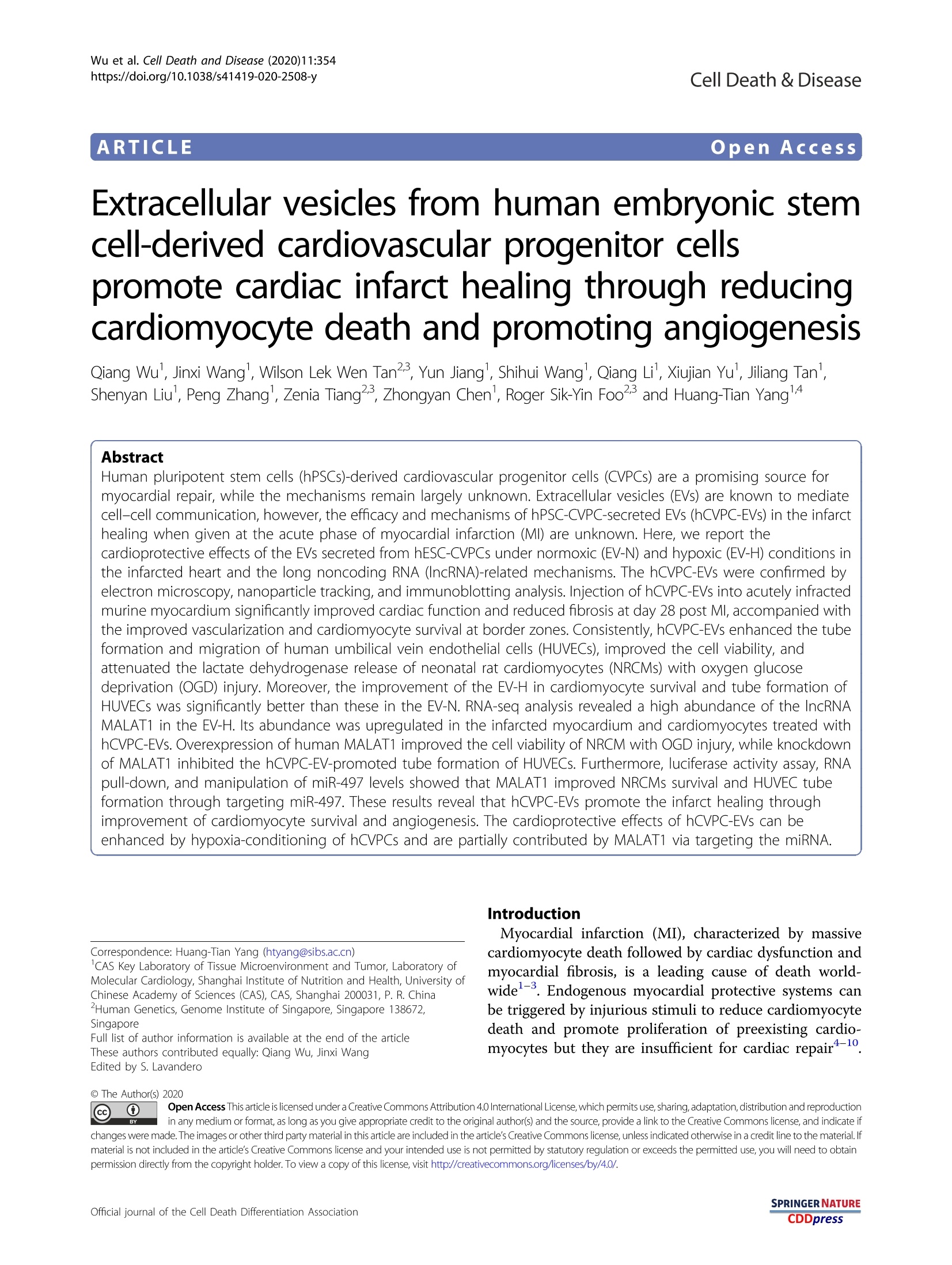
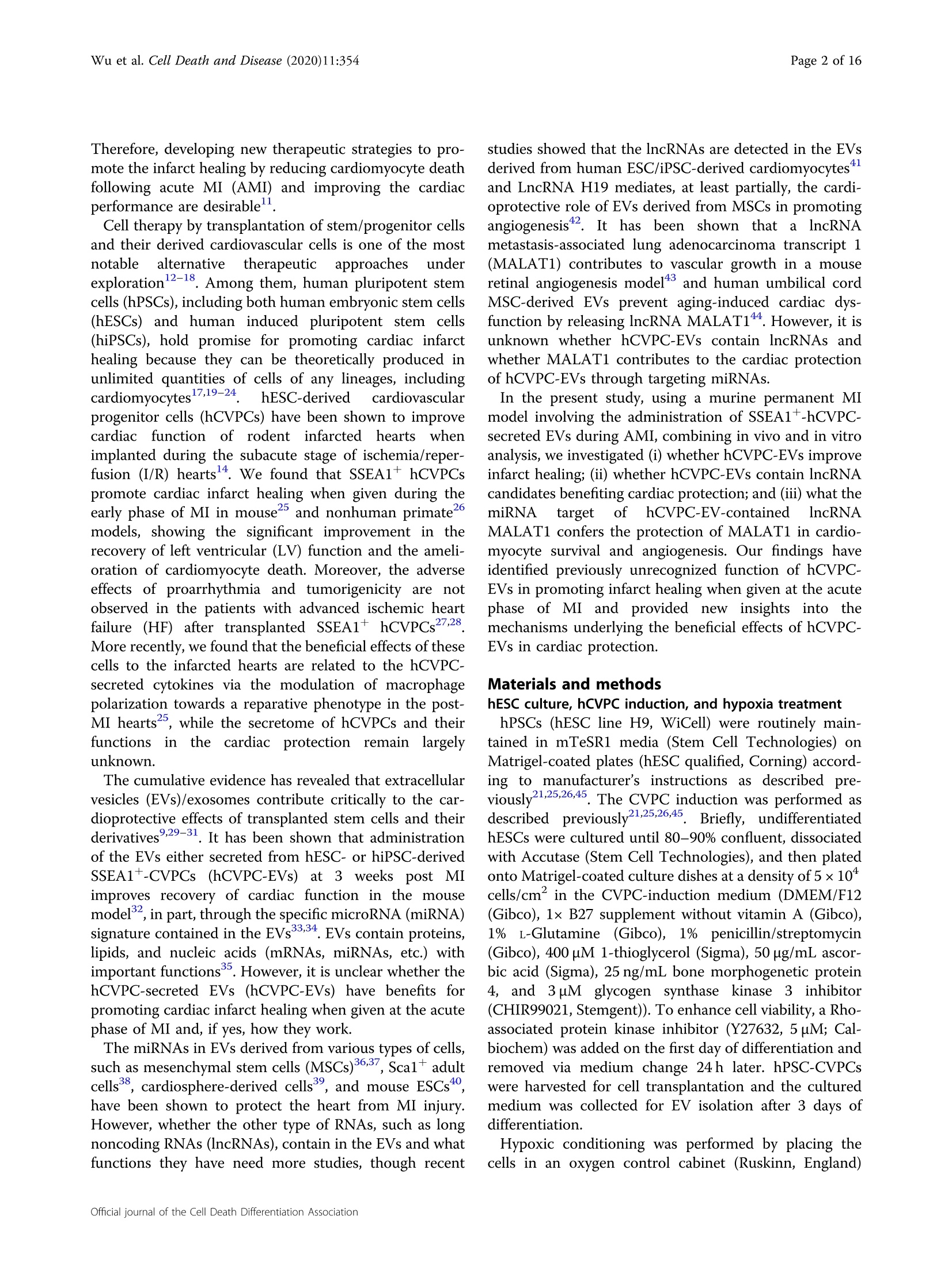
Wu et al. Cell Death and Disease (2020)11:354https://doi.org/10.1038/s41419-020-2508-yCell Death & DiseaseARTICLE Page 2 of 16Wu et al. Cell Death and Disease (2020)11:354 Open Access Extracellular vesicles from human embryonic stemcell-derived cardiovascular progenitor cellspromote cardiac infarct healing through reducingcardiomyocyte death and promoting angiogenesis Qiang Wu', Jinxi Wangl, Wilson Lek Wen Tan23, Yun Jiangl, Shihui Wang', Qiang Lil, Xiujian Yul, Jiliang Tan',Shenyan Liul, Peng Zhang, Zenia Tiang23, Zhongyan Chen, Roger Sik-Yin Fooand Huang-Tian Yang14 Abstract Human pluripotent stem cells (hPSCs)-derived cardiovascular progenitor cells (CVPCs) are a promising source formyocardial repair, while the mechanisms remain largely unknown. Extracellular vesicles (EVs) are known to mediatecell-cell communication,however, the efficacy and mechanisms of hPSC-CVPC-secreted EVs (hCVPC-EVs) in the infarcthealing when given at the acute phase of myocardial infarction (MI) are unknown. Here, we report thecardioprotective effects of the EVs secreted from hESC-CVPCs under normoxic (EV-N) and hypoxic (EV-H) conditions inthe infarcted heart and the long noncoding RNA (IncRNA)-related mechanisms. The hCVPC-EVs were confirmed byelectron microscopy, nanoparticle tracking, and immunoblotting analysis. Injection of hCVPC-EVs into acutely infractedmurine myocardium significantly improved cardiac function and reduced fibrosis at day 28 post MI, accompanied withthe improved vascularization and cardiomyocyte survival at border zones. Consistently, hCVPC-EVs enhanced the tubeformation and migration of human umbilical vein endothelial cells (HUVECs), improved the cell viability, andattenuated the lactate dehydrogenase release of neonatal rat cardiomyocytes (NRCMs) with oxygen glucosedeprivation (OGD) injury. Moreover, the improvement of the EV-H in cardiomyocyte survival and tube formation ofHUVECs was significantly better than these in the EV-N. RNA-seq analysis revealed a high abundance of the IncRNAMALAT1 in the EV-H. Its abundance was upregulated in the infarcted myocardium and cardiomyocytes treated withhCVPC-EVs.Overexpression of human MALAT1 improved the cell viability of NRCM with OGD injury,while knockdownof MALAT1 inhibited the hCVPC-EV-promoted tube formation of HUVECs. Furthermore, luciferase activity assay, RNApull-down, and manipulation of miR-497 levels showed that MALAT1 improved NRCMs survival and HUVEC tubeformation through targeting miR-497. These results reveal that hCVPC-EVs promote the infarct healing throughimprovement of cardiomyocyte survival and angiogenesis. The cardioprotective effects of hCVPC-EVs can beenhanced by hypoxia-conditioning of hCVPCs and are partially contributed by MALAT1 via targeting the miRNA. Correspondence: Huang-Tian Yang (htyang@sibs.ac.cn) ICAS Key Laboratory of Tissue Microenvironment and Tumor, Laboratory ofMolecular Cardiology, Shanghai Institute of Nutrition and Health, University ofChinese Academy of Sciences (CAS), CAS, Shanghai 200031, P. R. ChinaHuman Genetics, Genome Institute of Singapore, Singapore 138672,Singapore Full list of author information is available at the end of the article These authors contributed equally: Qiang Wu, Jinxi Wang Edited by S. Lavandero Introduction Myocardial infarction (MI), characterized by massivecardiomyocyte death followed by cardiac dysfunction andmyocardial fibrosis, is a leading cause of death world-wide-3. Endogenous myocardial protective systems canbe triggered by injurious stimuli to reduce cardiomyocytedeath and promote proliferation of preexisting cardio-myocytes but they are insufficient for cardiac repair+-10. @ The Author(s) 2020 Ocpen Access This article is licensed under a Creative Commons Attribution 4.0 International License, which permits use, sharing,adaptation, distribution and reproduction@ in any medium or format, as long as you give appropriate credit to the original author(s) and the source, provide a link to the Creative Commons license, and indicate ifchanges were made. The images or other third party material in this article are included in the article's Creative Commons license, unless indicated otherwise in a credit line to the material. Ifmaterial is not included in the article's Creative Commons license and your intended use is not permitted by statutory regulation or exceeds the permitted use, you will need to obtainpermission directly from the copyright holder. To view a copy of this license, visit http://creativecommons.org/licenses/by/4.0/. Therefore, developing new therapeutic strategies to pro-mote the infarct healing by reducing cardiomyocyte deathfollowing acute MI (AMI) and improving the cardiacperformance are desirable. Cell therapy by transplantation of stem/progenitor cellsand their derived cardiovascular cells is one of the mostnotableeaalternative:therapeutic approaches underexploration12-18. Among them, human pluripotent stemcells (hPSCs), including both human embryonic stem cells(hESCs) and human induced pluripotent stem cells(hiPSCs), hold promise for promoting cardiac infarcthealing because they can be theoretically produced inunlimited quantities of cells of any lineages, includingcardiomyocytes17,19-24. hESC-derived cardiovascularprogenitor cells (hCVPCs) have been shown to improvecardiac function of rodent infarcted heartss whenimplanted during the subacute stage of ischemia/reper-fusion (I/R) hearts4. We found that SSEA1+ hCVPCspromote cardiac infarct healing when given during theearly phase of MI in mouse25 and nonhuman primate6models, showing the significant improvement in therecovery of left ventricular (LV) function and the ameli-oration of cardiomyocyte death. Moreover, the adverseeffects of proarrhythmia and tumorigenicity are notobserved in the patients with advanced ischemic heartfailure (HF) after transplanted SSEA1+ hCVPCs27,28.More recently, we found that the beneficial effects of thesecells to the infarcted hearts are related to the hCVPC-secreted cytokines via the modulation of macrophagepolarization towards a reparative phenotype in the post-MI hearts, while the secretome of hCVPCs and theirfunctions inthe cardiacc protection remain largelyunknown. The cumulative evidence has revealed that extracellularvesicles (EVs)/exosomes contribute critically to the car-dioprotective effects of transplanted stem cells and theirderivatives ,29-31. It has been shown that administrationof the EVs either secreted from hESC- or hiPSC-derivedSSEA1+-CVPCs((hCVPC-EVs))at 3 weeks post MIimproves recovery of cardiac function in the mousemodel,in part, through the specific microRNA (miRNA)signature contained in the EVs33.34. EVs contain proteins,lipids, and nucleic acids (mRNAs, miRNAs, etc.) withimportant functions. However, it is unclear whether thehCVPC-secreted EVs (hCVPC-EVs) have benefits forpromoting cardiac infarct healing when given at the acutephase of MI and, if yes, how they work. The miRNAs in EVs derived from various types of cells,such as mesenchymal stem cells (MSCs)36,37, Sca1* adultcells, cardiosphere-derived cells, and mouse ESCs,have been shown to protect the heart from MI injury.However, whether the other type of RNAs, such as longnoncoding RNAs (lncRNAs), contain in the EVs and whatfunctions they have need more studies, though recent studies showed that the lncRNAs are detected in the EVsderived from human ESC/iPSC-derived cardiomyocytesand LncRNA H19 mediates, at least partially, the cardi-oprotective role of EVs derived from MSCs in promotingangiogenesis42. It has been shown thataaIncRNAmetastasis-associated lung adenocarcinoma transcript 1(MALAT1) contributes to vascular growth in a mouseretinal angiogenesis model+3 and human umbilical cordMSC-derived EVs prevent aging-induced cardiac dys-function by releasing IncRNA MALAT14. However, it isunknown whether hCVPC-EVs contain lncRNAs andwhether MALAT1 contributes to the cardiac protectionof hCVPC-EVs through targeting miRNAs. In the present study, using a murine permanent MImodel involving the administration of SSEA1+-hCVPC-secreted EVs during AMI, combining in vivo and in vitroanalysis, we investigated (i) whether hCVPC-EVs improveinfarct healing; (ii) whether hCVPC-EVs contain IncRNAcandidates benefiting cardiac protection; and (iii) what themiRNA targett(offhCVPC-EV-contained lncRNAMALAT1 confers the protection of MALAT1 in cardio-myocyte survival and angiogenesis. Our findings haveidentified previously unrecognized function of hCVPC-EVs in promoting infarct healing when given at the acutephase of MI andprovided new insightsinto themechanisms underlying the beneficial effects of hCVPC-EVs in cardiac protection. Materials and methods hESC culture, hCVPC induction, and hypoxia treatment hPSCs (hESC line H9, WiCell) were routinely main-tained in mTeSR1 media (Stem Cell Technologies) onMatrigel-coated plates (hESC qualified, Corning) accord-ing to manufacturer's instructions as described pre-viously2125,26,45. The CVPC induction was performed asdescribednreviausly21,25,26,45 Briefly, undifferentiatedhESCs were cultured until 80-90% confluent, dissociatedwith Accutase (Stem Cell Technologies), and then platedonto Matrigel-coated culture dishes at a density of 5×10*cells/cm’ in the CVPC-induction medium (DMEM/F12(Gibco), 1x B27 supplement without vitamin A (Gibco),1%L-Glutamine: (Gibco), 11% penicillin/streptomycin(Gibco), 400 uM 1-thioglycerol (Sigma), 50 ug/mL ascor-bic acid (Sigma), 25 ng/mL bone morphogenetic protein4,and3 uM glycogen synthase kinase3inhibitor(CHIR99021,Stemgent)). To enhance cell viability, a Rho-associated protein kinase inhibitor (Y27632, 5uM; Cal-biochem) was added on the first day of differentiation andremoved via medium change 24 h later. hPSC-CVPCswere harvested for cell transplantation and the culturedmedium was collected for EV isolation after 3 days ofdifferentiation. Hypoxic conditioning was performed by placing thecells in an oxygen control cabinet (Ruskinn, England) mounted within an incubator and equipped with oxygencontroller and sensor for continuous oxygen level mon-itoring for 48 h in DMEM basal medium. The oxygenconcentration in the cabinet was maintained at 1%, with aresidual gas mixture composed of 5% CO2 and balancedN2. Normoxic conditioning hCVPCsswere incubatedunder 21% O2 and 5% CO, for 48h in DMEM basalmedium. Both normoxic and hypoxic conditioned med-ium were collected for EV isolation. Flow cytometry analysis Flow cytometry wassperformed as ddescribed1 pre-viously. Cells to be examined were harvested, dis-sociated with Accutase (Stem Cell Technologies). Thesamples were stained for the presence of cardiac pro-genitor marker PE-conjugated SSEA1 (1:20; eBioscience),and then analyzed and quantified by flow cytometry(FACStar Plus Flow Cytometer, BD bioscience). Immunocytochemical staining analysis The cellss were immunostained as d ribed previously. Briefly, cells were fixed with 4% paraformalde-hyde, permeabilized in 0.3%Triton X-100 (Sigma),blocked in 10% normal goat serum (Vector Laboratories),and then incubated at 4℃ overnight with primary anti-bodies against MESP1 (1:100, Aviva Systems Biology),MEF2C (1:100, Cell Signaling), GATA4 (1:300, SantaCruz Biotechnology), ISL1 (1:100, Developmental StudiesHybridoma Bank), and NKX2-5 (1:200, Santa Cruz Bio-technology). Primary antibodies were detected withDyLight 549-conjugated secondary antibodies, and nucleiwere counterstained with DAPI (Sigma).The immunos-taining of isolated NRCMs were stained with a-actininantibody and terminal deoxynucleotidyl transferase-mediated dUTP nick end labeling (TUNEL) using the Insitu Cell Death Detection Kit (Roche Applied Science,Germany) to detect the cell death. EV isolation The EV-Nand EV-H were isolated from normoxic andhypoxic conditioned medium separately as describedpreviously4. Briefly, The collected hCVPC conditionalmedium was centrifugated at 300xg for 30 min followedby 2000xg for 30 min, 4℃ to remove cells and dead cells,and then centrifugated at 10,000xg for 30 min, 4℃ toremove celldebris,finallyy centrifugated twicee aat100,000xg for 70 min, 4℃ with a SW-41 rotor (BeckmanCoulter), followed by washing with phosphate-bufferedsaline (PBS). The final pellet containing EVs was resus-pended in PBS and analyzed by NanoSight NS300 (Mal-vern Panalytical), transmission electron microscope andWestern blot, or lysed with QIAzol reagent (#217084,Qiagen) for RNA analysis. Nanoparticle tracking analysis (NTA) The NTA was carried out to determine the EV size andconcentration by using NanoSight NS300) (MalvernPanalytical) on the isolated EVs as previously reported.The isolated EV pellet as described in the above EV Iso-lation method was resuspended in PBS, and then 10 uL ofit was used for NTA (the sample was diluted to 700 uLwith PBS), and 10 uL of it was used for Pierce BCA Pro-tein Assay. During NTA analysis, three 30 s video takenper sample were averaged as one value and five sampleswere examined in each group. The PBS was subtractedfrom particle number/mL after quantification. The ana-lysis was performed by using the NTA software (NTA 3.2Dev Build 3.2.16). Based on the measurement from NTAand Pierce BCA Protein Assay, the 1 ug EV protein had32.80±8.529×10°of particles in the EVs secreted fromhESC-CVPCs under normoxic cultivation (EV-N) groupand 34.60±11.76×10° of particles in the EVs secretedfrom hESC-CVPCs under hypoxic cultivation (EV-H)group as shown in Supplementary Fig. S1. Accordingly,the 20 ug EV protein contained about 485-827×10°particles in the EV-N group, and about 457-927×10°particles in the EV-H group (p>0.05,n=5). Western blot analysis The cells and the EVs were lysed with the radio-immunoprecipitation assay lysis buffer. Then, the proteinextracts were separated via gel electrophoresis, transferredto a polyvinylidene fluoride membrane, and blocked with5% bovine serum albumin for 1 h. The membranes wereincubated overnight with primary antibody against Alix(1:1000, Cell Signaling Technology), TSG101 (1:1000,Abcam), CD63 (1:200, Santa Cruz Biotechnology), CD81(1:500, Thermo Fisher Scientific), and CD9 (1:500,Thermo Fisher Scientific). Then, the membranes wereincubated with horseradish peroxidase-conjugated sec-ondary antibodies at room temperature for 2h andexposed via enhanced chemiluminescence. MI and EV treatment All surgical procedures described were performed injeiaccordance with the Guidelines for Care and Use ofLaboratory Animals published by the US NationalInstitutes of Health (NIH Publication, 8th Edition, 2011)and were approved by the Institutional Animal Care andUse Committee of Shanghai Institutes for BiologicalSciences. The male C57BL/6 mice aged 10-12 weekswere randomly divided into four groups: Sham PBS(n=8), MI+PBS (n=12), MI+EV-N (n=12), andMI+EV-H (n=15) after excluded the LV ejectionfraction (LVEF) values greater than 50% (2 in MI+PBS,and 1 in MI+EV-H) at day 2 post MI. The mice wereanesthetized via intraperitoneal injection of 50 mg/kg sodium pentobarbital,ventilateddwith avolume-regulated respirator (SAR830, Cwe Incorporated), andthe MI model was induced through the permanentligation of the left anterior descending (LAD) coronaryartery with a 10-0 Prolene suture as previously descri-bed25,40,47. Of 20 ug EVs, mixed with PBS in a totalvolume of 20 uL, were directly injected into two sites inthe border zones of infarcted myocardium (10 uL eachsite) after the ligation of LAD coronary artery, and theequal amount of PBS was injected into the sham hearttoo. Then the muscle and skin of the chest were thensutured with 6-0 and5-0 Prolene suture, respectively.The body temperature was maintained at 37℃ duringthe surgical procedure. Echocardiography Transthoracic echocardiography (Vevo 2100, VisualSonics) with a 25-MHz imaging transducer was per-formed on isoflurane anesthetized mice to measure theheart function as previously described.LVEF and LVsystolic dimensions (LVDs) were recorded and the aver-age values were collected from each of three consecutivecardiac cycles. Immunohistochemical staining Heart tissues were embedded in OCT (SAKURA)compound for histological analysis. were prepared at400 um intervals. For fluorescent immunohistochem-istry, Transversal frozen sections (5 um) were fixed with4% paraformaldehyde, permeabilized in 0.4% Triton X-100 (Sigma), and stained with anti-CD31, α-SMA, andcTnT antibody which were detected by fluorescentconjugated secondary antibodies. Nuclei were stainedwith DAPI (Sigma). Cell death was evaluated with an Insitu Cell Death Detection Kit (Roche Applied Science,Germany) for TUNEL staining as directed by the man-ufacturer’s instructions. The percentage of TUNELcardiomyocytes was quantified as the ratio of TUNELcTnT+ to total cTnT+ cells. The immunostaining ima-gesswere Eblindly capturedusinga Zeiss invertedmicroscope and processed using ZEN software. Threemicroscopic fields were quantified for each slice. TheImage-processing software (Image J) was used as theimage quantification software. Masson's trichrome staining For the Masson’s trichrome-stained images, each heartwas made five slices from the point of ligation to the apexof the heart. The morphometric parameters in the fiveslices of each heart including total LV area and scar areawere blindly analyzed, and the scar size was calculated asthe total scar area divided by the LV area as describedpreviously25,47 RNA extraction Total RNA was isolated from hCVPC-EVs using miR-Neasy Micro Kit (Qiagen) according to the manufacturer'sinstructions. The concentration of RNA fraction wasquantified using Nanodrop (Thermo Fisher Scientific)according to the manufacturer’s protocol. RNA-sequencing (RNA-seq) analysis Five EV samples from each of EV-N and EV-H groupsfrom H9 hESC origin were proceeded for RNA-seq ana-lysis as previously reported*148,49. Total RNA sequencingwas performed with the Truseq Stranded Total RNALibrary Prep kit (Illumina, RS-122-2201). This made useof Ribo-Zero to remove abundant cytoplasmic rRNA fromtotal RNA samples. Remaining intact RNA was frag-mented using a chemical mix, followed by first- andsecond-strand cDNA synthesis using random hexamerprimers.“End-repaired" fragments are ligated with uniqueIllumina proprietary adapters. All individually indexedsamples were pooled together and multiplexed forsequencing. Libraries were sequenced using the IlluminaHiseq 2500 sequencing system and paired-end 101 bpreads were generated for analysis. Fastq files were alignedagainst human referencee(hg19/hGRC37)) iusing: theTophat2. Duplicate reads were removed using MarkDu-plicates from Picard tools, and per gene read counts forEnsembl (v75) gene annotations were computed usinghtseq-count. Differential gene expression analysis wasperformed using R-package EdgeR. Principal componentanalysis was computed using R function prcomp. Hier-archical clustering and heatmaps were generated using R. Target prediction of MALAT1 The miRNA targets of MALAT1 were predicted bymiRcode (http://mircode.org/). The binding of MALAT1and miRNAs was also predicted by DianaTools (http://carolina.imis.athena-innovation.gr/diana_tools/web/index.php?r=lncbasev2%2Findex-predicted). Quantitative reverse transcription polymerase chainreaction (RT-qPCR) For miRNAs, total RNA was reverse-transcribed intocDNA by the miRcute Plus miRNA First-Strand cDNASynthesis Kit (#KR211-02, Tiangen Biotech) and subse-quently determined using a miRcute Plus miRNA qPCRDetection Kit (SYBR Green) (#FP411-02, Tiangen Bio-tech) with Qiagen predesigned primers. All kits were usedaccording to the manufacturer’s instructions. A U6 tran-script was used as an internal control to normalize RNAinput. For lncRNA, total RNA was reverse-transcribedinto cDNA by the ReverTra Ace (#TRT-101, TOYOBO)with random primers and subsequently determined usinga FastStart Universal SYBR Green Master ]Kit (#4913914001, Roche) with specific IncRNA primers. Allkits were used according to the manufacturer’s instruc-tions. IncRNA levels were normalized to glyceraldehyde3-phosphate dehydrogenase (GAPDH). Human MALAT1(hMALAT1) primer: hMALAT1 (forward): 5'-ctaggact-gaggagcaagcg-3; hMALAT1 (reverse): 5'-accaaatcgttagcgctcct-3'. Isolation, culture, and oxygen glucose deprivation (OGD)treatment of cardiomyocytes Isolation of neonatal rat cardiomyocytes (NRCMs) wereperformed as previously reported. Briefly, the NRCMswere prepared by enzymatic digestion of hearts obtainedfrom newborn (1 day old) Sprague-Dawley rat pups andplated on cell culture grade plates (coated with gelatin) ata density of 5×10 cells/cm’in DMEM/F12 medium andmaintained at 37C in humid air with 5% CO2. NRCMswere subjected to hypoxia in vitro in an oxygen controlcabinet (Ruskinn, England) mounted within an incubatorand equipped with oxygen controller and sensor forcontinuous oxygen level monitoring. A mixture of 85%nitrogen, 10% hydrogen, and 5% CO2 was utilized tocreate hypoxia and the O, in the chamber was monitoredand maintained at a level <0.1%. The NRCMs were treatedwith OGD injury by cultured in the culture mediumwithout serum and glucose and in hypoxic condition(<0.1%O2) for 6 h as previously reported. The EV-N orEV-H (1 ug/mL), miR-497-mimic (Ribobio, Guangzhou,China) or/and pcDNA3.1(-)-MALAT1 plasmid (ShanghaiIntegrated Biotech Solutions), MALAT1 GapmeR orcontrol (Guangzhou Epibiotek) with Lipofectamine 3000(Thermo Fisher Scientific) were added into the culturemedium of the NRCMs. Then, the cardiomyocytes werestained by TUNEL, cTnT, and DAPI to evaluate the levelofcell death in the NRCMs. The Image-processing soft-ware (Image J) was used to quantify the TUNELnucleusand total nucleus. The Cell Counting Kit-8 (Sigma-Aldrich) was applied and absorbance of formazan dyeproduced by living cells was measured in an Infinite M200Microplate Reader (Tecan, Maennedorf, Switzerland).The culture medium from various groups was collected tomeasure the activity of lactate dehydrogenase (LDH)release with a LDH Release Assay Kit (Beyotime, China). Isolation of adult mouse cardiomyocytes (AMCMs)were performed by a langendorff-based approach as pre-viously reported2. Briefly, male C57BL/6 mice aged10-12 weeks were anesthetized with1 pentobarbital(50 mg/kg body weight, i.p.), then the chest was opened tolift the heart and cut the aorta. The heart was thenattached to the perfusion cannula through the aorta andperfused with the digestion buffer (Collagenase II 0.5 mg/mL (Worthington, USA), Protease XIV 0.05 mg/mL(Sigma-Aldrich, Singapore)) for 10-15 min at the flowrate of 4 ml/min following 3 min of perfusion in the perfusion buffer. Once appeared flaccid, the heart wasremoved from the cannula and the ventricle was cut intosections followed by dissociated the cells with a plasticpipette in digestion buffer. Further, the cell suspensionunderwent gravity settling for four rounds within Careintroduction buffers. The cells resuspended with aplating medium were plated onto laminin (5 ug/mL,Thermo Scientific, Singapore) coated culture dish afterpassed through a 100-um filter, in a humidified tissueculture incubator (37°℃, 5% CO2), and after 1 h, the cellswere cultivated in the culture medium. The AMCMswere treated with hCVPC-EVs (1 ug/mL) and PBS (equalvolume) for 24 h, and then were washed twice with PBSbefore RNA extraction and RT-qPCR. Migration and tube formation assays Human umbilical vein endothelial cells (HUVECs)(Shanghai Zhongqiao Xinzhou Biotechnology) were cul-tured with the endothelial cell medium (ECM) (Sciencell,#1001). The ECM consists of basal culture medium,supplemented with 5% fetal bovine serum (FBS, Sciencell,#0025), 1% endothelial growth factor (ECGS, Sciencell,#1052), and 1% penicillin/streptomycin solution (P/S,Sciencell, #0503). Cell migration assay was performed aspreviouslyyreported54.55, with 10 ug/mL mitomycin(Sigma M0503)) treated for 2 h, the 100% confluentmonolayers of HUVEC were scratched and washed threetimes with PBS. Then the hCVPC-secreted EV-N or EV-H(1ug/mL), or PBS (equal volume to the EVs) were addedinto the 12-well plates seeded with HUVECs. The markedareas were captured by using a Zeiss inverted microscopebefore and after different treatments for 24h3 For tube formation assay, 48-well plates were coatedwith 200 uL of Growth Factor Reduced Matrigel (Corn-ing) per well and incubated at 37℃ for 30 min. HUVECswere seeded at a density of 5×10 cells/cm’ in theDMEM Basal Medium and maintained at 37°C in humidair with 5% CO2 for 8 h. The EV-N or EV-H (1 ug/mL),miR-497-mimic (Ribobio, Guangzhou, China) or/andpcDNA3.1(-)-MALAT1 plasmid (Shanghai IntegratedBiotechhsSolutions), jMALAT1.GapmeGarRorcontrol(Guangzhou Epibiotek) with Lipofectamine 3000(Thermo Fisher Scientific) were added into the culturemedium of the HUVECs. Capillary-like structure forma-tion by HUVECs was recorded using a Zeiss invertedmicroscope. Thee IImageJ was used to quantifythetubelength4. Luciferase activity To verify the direct interactions between MALAT1 andmiR-497, the hMALAT1 cDNA full length (8779 bp) wascloned into psiCHECK2 (Promega, Madison, WI) fromthe pcDNA3.1(-)-MALAT1 plasmid(Shanghai Inte-grated Biotech Solutions) between the XhoI and NotI restriction endonuclease sites. The map of psiCHECK2-MALAT1 plasmid was shown in Supplementary Fig.S2.The renilla luciferase, hRluc, was used to monitor changesin expression as the result of MALAT1 and miRNAinteraction. Synthetic firefly luciferase gene (hluc+) wasthe control luciferase. SV40 promoter was the promotorof hRluc and MALAT cDNA full length sequence. Lipo-fectamine 3000 (Thermo Fisher Scientific) was used tocotransfect the plasmids together with synthetic negativecontroll ormiR-150-Or miR-497-mimic(Ribobio,Guangzhou, China) into HEK293T cells growing in 48-well plates. Twenty-four hours later, the cells were lysedin 1x passive lysis buffer, and the activities of Renillaluciferase and firefly luciferase were determined with theDual Luciferase Reporter Assay System (Promega). RNA pull down assay The biotin labeled has-miR-497-5p (bio-miR497-5p)was purchased from Guangzhou RiboBio. Bio-miR-497-5p (100nM) was transfected into HUVECs (about 50%confluent) with Lipofectamine 3000 (Thermo). After 24 hof transfection, the cell lysate was collected and pulleddown ttheeRNA withnStreptavidinnNMagnetic Beads(Thermo, Cat:88816)56, then measured relative abundanceof hMALAT1 by RT-qPCR. Statistics Data are expressed as mean±SEM. Statistical sig-nificance was analyzed by using the unpaired Student’s ttest or one-way analysis of variance (ANOVA) followedwithiBonferroni’s multiple as appropriate. Two-wayANOVA was applied with Tukey’s multiple comparisonfor analysis of echocardiographic data. Statistical analyseswere performed with Graphpad Prism software (version6.1). A p value <0.05wasconsidered statisticallysignificant. Results Characterization of hCVPC-secreted EVs SSEA1+-hCVPCs were generated from hESC line H9(WiCell) as previously reported21,25,26,45. The generatedcells expressed SSEA1, a surface marker of hCVPCs 58in 96.8-97.8% purity analyzed by flow cytometry (Sup-plementary Fig. S3a) and displayed early CVPC markersMESP1, ISL1, MEF2C,GATA4, and NKX 2-5 detected byimmunostaining (Supplementary Fig. S3b). Transmissionelectron micrographs of hCVPCs demonstrated the pre-sence of EV-like vesicles within multivesicular bodies(MVBs) in the cytoplasmic area (Fig. 1a). The secretedEVs were isolated from hCVPCs and showed a double-membrane-bound, cup-shaped typical shape (Fig. 1b).Nanoparticle tracking analysis (NTA) confirmed themode size of secreted EVs from hCVPCs was around118 nm in the EV-N and 110 nm in the EV-H (Fig. 1c), with the particle concentrations around 0.82×10°/mL inthe original hCVPC supernatant and 0.95×10/mL in thehypoxia-treated hCVPC supernatant (Fig. 1d). The Wes-tern blot analysis further confirmed the EV marker pro-teins ALG-2 interactingprotein X (ALIX), tumorsusceptibility gene 101 protein (TSG101), CD63, CD9,andCD81 in the hESCs,hCVPCs and the EV preparationsderived from these cells (Fig. 1e). These data demonstratethe successful isolation of hCVPC-secreted EVs. Intramyocardial delivery of hCVPC-EVs improves post-MIcardiac function and reduces scar size To assess the therapeutic efficacy of hCVPC-EVs whendelivered at the acute phase of the murine MI model, theEVs secreted from hCVPCs under normoxic (EV-N) andhypoxic (EV-H) culture conditions were intramyocardiallyinjected in mice with AMI. The functional outcome wasassessed at 2, 7, 14, and 28 days post MI as indicated inFig. 2a. The establishment of MI model by permanentligation of the LAD coronary artery was confirmed byechocardiography analysis (Fig. 2b). Changes in LVEF andLVDs (Fig. 2b) were similar among the vehicle PBS con-trol, EV-N and EV-H groups at day 2 post MI, indicating acomparable infarcted area among the groups when the MImodel was undertaken. However, MI-worsened LVEF andLVDs in the PBS control groupwere significantlyimproved in the EV-N and EV-H groups during theobservation time up to 28 days post MI, with the EV-Hshowed a better recovery tendency (Fig.2b). Consistently,Masson’s trichrome-staining analysis showed that the scararea/LV area was significantly smaller in the EV-N andEV-H groups than that in the PBS group at day 28 postMI (Fig. 2c). Micro-PET analysis further showed theuniform uptake of fludeoxyglucose (FDG), demonstratingnormal myocardial viability in the heart of Sham mouse,a smaller FDG-uptake area in the MI heart, and partiallyrestored areas in the EV-N and EV-H groups at day 28post MI (Fig. 2d). Taken together, these results demon-strate that the hCVPC-secreted EVs have cardioprotectiveeffects ini augmentingcardiac functionandl limitingfibrosis formation. Moreover, the EVs secreted fromhCVPCs under the hypoxia-culture condition appear tohave a better benefit in improving cardiac function of theinfarcted hearts. hCVPC-EVs improve cardiomyocyte survival in vivo andin vitro To determine whether the inhibition of cardiomyocytedeath is one of mechanisms underlying the cardiopro-tective effects of hCVPC-EVs, we examined the numberof TUNEL+-cardiomyocytes in the infarcted hearts. TheTUNEL+cardiomyocytes in the border zone of day 3post MI hearts were significant reduced in the EV-N andEV-H groups and, notably, the number of TUNEL Fig. 1 Identification of extracellular vesicles (EVs) secreted by hESC-CVPCs. a Transmission electron microscope revealed that hESC-CVPCscontain multivesicular bodies (MVBs). b Extracellular vesicles isolated from hESC-CVPC-conditioned medium showed typical cup-shaped shapes atdiameter ~30-150 nm. c, d The nanoparticle tracking analysis of the mode size c and the particle concentrations for the EVs secreted from hCVPCswith (EV-H) and without hypoxia treatment (EV-N) in the culture supernatant of hCVPCs (d). n=5. e Western blot analysis of the EV markers, Alix,TSG101,CD63,CD9, and CD81. Scale bar, 100 nm. cardiomyocytes in the EV-H group were lesser than thatin the EV-N (Fig. 3a), indicating that the EVs secretedfrom hypoxia-conditioned hCVPCs have a better effecton the inhibiting of cardiomyocyte death. These find-ings suggest that hCVPC-EVs delivery can protectcardiomyocytes from MI-induced cell death, and thiseffect can be enhanced by hypoxia-conditioning ofhCVPCs. To determine the direct effect of hCVPC-EVs on thecardiomyocytes, we examined the role of hCVPC-secretedEVs in the NRCMs with the OGD injury. Consistent withthe in vivo data, the OGD treatment significantly reducedthe cell viability, while the reduction was significantlyattenuated in the EV-N- and the EV-H-treated cells, andthe latter showed a better protective effect than that in theEV-N (Fig. 3b). In addition, the OGD-enhanced LDHactivity in the culture medium of NRCMss was sig-nificantly reduced in the EV-N and EV-H groups (Fig. 3c).These results suggest that both EV-N and EV-H canimprove the survival of cardiomyocytes during in vivo MIand in vitro OGD injury. hCVPC-EVs promote angiogenesis in vivo and in vitro Angiogenesis can also contribute to the reduction of celldeath and fibrosis size in the infarcted hearts. We there-fore examined the blood vessel density in the infarctedhearts. Immunohistological chemistry analysis showedthat the number of CD31+vessels (Fig. 4a) and a-SMA+vessels (Fig. 4b) at the border zone of day 28 post MIhearts was significantly increased in the EV-N and EV-Hgroups compared with these in the PBS control ones. Theeffect of hCVPC-EVs on endothelial cells was furtherexamined by tube-formation assay using HUVECs. Thetube length of HUVECs treated with hCVPC-EV-N orhCVPC-EV-H for 8 h was significantly increased com-pared with that in the PBS control group, with a bettereffect in the hCVPC-EV-H than that in the hCVPC-EV-N(Fig. 4c). The HUVEC migration after the inhibition ofproliferation by 10 ug/mL mitomycin was promoted inthe hCVPC-EV-treated groups (Fig. 4d). These datademonstrate that the beneficial effect of hCVPC-EVs inendothelial cells may contribute to the promoted angio-genesis seen in the hCVPC-EV-treated infarcted hearts. Fig. 2 Cardioprotective effects of hCVPC-EVs delivered to acutely infarcted murine hearts by permanent ligation of the LAD coronaryartery. a Schematic of treatment and analysis using hCVPC-extracellular vesicles. b Echocardiographic analysis of LVEF and LVDs. n =8 (PBS),8 (EV-N),and 11 (EV-H). c Representative cross-sectional images and quantitative data of hearts stained with Masson's trichrome at day 28 post MI. n=6-8.Scale bars, 1 mm. d In vivo PET/CTimages at day 28 post MI. *p<0.05, **p<0.01,***p<0.001. To understand the mechanism of the cardioprotectiveeffect of hCVPCs-Evs, we performed RNA-seq analysis onthe EVs isolated from hCVPCs with or without hypoxia-conditioning and then aligned them to a database forRNA annotations (Fig. 5a). Some mRNAs with known functions were detected in both EV-N and EV-H groups,such as insulin like growth factor 2 (IGF2) and superoxidedismutase 2 (Sod2), which were reported to improvecardiomyocyte survival47.60. Similarly, ferritin heavy chain1 (FTH1) and endothelial PAS domain protein 1 (EPAS1)mRNA detected in the EV-Nand EV-H have also been Fig. 3 hCVPC-extracellular vesicles reduce cell death of cardiomyocyte. a Representative and quantification of IHC staining for TUNEL+ cells inthe border zone of infarcted hearts at day 3 post MI and sham hearts.n= 12 slices from 4 hearts each group. Scale bar, 100 um. b Cell viability ofNRCMs under OGD injury treated with the EV-N and the EV-H, n=6. Scale bar, 100 um. c The activity of LDH in the culture medium of NRCMssubjected to a 6-h of OGD injury with and without the treatment of 1 ug EV-N or EV-H. n=5.*p<0.05,**p<0.01, ***p<0.001. cTnT cardiac troponinT, TUNEL terminal deoxynucleotidyl transferase dUTP nick end labeling, OGD oxygen glucose deprivation. shown to promote angiogenesis1-63. The differentialanalysis using corrected counts per million (CPM) iden-tified five genes with significant changes between the EV-N and the EV-H groups that were segregated by hier-archical clustering (Fig. 5b). Among them, four mRNAs,FTH1, TPT1, BLOC1S6, MT-ATP6, and one lncRNAMALAT1 were differentially expressed between the twogroups (Fig. 5b). The CPM values of the sequencingresults showed that MALAT1 abundance in the EV-H wassignificantly higher than that in the EV-N (Fig. 5c). Thedifferential expression for MALAT1 was further con-firmed by RT-qPCR analysis (Fig.5d). These data suggestthat the MALAT1 seems exist in the hCVPC-EVs andmight be involved in the beneficial effects of the hCVPC-EVs in the cardioprotection. LncRNA MALAT1 in the hCVPC-EVs contributes tocardiomyocyte protection and tube formation To determine whether MALAT1 contributes to cardi-omyocyte survival and tube formation of endothelial cells,we performed experiments with knockdown and over-expression of hMALAT1. The hMALAT1 abundance wassignificantly upregulated in the MI myocardium injectedwith the hCVPC-EVs compared with these in the Shamand the MI groups (Fig. 6a). Similarly, the hMALAT1abundance was enhanced in the NRCMs (Fig. 6b) and theAMCMs(Supplementary Fig. S4)treated withth0hCVPC-EVs. Overexpression of hMALAT1 was con-firmed to improve the viability of NRCMs during OGDinjury (Fig. 6c). In addition, the hMALAT1 was knockeddown by~90% with MALAT1 GapmeR in the recipient post MI. n= 12 slices from 4 hearts each group. c Tube formation of HUVECs treated with EV-N and EV-H(n=6). d Migration of HUVECs treated with the EV-N and the EV-H after inhibition of proliferation with 10 ug /mL mitomycin for 2 h (n =5). Scale bar, 100 um.*p<0.05, **p <0.01,***p<0.001. HUVECs (Fig. 6d). The hCVPC-EV-promoted tube for-mation of HUVECs was significantly inhibited by theknockdown of hMALAT1with hMALAT1 GapmeR(Fig. 6e). MALAT1 exerts cardioprotective effects by targeting miR-497 Next, we investigated the potential molecular target ofMALAT1 in the cardioprotection. The potential binding ofMALAT1 to miR-497 was predicted by using miRcodeWebsite (mircode.org) and DianaTools Website (http://carolina.imis.athena-innovation.gr/diana_tools/web/index.php?r=lncbasev2%2Findex-predicted). The analysis showedthat there were six predicted binding sites of MALAT1 withmiR-497 (Supplementary Fig. S5)..The expression ofmiR-497 was detected in the mouse ventricles of Sham, MI+PBS, and MI+hCVPC-EV groups (SupplementaryFig. S6a) as well as in the NRCMs (Supplementary Fig. S6b).To confirm it, we constructed luciferase vectors carryingthe hMALAT1 cDNA full length sequence to examine thepotential direct bindings between the MALAT1 and miR-497, and miR-150 was used as a negative control. As shownin Fig. 7a, the luciferase activity was significantly decreasedin the miR-497- but not the miR-150-mimic transfectedcells compared with that of the control-mimic transfectedcells. The RNA pull down assay further confirmed that thehMALAT1 was enriched about the 8.2-fold in the Bio-miR-497-5p group (biotin-labeled miR-497-5p) compared withthe control group (Bio-NC, biotin labeled scramble RNA) inthe HUVEC, indicating that MALAT1 could bind withmiR-497-5p (Fig.7b). Next, we examined the function ofMALAT1 and miR-497 on cardiomyocytes and endothelial Fig.5 The analysis of total RNAs in hCVPC-EVs. a, b EV-N and EV-Hsamples were shown to segregate in PCA (a), and based significantlyon the expression of five genes shown by hierarchical clustering (b).c The CPM values of the sequencing results showed that MALAT1abundance in EV-H was significantly higher than that in EV-N.d MALAT1 expression was verified by RT-qPCR. n=5. *p<0.05. CPMcounts per million, PCA principle component analysis. cells. In NRCMs, OGD injury-induced reduction of cardi-omyocyte viability (Fig.7c) and increased TUNELcardi-omyocytes (Fig.7d) were aggravated by miR-497-mimic andthe protective effects of MALAT1 were also reversed bymiR-497-mimic. Similarly, the tube formation of HUVECswas impaired by miR-497-mimic and MALAT1 promotedtube formation was also abrogated by miR-497-mimic(Fig. 7e). These results indicate that MALAT1 fromhCVPC-EVs may exert its cardioprotective effects by tar-geting miR-497. Discussion In this study, we report that the hESC-CVPC-secretedEVs protect acutely infarcted hearts from progressiveworsening of cardiac function and reduce scar formationwhen delivered at the early phase of MI. Our results pointto the protective effects being associated with theimprovement in the existing cardiomyocyte survival andvascularization. Further analysis shows that the cardio-protective effects of hCVPC-EVs appear to be partiallymediated by the hCVPC-EV-contained IncRNA MALAT1via targeting miR-497 and the abundance of MALAT1 intheehCVPC-EVs is upregulatediin hypoxia-treatedhCVPCs (Fig. 8). These findings reveal the previously unrecognized role of the hCVPC-secreted EVs in cardiacprotection when given during the acute phase of MI andprovide new insights into the RNA content of hCVPC-EVs and how a lncRNA mediator exerts cardioprotectiveeffects. EVs secreted from stem cells and hPSC-derived cardiaclineage cells hold tremendous promise for cardioprotec-tion, while the secreted EVsare not equal amongcells,30,40,64,65. Here, we demonstrate for the first timethat the hCVPC-EV delivery administrated at the acutephase of MI improves cardiac function and reduces scartissue, which recapitulates the beneficial effects of theirparent cells in the treatment of AMI in the murineandnonhuman primate model25,26. These findings, togetherwith the observations of the cardioprotective effects fromvarious stem cells and their secreted EVs when delivery atthe acute phase of MI39,40,65-68cor during early reperfu-sion,indicate the acute phase of MI is a critical windowfor promoting infarct healing. Our findings are consistent with the observation of theimprovement of cardiac function when the delivery of EVseither secreted from SSEA1+-hESC- or hiPSC-derivedCVPCs(hCVPC-EVs)s)into the murine hearts after2-3 weeks of post-MI chronic HF32.33. Moreover, thehCVPC-EVs recapitulate the beneficial effects of theirparent cells in the treatment of either AMI25,26andchronic post-infarct HF murine model, suggesting thatthe EVs are active elements of secretome of hPSC-derivedcardiac lineage cells, playing major roles in the salvage ofinfarcted myocardium. Similar observations are obtainedfrom the EVs secreted by hPSC-derived cardiomyo-cytes41,65,68,70. Considering the allogeneic EVs do notinduce significant immune responses of the parent cellsafter repeated dosing, EVs secreted by stem cells andhPSC-derived cardiovascular cells might be used astherapeutic agents, and these vesicles need to be thor-oughly dissected to explore the potential use of a cell-derived but cell-free therapy for cardioprotection. Another finding here is that the EVs from hypoxichCVPCs have better benefits in the improvement of car-diomyocyte survival and the promotion of vascularizationthan these in the normoxic ones. Similar results showingthe enhanced cardioprotective effects from the EVssecreted by hypoxic c-kit cells59and MSCswereobserved in a rat model of I/R and a AMI model ofnonhuman primates. These observations suggest that thesecretome of implanted stem/progenitor cells would alterin the postinfarct tissue and a certain degree of hypoxia-preconditioning of the cells before the implantation wouldbe a way to enhance the therapeutic effect of both cellsand secreted vesicles. It has been shown that the EVs communicate with thetarget cells directly and transfer the RNAs and proteinsrepresenting their parentalccells37,51,71. Cumulated Fig. 6 MALAT1 promotes HUVEC tube formation and cardiomyocyte survival. a The abundance of exogenous MALAT1 was significantlyincreased in cardiac tissue of the MI + EV group (n=7) compared with the sham group (n=4) and the MI group (n=5). b The MALAT1 abundancewas significantly upregulated in the NRCMs treated with hCVPC-EVs. n=3. c Cell viability in the MALAT1-overexpressing NRCMs with and withoutOGD injury. n=9. d MALAT1 was knocked down in the HUVECs with the MALAT1 GapmeR. n=5. e MALAT1 GapmeR inhibited hCVPC-EV-promotedHUVECs tube formation n=3 each. Scale bar, 100 um. *p<0.05, **p<0.01, ***p<0.001. evidence has shown that the miRNAs in the stem cell-secreted EVs contribute critically to the vesicle-mediatedbenefits for cardioprotection40,69,72..The hCVPC-EVshave been shown containing the specific miRNA sig-nature associated with the tissue-repair pathways, whilelittle is known whether lncRNAs in the EVs from hPSC-derived cardiac lineage cells. Here, we identified theexistence of lncRNA MALAT1 in the hCVPC-EVs. This issupported by the following observations: (i) the MALAT1is detected in the RNAseq analysis from the EVs secretedby hCVPC-EV-N and its abundance is higher in the EVssecreted from hypoxic treated hCVPCs than in thehCVPC-EV-N (Fig. 5a-c); (ii) these results are confirmedby RT-qPCR analysis (Fig. 5d); (iii) the high abundance ofexogenous hMALAT1 is detected in the cardiac tissue ofthe infarcted hearts injected with hCVPC-EVs comparedwith that in the Sham group and the MI group (Fig. 6a);and (iv) the hMALAT1 abundance is significantly upre-gulated in the cultured NRCMs (Fig. 6b) and the AMCMs(Supplementary Fig. S4) treated with the hCVPC-EVs. Supportively, the IncRNAs are detected in the EVssecreted from hESC/iPSC-derived cardiomyocytes We further confirmed that the MALAT1 protects thecardiomyocytes from OGD injury and promotes tubeformation of endothelial cells. These effects may partiallycontribute to the cardioprotection of hCVPC-EVs as EVscontain many kinds of nucleic acids and proteins. Con-sistently, MALAT1 has been detected in the EVs fromhuman umbilical cord MSCs and shown to prevent aging-induced cardiac dysfunction44. It also contributes thevascular growth in mouse retinal angiogenesis and hin-dlimb ischemia model43. Other kinds of cardioprotectivecomponents need to be investigated in further research. Mechanistically, we identified that MALAT1 exerts itsbeneficial effects through targeting miR-497. This issupported by the following observations: (i) the luciferaseactivity assay suggests that miR-497 might be a target ofMALAT1 (Fig. 7a); (ii) the miR-497-mimic aggravates thecardiomyocyte injury and canceled the protective effectof1MALAT11iinnthe cardiomyocytes (Fig.7c,. d); Fig. 7 MALAT1 cancels the pro-cardiomyocyte-death effect and suppress-angiogenesis effect of miR497. a Luciferase activity of psiCHECK2-MALAT1 upon transfection of indicated miRNA mimics in 293T cells. b The RNA pull down assay in the HUVECs transfected with biotin-labeled miR-497-5p (Bio-miR-497-5p). The MALAT1 combined with Bio-miR-497-5p was pulled down with PierceStreptavidin Magnetic Beads. c The cell viabilityin the NRCMs under OGD injury after transfection of pcDNA3.1-MALAT1 and/or miR-497.d Representative and quantification of IHC staining forTUNEL+ NRCMs after OGD injury. e HUVEC tube formation after the transfection of pcDNA3.1-MALAT1 and/or miR-497. TUNEL terminaldeoxynucleotidyl transferase dUTP nick end labeling, OGD oxygen glucose deprivation. Scale bar, 100 um. n=5 each for (a-e). *p<0.05, **p<0.01,***p<0.001. (iii) MALAT1-promoted the tube formation of endothe-lial cells is abrogated by miR-497 (Fig. 7e); and (iv) theRNA pull down assay showed that MALAT1 has aninteraction with miR-497 in the HUVECs (Fig.7b). Thesedata reveal a new target of MALAT1 in the cardiopro-tection. The deteriorative effects of miR-497 in cardio-myocytes is consistent with the observation showing thatthe inhibition of miRNA-497 ameliorates anoxia/reox-ygenation-induced cardiomyocyte injury by suppressingaov73 Althcell apoptosis and enhancing autophagy. Although ourresults identify the binding of MALAT1 to miR-497, it isunclear how many MALAT IncRNA need to be delivered to infarcted hearts to sufficiently bind and inhibit miR497.Taken together, the MALAT1 in hCVPC-EVs appears tobe one of the mechanisms partially contribute to thecardioprotective effect of hCVPC-EVs viaa promotingangiogenesis and inhibiting cardiomyocyte death by tar-geting miR-497. The effects of MALAT1 and miR-497 inthe infarcted hearts need to be identified and themechanisms of hCVPC-EVss in cardioprotectionarerequired to be thoroughly elucidated. Limitations: The cardiomyocytes we used for thein vitro injury model are NRCMs (Fig. 7), which do notalways represent the response in the adult Fig. 8 Schematic representation of the therapeutic effect ofhCVPC-secreted extracellular vesicles in the infarcted hearts.Delivery of hCVPC-EVs during early phase of MI has many beneficialeffects of promoting cardiac function, reducing fibrosis, improvingcardiomyocyte survival, and promoting angiogenesis. These beneficialeffects are enhanced by using the extracellular vesicles secreted fromhypoxic hCVPCs. Moreover, the hCVPC-EVs contain IncRNA MALAT1,which appear to contribute to the inhibition of cardiomyocyte deathand promoting of angiogenesis via targeting miR-497, and theMALAT1 abundance in the hCVPC-EVs can be up-regulated byhypoxia-conditioning of hCVPCs. cardiomyocytes. The cardioprotective effects of MALAT1and the underlying mechanisms need to be verified in theinfarcted hearts. In conclusion, we have shown that the delivery ofhCVPC-EVs during early phase of AMI can promoterecovery of cardiac function and reduce fibrosis forma-tion. These beneficial effects are enhanced by using theEVs secreted from hypoxia-treated hCVPCs and areassociated with the improvement of existing cardiomyo-cyte survival at the acute phase of infarcted hearts and the.promotion of angiogenesis. Moreover, the hCVPC-EVscontain lncRNA MALAT1, which seems partially con-tribute to the inhibition of cardiomyocyte death andpromotion of angiogenesis via targeting miR-497. Inaddition, the MALAT1 abundance in the hCVPC-EVs canbe upregulated by hypoxia-conditioning of hCVPCs. These findings provide new insights into the cardiopro-tective mechanisms ofhCVPCs and the cell-secreted EVs,and suggest that hCVPC-EVs might be used as a tool tounderstand the mechanism of infarct healing and pro-mote healing of infarcted hearts. Acknowledgements This work was supported by grants from National Natural Science Foundationof China (81520108004 and 81470422 to Y.H.T), the Strategic Priority ResearchProgram of the Chinese Academy of Sciences (No.XDA16010201 to Y.H.T.), andNational Key R&D Program of China (2017YFA0103700 to Y.H.T. and C.Z.Y.,2016YFC1301204 to Y.H.T.). The authors thank WiCell Research Institute forproviding the H9 hESCs. We thank Prof. Tiansheng Chen (HuazhongAgricultural University) for his constructive suggestions and Dr. Huajun Bai(Shanghai Institute of Nutrition and Health) for her assistance in hESC culture Author details CAS Key Laboratory of Tissue Microenvironment and Tumor, Laboratory ofMolecular Cardiology, Shanghai Institute of Nutrition and Health, University ofChinese Academy of Sciences (CAS), CAS, Shanghai 200031, P. R. ChinaHuman Genetics, Genome Institute of Singapore, Singapore 138672,Singapore.Cardiovascular Research Institute, Yong Loo Lin School ofMedicine, National University of Singapore, Singapore 117599, Singapore“Institute for Stem Cell and Regeneration, CAS, Beijing 100101, P. R. China Conflict of interest The authors declare that they have no conflict of interest. Publisher's note Springer Nature remains neutral with regard to jurisdictional claims in. published maps and institutional affiliations. Supplementary Information accompanies this paper at (https://doi.org/10.1038/s41419-020-2508-y). Received: 17 September 2019 Revised: 12 April 2020 Accepted: 14 April2020 Published online: 11 May 2020 References 1Roger, V. L. Epidemiology of heart failure. Circ. Res. 113, 646-659 (2013).2 Takemura, G., Kanoh, M., Minatoguchi,S. & Fujiwara, H. Cardiomyocyteapoptosis in the failing heart-a critical review from definition and classifi-cation of cell death. Int.J. Cardiol.167, 2373-2386 (2013) 3.Davidson, S. M. et al. Multitarget strategies to reduce myocardial ischemia/reperfusion injury: JACC review topic of the week. J. Am. Coll. Cardiol. 73,89-99(2019). 4.Senyo, S. E. et al. Mammalian heart renewal by pre-existing cardiomyocytes.Nature 493, 433-436 (2013). 5.Beltrami, A. P. et al. Evidence that human cardiac myocytes divide aftermyocardial infarction. N. Engl. J. Med.344, 1750-1757 (2001). 6. Asemu, G., Papousek, F., Ostadal, B. & Kolar, F. Adaptation to high altitudehypoxia protects the rat heart against ischemia-induced arrhythmias. Invol-vement of mitochondrial K(ATP) channel.J.Mol. Cell. Cardiol. 31, 1821-1831(1999). 7.. Hausenloy, D. J., Lecour, S. & Yellon, D. M. Reperfusion injury salvage kinaseand survivor activating factor enhancement prosurvival signaling pathways inischemic postconditioning: two sides of the same coin. Antioxid. Redox Signal.14,893-907 (2011). 8.Wang, Z. H., Liu, J. L., Wu, L, Yu, Z. & Yang, H. T.Concentration-dependent.wrestling between detrimental and protective effects of H202 during myo-cardial ischemia/reperfusion. Cell Death Dis. 5, e1297 (2014). 9.Sahoo, S. & Losordo, D. W. Exosomes and cardiac repair after myocardialinfarction. Circ. Res. 114, 333-344 (2014). 10 Nakada, Y. et al. Hypoxia induces heart regeneration in adult mice. Nature 541,222-227 (2017). 11 Cahill, T.J., Choudhury, R. P. & Riley, P. R. Heart regeneration and repair aftermyocardial infarction: translational opportunities for novel therapeutics. Nat.Rev. Drug Discov. 16,699-717(2017). 12 Mauritz, C. et al. Induced pluripotent stem cell (iPSC)-derived Flk-1 progenitorcells engraft, differentiate, and improve heart function in a mouse model ofacute myocardial infarction. Eur. Heart J. 32, 2634-2641 (2011). .13 Hu, X. et al. A large-scale investigation of hypoxia-preconditioned allogeneicmesenchymal stem cells for myocardial repair in nonhuman primates: para-crine activity without remuscularization. Circ. Res. 118, 970-983 (2016). .14 Fernandes, S. et al. Comparison of human embryonic stem cell-derived car-diomyocytes, cardiovascular progenitors, and bone marrow mononuclearcells for cardiac repair. Stem Cell Rep. 5, 753-762 (2015). 15 Shiba, Y. et al. Allogeneic transplantation of iPS cell-derived cardiomyocytesregenerates primate hearts. Nature 538, 388-391 (2016). 16 Liu, Y. W.et al. Human embryonic stem cell-derived cardiomyocytes restorefunction in infarcted hearts of non-human primates. Nat. Biotechnol. 36,597-605 (2018). 17 Ye, L. et al. Cardiac repair in a porcine model of acute myocardial infarctionwith human induced pluripotent stem cell-derived cardiovascular cells. Cell. Stem Cell 15, 750-761 (2014). 18 Berry, J. L. et al. Convergences of life sciences and engineering in under-standing and treating heart failure. Circ. Res. 124, 161-169 (2019). 19 Burridge, P. W., Keller, G., Gold, J. D. & Wu, J. C. Production of de novocardiomyocytes: human pluripotent stem cell differentiation and directreprogramming. Cell Stem Cel/ 10, 16-28 (2012). 20 Mummery, C. L. et al. Differentiation of human embryonic stem cells andinduced pluripotent stem cells to cardiomyocytes: a methods overview. Circ.Res. 111, 344-358(2012). 211. Cao, N. et al. Highly efficient induction and long-term maintenance of mul-tipotent cardiovascular progenitors from human pluripotent stem cells underdefined conditions. Cell Res. 23, 1119-1132 (2013). 22 Huang, J. et al. Coupling switch of P2Y-IP3 receptors mediates differentialCa(2+) signaling in human embryonic stem cells and derived cardiovascularprogenitor cells. Purinergic Signal. 12, 465-478 (2016). .23 Birket, M. J. & Mummery, C. L. Pluripotent stem cell derived cardiovascularprogenitors-a developmental perspective. Dev. Biol.400,169-179 (2015). 24 Shiba, Y. et al. Human ES-cell-derived cardiomyocytes electrically couple andsuppress arrhythmias in injured hearts. Nature 489, 322-325 (2012). 25 Wang, J. et al. Human embryonic stem cell-derived cardiovascular progenitorsrepair infarcted hearts through modulation of macrophages via activation ofSTAT6. Antioxid. Redox Signal. 31, 369-386 (2019). .26 Zhu, K. et al. Lack of remuscularization following transplantation of humanembryonic stem cell-derived cardiovascular progenitor cells in infarctednonhuman primates. Circ. Res. 122, 958-969 (2018). 27 Menasche, P. et al. Human embryonic stem cell-derived cardiac progenitorsfor severe heart failure treatment: first clinical case report. Eur. Heart J. 36,2011-2017 (2015). .28 Menasche, P. et al. Transplantation of human embryonic stem cell-derivedcardiovascular progenitors for severe ischemic left ventricular dysfunction. J.Am. Coll. Cardiol. 71, 429-438 (2018). 29 Kishore, R. & Khan, M. More than tiny sacks: stem cell exosomes as cell-freemodality for cardiac repair. Circ. Res. 118, 330-343 (2016). .30 Barile, L., Moccetti, T., Marban, E. & Vassalli, G. Roles of exosomes in cardio-protection. Eur. Heart J. 38, 1372-1379 (2016). ( 31 B oulanger, C. M., Loyer, X., R autou, P. E . & A mabile, N . E x tracellul a r v esicles in coronary artery disease. Nat. Rev. Cardiol. 14, 259-272 ( 2017). ) ( 32 Kervadec, A. et a l . C a rdiovascular pr o genitor-derived extracellular ves i clesrecapitulate t he b e neficial e f fects o f t h eir p a rent ce l ls in th e treatment ofchroni c h eart failure. . Heart Lung T ransplant. 35, 795-807 ( 2 016). ) ( 33 E l Harane, N. e t a l . Acellular t herapeutic a p proach for hea r t f ailure: in vitroproduction of extracellular vesicles from h u man c a rdiovascular progenitors. Eur. Heart J. 39 , 1835 - 1847 (2018). ) 34 Colombo, M., Raposo, G. & Thery, C. Biogenesis, secretion, and intercellularinteractions of exosomes and other extracellular vesicles. Annu. Rev. Cell Dev.Biol. 30,255-289 (2014). 35 Valadi, H. et al. Exosome-mediated transfer of mRNAs and microRNAs is anovel mechanism of genetic exchange between cells. Nat. Cell Biol. 9,654-659(2007). ( 36 Mayourian, J . e t al. Exosoma l microRNA-21-5p mediates mesenchymal stemcell paracrine e ffects on h uman car d iac tis s ue con t ractility. Circ. Res. 1 22, 933-944 ( 2018). ) 377.. XXiao, C. et al. Transplanted mesenchymal stem cells reduce autophagic flux ininfarcted hearts via the exosomal transfer of mir-125b. Circ. Res. 123, 564-578(2018). .38 Xiao, J. et al. Cardiac progenitor cell-derived exosomes prevent cardiomyo-cytes apoptosis through exosomal miR-21 by targeting PDCD4. Cell Death Dis.7, e2277 (2016). ( .39 I brahim, A. G.-E., Ch e ng, K . & Ma r ban, E. Exo s omes as critic al agents of cardiacregeneration t riggered by cell t herapy. Stem Cell Rep. 2, 606- 6 19 (2014). ) .40 Khan,M. et al. Embryonic stem cell-derived exosomes promote endogenousrepair mechanisms and enhance cardiac function following myocardialinfarction. Circ. Res.117,52-64(2015). 41 Lee, W. H. et al. Comparison of non-coding RNAs in exosomes and functionalefficacy of human embryonic stem cell- versus induced pluripotent stem cell-. derived cardiomyocytes. Stem Cells 35, 2138-2149 (2017). 42 Huang, P. et al. Atorvastatin enhances the therapeutic efficacy of mesench-ymal stem cells derived exosomes in acute myocardial infarction via up-regulating long non-coding RNA H19. Cardiovasc. Res. 116, 353-367 (2019). .43 Michalik, K. M. et al. Long noncoding RNA MALAT1 regulates endothelial cellfunction and vessel growth. Circ. Res. 114, 1389-1397 (2014). .44 Zhu, B. et al. Stem cell-derived exosomes prevent aging-induced cardiacdysfunction through a novel exosome/lncRNA MALAT1/NF-kappaB/TNF-alphasignaling pathway. Oxid. Med. Cell. Longev.2019,9739258 (2019). 45. Cao, N., Liang, H. & Yang, H. T. Generation, expansion, and differentiation ofcardiovascular progenitor cells from human pluripotent stem cells. MethodsMol. Biol. 1212, 113-125 (2015) .46 Thery, C., Amigorena, S., Raposo, G. & Clayton, A. Isolation and characterization, of exosomes from cell culture supernatants and biological fluids. Curr. Protoc.Cell Biol. Chapter 3, Unit 3 22 (2006). 47 Zacchigna, S. et al. Paracrine effect of regulatory T cells promotes cardio-myocyte proliferation during pregnancy and after myocardial infarction. Nat.Commun. 9,2432 (2018). .48 Tan, W. L. et al. A landscape of circular RNA expression in the human heart.Cardiovasc. Res. 113, 298-309 (2017). .49. Jonsson, M. K. B. et al. A transcriptomic and epigenomic comparison of fetaland adult human cardiac fibroblasts reveals novel key transcription factors inadult cardiac fibroblasts. JACC Basic Transl. Sci. 1, 590-602 (2016). .50 Vandergriff, A. C., Hensley, M. T. & Cheng, K. Isolation and cryopreservation ofneonatal rat cardiomyocytes. J. Vis. Exp. 98, e52726 (2015) ( . 5 1 . Feng, Y . e t a l. H e at s hock i m proves sc a -1 stem cel l s survival an d dir e ctsischemic c ardiomyocytes t owards a p r osurvival p henotype v i a e x osomal transfer : a critic a l ro l e f o r HSF1/mi R -34a / HSP70 pathway. Stem C e lls 3 2 462-472 (2013). ) .52 Liao, R. & Jain, M. Isolation, culture, and functional analysis of adult mousecardiomyocytes. Methods Mol. Med. 139,251-262 (2007). 53 Ackers-Johnson, M. et al. A Simplified, Langendorff-Free Method for Con-comitant Isolation of Viable Cardiac Myocytes and Nonmyocytes From theAdult Mouse Heart. Circ. Res. 119, 909-920 (2016). ( 54 Glenn, H . L., Messner, J . & Meldrum, D. R . A s i mple non-perturbing cellmigration assay i n sensitive to proliferation e ffects. Sci . Rep. 6 , 31694 (2016). ) .55 Simpson, M. J. et al. Quantifying the roles of cell motility and cell proliferationin a circular barrier assay. J.R. Soc.Interface 10, 20130007 (2013). 56 Wang, H. et al. STAT3-mediated upregulation of IncRNA HOXD-AS1 as aceRNA facilitates liver cancer metastasis by regulating SOX4. Mol. Cancer 16,136 (2017). ( 57. B lin, G. et a l . A purified population o f m ultipotent c a rdiovascular progenitorsderived f rom primate plu r ipotent s tem c e lls e ngrafts i n p ostmyocardialinfarcted nonhuman primates. J. Cli n . In v est. 120, 11 2 5-1139 (201 0 ). ) ( 58 Leschi k , J. , Stefanovic, S. , Brino n , B . & Puceat, M. C ardiac c ommitme n t ofprimate embryonic s t e m cel l s. Nat. Protoc. 3 , 1 3 81-1387 (2008). ) ( .59 Ghesani, M ., D epuey, E. G . & Rozanski, A. Role of F -18 FDG positron emissiontomography (P E T) in t h e assessment of myocardial viability. Echocardiography .22,165- 1 77 (2005). ) ( .60 Liu, A. J. e t al. Sirtu i n 1 mediates hydrogen sulfide-induced cytoprotectioneffects in neonatal mouse c a rdiomyocytes. Chin. M e d. J. 130, 23 4 6- 2 35 3 (2017). ) ( 61. T akeda, N . et al. Endothel i al PAS domain protein 1 g e ne promotes angio-genesis through the transactivation o f both vascular endothelial growth factor and i t s r eceptor, Flt- 1 . Circ. Res. 95, 146-153 ( 2004). ) 62 Siegert, I. et al. Ferritin-mediated iron sequestration stabilizes hypoxia-induciblefactor-1alpha upon LPS activation in the presence of ample oxygen. Cell Rep.13,2048-2055 (2015) ( .63 Yi, F.et al.MicroRNA-193-5p m odula t es a n giogenesis through IGF2 in type 2 diabetic cardiomyopathy. B iochem. Biophys. R es. C ommun. 49 1 , 876 - 882 ( 2017). ) ( .64 H ea l len, T . R . & M a rtin, J. F . H e a rt r e pair via cardiomyocyte-secreted vesicl e s N at. B iomed. E ng. 2 , 2 71-272 ( 2 018). ) ( .65 T achibana, A . et al. P aracrine effects of t he pluripotent s t em cell-derived ca r -diac myocytes salvage the injured myocardium. Circ. Res. 121, e22 - e36 (201 7 ) ) ( 66 Bartulos, O. et al. ISL1 cardiovascular p rogenitor c e lls for cardiac r e pair aftermyocardial i n farct i on. J CI I nsight 1,10 (2016). ) ( . 67 B arile, L. et al . Extracell u lar vesi c les from h uman cardiac progenitor cells inhi b i tcardiomyocyte a poptosis a nd i m prove ca r diac f u nction a f ter myocardial infarction. Cardiovasc. Res. 103, 5 30-541 (2014). ) ( 68 L i u, B . et a l. Cardiac r ecovery vi a ex t ended cell- f ree deli v ery of extra c ellularvesicl e s secreted by cardiomyocytes derived from i n duced pl u ripotent stem cells.Nat. Biomed. Eng. 2, 293-303 (2018). ) 69. Gray, W. D.et al. Identification of therapeutic covariant microRNA clusters inhypoxia treated cardiac progenitor cell exosomes using systems biology. Circ.Res. 116, 255-263 (2014) 70. Jung, J. H, Fu, X. &Yang, P. C. Exosomes generated from iPSC-derivatives: newdirection for stem cell therapy in human heart diseases. Circ. Res. 120, 407-417(2017). .71 Ong, S. G. et al. Cross talk of combined gene and cell therapy in ischemicheart disease: role of exosomal microRNA transfer. Circulation 130, S60-S69(2014) 72. Adamiak, M. et al. Induced pluripotent stem cell (iPSC)-derived extracellularvesicles are safer and more effective for cardiac repair than iPSCs. Circ. Res. 122,296-309(2017). .73. Li, X. et al. Inhibition of microRNA-497 ameliorates anoxia/reoxygenation injuryin cardiomyocytes by suppressing cell apoptosis and enhancing autophagy.Oncotarget 6, 18829-18844 (2015). SPRINGER NATURECDDpressOfficial journal of the Cell Death Differentiation Association Official journal of the Cell Death Differentiation Association 文章题目:Extracellular vesicles from human embryonic stem cell-derived cardiovascular progenitor cells promote cardiac infarct healing through reducing cardiomyocyte death and promoting angiogenesis人胚胎干细胞来源的心血管祖细胞的细胞外囊泡通过减少心肌细胞死亡和促进血管生成来促进心肌梗死的愈合文章出处:Cell Death Dis, 2020, 11: 354. 中国科学院组织微环境与肿瘤重点实验室,分子心脏病实验室工作站使用情况:Ruskinn Hypoxic workstation使用气体浓度:低氧(1% O2,<0.1%)摘要:人类多能干细胞(hPSCs)衍生的心血管祖细胞(CVPCs)是心肌修复的一种有前途的来源,但其机制仍在很大程度上未知。细胞外囊泡(ev)已知介导细胞-细胞通讯,然而,在心肌梗死(MI)急性期给予hPSC - CVPC 分泌的EV(hCVPC- EV 在梗死愈合中的作用和机制尚不清楚。本实验研究了在梗死心脏常氧(EV-N)和低氧(EV-H)条件下hESC-CVPCs 分泌的EV 的心脏保护作用和长链非编码RNA (lncRNA)相关机制。研究发现,急性心肌梗死小鼠心肌注射hCVPC- EV 可显著改善心肌功能,减少心肌纤维化,改善边缘区血管化和心肌细胞存活。同时,hCVPC- EV 增强了人脐静脉内皮细胞(HUVECs)的导管形成和迁移,提高了细胞活力,并减弱了氧葡萄糖剥夺(OGD)损伤的新生大鼠心肌细胞(NRCMs)的乳酸脱氢酶释放。EV-H 对HUVECs 心肌细胞存活和管形成的改善明显优于EV-N。RNA-seq 分析显示在EV-H 中存在大量的lncRNA MALAT1。在经hCVPC - EV 处理的梗死心肌和心肌细胞中,其丰度上调。过表达人MALAT1 可改善OGD 损伤的NRCM 细胞活力,而敲低MALAT1 可抑制hCVPC - EV 促进的HUVECs 管的形成。此外,研究表明,MALAT1 通过靶向miR-497 改善了NRCMs 的生存和HUVEC 管的形成。这些结果说明,hCVPC - EV 通过改善心肌细胞存活和血管生成促进梗死愈合。hCVPC- EV 的心脏保护作用可以通过hCVPC 的缺氧调节而增强,并部分由MALAT1 通过靶向miRNA 发挥作用。Fig. 2 Cardioprotective effects of hCVPC-EVs delivered to acutely infarcted murine hearts by permanent ligation of the LAD coronary artery. a Schematic of treatment and analysis using hCVPC-extracellular vesicles. b Echocardiographic analysis of LVEF and LVDs. n = 8 (PBS), 8 (EV-N), and 11 (EV-H). c Representative cross-sectional images and quantitative data of hearts stained with Masson’s trichrome at day 28 post MI. n = 6–8. Scale bars, 1 mm. d In vivo PET/CT images at day 28 post MI. *p < 0.05, **p < 0.01, ***p < 0.001.Fig. 4 Effects of hCVPC-EVs on HUVECs migration, tube formation and NRCMs protection. a, b Representative and quantification of immunohistochemical (IHC) staining for CD31+ endothelial cells (a) and α-SMA+ blood vessels (b) in the border zone of infarcted hearts at day 28 post MI. n = 12 slices from 4 hearts each group. c Tube formation of HUVECs treated with EV-N and EV-H (n = 6). d Migration of HUVECs treated with the EV-N and the EV-H after inhibition of proliferation with 10 μg /mL mitomycin for 2 h (n = 5). Scale bar, 100 μm. *p < 0.05, **p < 0.01, ***p < 0.001. 为了评估hCVPC-EVs 在小鼠心肌梗死模型的急性期给药时的治疗效果,在常氧(EV-N)和低氧(EVH)培养条件下hCVPC-EVs 在患有心肌梗死的小鼠中进行心肌内注射;研究表明hCVPC-EV 在增强心功能和限制纤维化形成方面具有心脏保护作用,而低氧培养的hCVPCs-EV 对改善梗死心脏的功能有更好的益处(图2); 血管新生有助于减少梗死心脏的细胞死亡和纤维化;免疫组织化学分析显示,MI 后28 天,EV-N和EV-H 组心肌边缘区CD31+血管(图4a)和α-SMA+血管(图4b)数量显著增加;hCVPC-EV-N 或hCVPC-EV-H 处理后的HUVECs 管长明显增加,且hCVPC-EV-H 处理效果优于hCVPC-EV-N 处理(图4C);hCVPC-EV 处理也促进了HUVEC 的迁移(图4d)。揭示hCVPC-EV 可促进血管生成,而低氧培养的hCVPC-EV 效果更明显。
关闭-
1/16

-
2/16

还剩14页未读,是否继续阅读?
继续免费阅读全文产品配置单
北京隆福佳生物科技有限公司为您提供《心血管祖细胞的细胞外囊泡中促进心肌梗死的愈合检测方案(其它培养箱)》,该方案主要用于其他中生化检验检测,参考标准《暂无》,《心血管祖细胞的细胞外囊泡中促进心肌梗死的愈合检测方案(其它培养箱)》用到的仪器有null。
我要纠错
相关方案


 咨询
咨询


