

方案详情文
智能文字提取功能测试中
Biochemical Pharmacology 175 (2020)113915Contents lists available at ScienceDirect Biochemical Pharmacology 175 (2020)113915H. Zhang, et al. Biochemical Pharmacology journal homepage: www.elsevier.com/locate/biochempharm Necroptosis mediated by impaired autophagy flux contributes to adverseventricular remodeling after myocardial infarction Haining Zhanga,,*, Yuan Yin, Yumei Liu , Gangling Zou , Hao Huang, Peipei Qian, Guiping Zhang, Jinxin Zhange* Department of Pharmacology, School of Pharmaceutical Sciences and the Fifth Affiliated Hospital, Guangzhou Medical University, Guangzhou 511436, PR China" Affiliated Guangxi International Zhuang Medical Hospital, Guangxi University of Traditional Chinese Medicine, Nanning 530021, PR ChinaMedical College of Jiaying University, Meizhou 514031, PR China“Nanhai Mental Health Center, People’s Hospital of Nanhai District, Foshan 528200, PR China°Department of Medical Statistics and Epidemiology, School of Public Health, Sun Yat-sen University, Guangzhou 510080, China A RTICLEINF O ABSTRA C T Keywords:Myocardial ischemiaLoss of cardiomyocytesNecroptosisRIP3AutophagyCardiac remodeling Loss of functional cardiomyocytes by cell death after myocardial infarction is most critical for the subsequent leftventricular remodeling, cardiac dysfunction and heart failure. Numerous studies have implicated that dysre-gulation of autophagy might contribute to cardiomyocyte death. However, the underlying mechanisms by whichautophagy dysregulation-mediated cell death remains to be elusive. Herein, we showed that, in response tomyocardial ischemic damage in vivo and in vitro, autophagy activity was increased quickly but followed by theprocess of impaired autophagic degradation as evidenced by the sustained higher level of beclin1 until 12 weeksafter myocardial infarction,while, increased accumulation of LC3 and p62. The results from both tandem mRFP-GFP-LC3 adenovirus and lysosomal inhibitor chloroquine supported defective autophagy induction by ischemiainjury. Importantly, we found that the impaired autophagy flux, induced not only pharmacologically by CQ butalso genetically by beclin1 knockdown, upregulated the expression of RIP3 and aggravated OGD-induced ne-croptotic cardiomyocyte death and cardiac dysfunction. While, upregulation of autophagy by cardiac-specificbeclin1 overexpression partially ameliorated cardiac dysfunction after MI. Furthermore, constitutive activationof necroptosis by forced cardiac-specific overexpression of RIP3 aggravated necrotic cardiomyocyte death,post-MI cardiac remodeling and cardiac dysfunction, but all of which could be ameliorated by inhibition of ne-croptosis by RIP3 knockdown. In conclusion, these results suggested that autophagy dysfunction-mediated ne-croptosis mechanistically contributed to loss of cardiomyocytes, adverse ventricular remodeling and progressiveheart failure after myocardial Infarction. Inhibition of necroptosis might be the potential target for preventingpost-infarction cardiac remodeling and heart failure. 1. Introduction Myocardial infarction (MI) is a common complication of coronaryartery disease and the leading cause of death worldwide [1]. FollowingMI, maladaptive cardiac remodeling with sustained loss of cardio-D-myocytes by cell death accounts for the deteriorated cardiac perfor-mance and ultimately clinical heart failure or sudden death. Despiterecent advances in treatment and increased availability of heart trans-plants, the progressive heart failure after MI remains challenging to betreated, and approximately half of the patients that developed heartfailure die within 5 years of diagnosis [2]. Therefore, the developmentof new therapeutically promising strategies to prevent the loss of cardiomyocytes and cardiac remodeling after myocardial infarction ispotentially amenable for control and therapeutic intervention of heartfailure. It is increasingly apparent that autophagy, as a lysosome-dependentdegradation pathway, plays indispensable roles in the maintenance ofcardiac homoeostasis and performance by preventing accumulation ofdamaged proteins and organelles, either under normal condition or inresponse to stresses such as nutrient deprivation, growth factor with-drawal, hypoxia, and infection [3]. As such, autophagy machinery isfunctionally protective from cell death and supports cell survivalespecially under stressful conditions. However, accumulating evidencerevealed that the excessive or insufficient autophagic activity can each ( * Corresponding authors. ) ( E-mail addresses : z h a n ghn @g zh m u. e du. c n ( H . Z h ang), z h ji n x@m a i l. s y s u . e d u.c n (J . Zhang). ) ( H a ining Zhang and Yuan Yin contributed eq u ally to this work. ) ( Received 22 October 2019; Accepted 1 1 M arch 2020 ) contribute to cell death either in autophagic cell death, or apoptosis, orin non-apoptotic, including necrotic cell death [4], which might beassociated with the pathogenesis of cardiovascular diseases, includingheart failure [5,6], hypertrophic and dilated cardiomyopathy and is-chemic heart disease [7,8].But to date, it remains controversial overthe role of autophagy in post-MI cardiac remodeling. Also, the under-lying mechanism by which the dysregulated autophagy involved in celldeath under conditions of ischemic stress has not been fully char-acterized. Numerous studies have demonstrated that necrotic cardiomyocytedeath induced by acute myocardial infarction is an important compo-nent in the loss of cardiomyocytes [9-11]. However, the role of necrosisin the long-term maladaptive cardiac remodeling and in the progressionof heart failure after MI was largely ignored since necrosis was believedto be an unregulated, passive form of cell death. Emerging evidencesuggests that necrosis may itself be a regulated cellular process. Ne-croptosis, the best-characterized form of regulated necrotic cell death,exhibits the morphological features of necrosis, but is controlled bydefined signaling mechanisms. The receptor-interacting protein kinase1 (RIP1) and RIP3 are the key signal molecules in regulating and ac-tivating of necroptosis. While mixed lineage kinase domain-like (MLKL)has been suggested to act as the executor following the activation ofnecroptosis [12]. As a new form of programmed cell death, necroptosishas been implicated in a number of pathological conditions such ascancer, neurodegenerative diseases, viral infection and inflammation[13-16].Recent studies also reported that RIP3-mediated necroptosis isrelated to the post-ischemic cardiac dysfunction [17,18]. Necrostatin,the inhibitor of the kinase activity of RIP1, could reduce infarct size inresponse to ischemia-reperfusion (I/R) [19], indicating the pathologicalrole of necroptosis in the post-ischemic injury. However, the role ofnecroptosis in the sustained loss of cardiomyocytes and underling reg-ulatory mechanism in the context of myocardial infarction remains tobe elusive. In this study, we showed that autophagy flux in the heart tissue wasimpaired by myocardial ischemic damage in vivo and in vitro.Importantly, we found that autophagy dysfunction could mediate ne-croptosis, thereby contribute to the cardiomyocyte loss. Inhibition ofnecroptosis might be the potential target for preventing post-infarctioncardiac remodeling and heart failure. 2. Materials and methods 2.1. Reagents MTT (3-(4,5-dimethylthiazou-2-yl)-2,5-diphenyltrtrazolium-bro-mide) and Chloroquine (CQ) were purchased from Sigma-Aldrich (St.Louis. MO, USA)..Antibodyagainst LC3(Cat#3868),,Beclin-1(Cat#3738), p62(Cat#5114) were from Cell Signaling Technology(Beverly, MA, USA). Antibody against RIP1 (Cat#17519-1-AP) andRIP3 (Cat#17563-1-AP) were from Proteintech (Rosemont, IL, USA).MLKL antibody (Cat#sc-165025) and B-actin antibody (Cat#BS6007M)were from Santa Cruz Biotechnology (California, CA, USA) andBioworld Technology (St. Louis Park, MN, USA), respectively. Hoechst-33342 and propidium iodide (PI) were form KeyGEN BioTECH, China. 2.2. Animal model and treatment C57BL/6 mice (22-25 g body weight) were obtained from the ex-perimental animal center of Guangdong Province (Guangzhou, China).Mice received a standard diet and water ad libitum and were treatedaccording to the Guide for the Care and Use of Laboratory Animals,Eighth Edition, (2011, published by The National Academies Press). Allexperimental procedures were approved by Institutional Animal Careand Use Committee of Guangzhou Medical University. Myocardial infarction (MI) was induced by permanent ligation ofthe left anterior descending (LAD) coronary artery. Briefly, mice were anesthetized by sodium pentobarbital (50 mg/kg, ip) and artificiallyventilated with an animal ventilator (DH-140, Zhejiang, China). Afterthoracotomy at the third or fourth intercostal space, the anterior des-cending branch of left coronary artery was ligated.Mice underwent thesame surgical procedure without ligating LAD coronary artery served assham control. CQ (10 mg/kg/day) was injected intraperitoneally oneday after MI and given once a day for 4 weeks. 2.3. Echocardiography Cardiac function was evaluated by echocardiography with Vevo2100, a high resolution imaging system (Vevo 2100; VisualSonics Inc.,Ontario, Canada) equipped with a 25 MHz imaging transducer. Two-dimensional echocardiographic views of parasternal long-axis andshort-axis as well as the apical four chamber were obtained. Cardiacfunction parameters, including left ventricular internal dimension indiastole (LVIDd), left ventricular internal dimension in systole (LVIDs),the cardiac output (CO), left ventricle ejection fraction (LVEF), leftventricle fractional shortening (LVFS) and other parameters were ana-lyzed according to the instruction of the Vevo 2100. 2.4. Determination of infarct size The mice were sacrificed and the ventricles were sliced transversely.The slices were incubated with 1%2,3,5-triphenyl tetrazolium chloride(TTC, pH 7.4) for 20 min at 37℃. The infarct area was shown as that ofTTC-negative. 2.5. Histologic analysis Mice were sacrificed and the hearts were harvested. After fixed in4% paraformaldehyde, the tissue blocks were embedded by paraffinand sectioned. Following routine dewaxing, hematoxylin and eosin (H&E) staining and Masson trichrome staining were carried out and themicroscopic findings were captured with appropriate objective lenses.Fibrosis in the infarct border zone was quantified using ImageJ software(NIH, version 1.30,http://rsb.info.nih.gov/ij/), and expressed as apercentage of area of fibrotic tissue (blue = collagen) over LV area(above background). More than five fields in three different sectionswere examined for each mouse by the researcher who was blinded tothe treatments. 2.6. Electron microscopy LV myocardium (1 mm) in the border zone of the ischemic heartwere pre-fixed in 2.5% glutaraldehyde and 1% osmium tetroxide, fol-lowed by incubation with 1% OsO4 for 3 h at 4℃, and then dehydratedin graded series of ethanol, and flat embedded in epoxy resin. Ultrathinsections were stained with uranyl acetate and lead citrate, and observedunder a transmission electron microscope (HITACHI H-600, Japan). 2.7. Cell culture and treatment H9C2 cells, a subclone of the original clonal cell line derived fromembryonic BD1X rat heart tissue, were purchased from the AmericanType Culture Collection (ATCC, Rockville, MD), and maintained inDulbecco’s modified Eagles’s medium (DMEM, Gibco, USA) supple-mented with 10% fetal calf serum, penicillin (100 U/mL) and strepto-mycin (100 ug/mL). The cells were cultured at 37C in a humidifiedatmosphere with 5% CO2 and 95% air and passaged by trypsin whenconfluent. For oxygen-glucose deprivation (OGD), the culture mediumof plating cells was changed into DMEM without glucose and serum,and then cultured in an anoxia chamber (InVivo 500, Ruskinn LifeScience) saturated with 94%N2/5%CO2/1%O2 for indicated time. CQ(10mol/L) was added at 2 h before OGD. 2.8. Cell viability assay Cell viability was estimated using a colorimetric assay based ontetrazoliumdye(MTT(3-(4,5-dimethylthiazou-2-yl)-2,5-diphenyltr-trazolium-bromide)) conversion intoablueformazanproduct.Following OGD, cells were incubated with 10 uL of MTT solution(5 mg/mL). The crystals of formazan precipitate were dissolved using150 uL of DMSO, and the absorbance was then detected using aMicroplate Reader (Bio-Rad Laboratories Inc., Hercules, CA, USA). 2.9. Necrosis analysis Following OGD, cells were stained with Hoechst-33342 (5 ug/mL)and propidium iodide (PI, 5 ug/mL), and imaged by use of a fluores-cence microscope (Olympus 1× 2-UCB-2). Necrotic cells were identi-fied as having PI-stained nuclei. 2.10. Quantitation of autophagy with mRFP-GFP-LC3 adenovirus After infected with mRFP-GFP-LC3 adenoviral particles (50 MOI,Hanbio Biotechnology Co., Ltd. Shanghai, China) for 24 h, the cellssubjected to OGD for indicated time. Fluorescent signals were capturedwith the confocal laser scanning microscopy (Nikon America Inc.,Melville, NY). The number of autolysosomes and autophagosomes wasdetermined by counting of red puncta or yellow puncta, respectively.Thirty randomly selected cells per experimental group were analyzed. 2.11. Immunostaining of cardiomyocytes After treatment, the cells were fixed with 4% paraformaldehyde andthe cell membrane was labeled with Dil (red) (Invitrogen, Grand Island,NY). The labeled cell was then permeabilized with 0.1% TritonX-100and blocked with PBS 2% fat-free milk. Immunofluorescent stainingwas performed using primary antibody against MLKL (1:100) and thesecond antibody conjugated to Alex-488 (green) (1:1000, Santa Cruz,CA, USA). Nuclei were co-stained with DAPI (blue) (Invitrogen, GrandIsland, NY). Cells were observed and imaged by use of confocal laserscanning microscopy (Olympus 1 ×2-UCB-2). 2.12. Generation of RIP3 and Beclin1 construct shRNA against RIP3 or Beclin1 was constructed into pLKO.1 lenti-viral vector (Open Biosystems, Ottawa, Canada) following the manu-facturer's instruction. RIP3 and Beclin1 (NM_139342 and BC074011)were amplified from rat cDNA and cloned into pCDH lentivirus vector(Open Biosystems, Ottawa, Canada). All of constructs were verified byDNA sequence analysis. 2.13. Lentiviral preparation and infection in vitro Lentiviruses were prepared as previously described. Lentiviralplasmid containing no targeted sequences or scramble sequences servedas vector control and non-silencing control, respectively. Briefly, len-tiviral plasmids carrying cDNAs or shRNAs of RIP3 or Beclinl were co-transfected with lentiviral packing plasmids into HEK-293 T cells usingFuGENE6 reagent (Roche, Indianapolis, IN, USA). High-titer lentiviralstock was produced in HEK-293 T cells 48 h after transfection.Myocytes were infected for 24 h by application of 50 MOI of the len-tivirus to the culture medium, and then cultured for a further 72 h priorto treatments and further experimentation. Overexpression or knock-down was confirmed by western blotting. 2.14. In vivo cardiac-specific gene manipulation by intramyocardialinjection in mice In vivo cardiac-specific gene delivery is achieved as described previously [20]. Briefly, under anesthesia, the mouse heart was quicklyexposed via a left thoracotomy at the fifth intercostal space. Lentiviruscarrying RIP3/Beclin1(~2 ×10 PFU) or RIP3/Beclin1shRNA(~2 ×10PFU) was delivered via three separate intramyocardial in-jections into the left ventricular free wall. Myocardial target gene ex-pression was analyzed 4 days after virus injection by western blotting. 2.15. Western blotting analysis After treatment, the proteins extracted from heart tissues or cardi-omyocytes were separated by SDS-PAGE and transferred to PVDFmembranes (Roche Molecular Biochemicals, Mannheim, Germany). Themembranes were blocked and detected with anti-LC3 antibody(1:1000), anti-Beclin1 antibody (1:1000), anti-p62 antibody (1:1000),anti-RIP3 antibody (1:1000), anti-RIP1 antibody (1:1000) or anti-p-actin antibody (1:4000), respectively. The density of target bands wasaccurately determined by the computer-aided Quantity One analysissystem. In order to avoid variability from different treatment time, miceor the proteins for detection per se, the expression of proteins in theinfarct boarder zone in MI mice was normalized to that of the shammice in each time point, and p-actin served as a loading control. 2.16. Statistical analysis All yvalues were expressed asas 1 mean ±standard deviation.Differences between two groups were analyzed by Student t test, whiledifferences among two more groups were evaluated by one-wayANOVA for independent samples or by ANOVA for repeated measure-ments followed by Tukey post-hoc test using software of SPSS 11.5(SPSS Science, Chicago, IL, USA). A value of P< 0.05 was taken asstatistically significant. 3. Results 3.1. Cardiac dysfunction and heart failure were induced by myocardialinfarction in mice A permanent ligation of the left anterior descending (LAD) coronaryartery was used to induce myocardial infarction in mice. The heart-to-body weight ratio (HW/BW) of mice was calculated and the myocardialinfarction area was detected by TTC staining. As shown in Fig. 1A andB, both the HW/BW and the myocardial infarction area were increasedover time after MI as compared to sham mice. Following MI, the heartsin MI mice exhibited growing thinned left ventricular anterior wall(LVAW) and left ventricular posterior wall (LVPW), but enlarged LVdimension (LVID) either at the end of diastole or at the end of systole.The fractional shortening (FS) and ejection fraction (EF) of left ven-tricular in MI mice were also significantly decreased from 1 week afterMI, indicating persistent impaired systolic function in MI mice from1 week. The transmitral filling pattern obtained by echocardiographyshowed there was no significant difference concerning the ratio of E-wave velocity to A-wave velocity (E/A) between MI mice and shammice from 1 to 4 weeks. However, isovolumetric relaxation time (LVRT)and isovolumetric constriction time (LVCT) were prolonged in MI micefrom 2 weeks, and the early diastolic mitral annulus velocity/late dia-stolic mitral annulus velocity (e’/a’) was significantly reduced from4 week, implying the gradually impaired diastolic function in MI micefrom 2 weeks and remarkable diastolic dysfunction in MI mice from8 weeks as reflecting by significant increase of the ratio of E/A. By weekeight after MI, the ejection time (ET) was markedly prolonged with thecardiac output (Co) concomitantly decreased in MI mice than that ofsham mice (Fig. 1C and D), indicating that MI mice progressed to theheart failure with both of systolic and diastolic dysfunction. A B apex cordis→→ basis cordis Fig. 1. Cardiac dysfunction induced by myocardial infarction (MI).Myocardial infarction was induced by permanent ligation of the left anteriordescending (LAD) coronary artery of mice. (A) The ratio of heart weight to bodyweight (HW/BW) was analyzed at indicated time point after ligation surgery ineach experimental group (Student t test, n = 6,*P <0.05 vs. sham). (B)Representative images of heart sections with TTC staining of the infarcted area.(C) Representative M-mode echocardiograms and the analyzed results of car-diac function obtained from mice in each experimental group (ANOVA for re-peated measurements,n =6, *P < 0.05 or **P < 0.01 vs. sham). (D)Representative transmitral flow and tissue Doppler echocardiograms and theanalyzed results of cardiac function obtained from mice in each experimentalgroup (ANOVA for repeated measurements, n= 6,*P < 0.05 or **P <0.01vs. sham). shown in Fig. 2A, myocardial infarction induced the significant upre-gulation of beclin1 in the border zone of infarcted heart from 1 day, andremained higher until 12 weeks as compared to the sham mice. Thevalue of LC3 II/I was also increased from 1 day to 3 days, but decreasedquickly from 1 week after MI. On the contrary, the expression of p62was reduced at 1 day after MI, but increased persistently until12 weeks. These results indicated that autophagy activity was inducedby MI at the very early stage, but autophagy flux (the entire process ofautophagy) was hampered with the progression of cardiac dysfunction. The autophagic activity in H9C2 cardiomyocytes subjected to OGDin vitro was also examined. The results showed that OGD treatmenttime-dependently upregulated expression of beclin1 and LC3II/I(Fig. 2B). The expression of p62 was downregulated within 3 h afterOGD but showed a marked accumulation after 6 h. As the increase inLC3II/I and accumulation of p62 could be from either increased in-duction of autophagy or, on the contrary, due to inefficient autophagicdegradation of the cargo, we subsequently examined the effects ofchloroquine (CQ), a lysosomal inhibitor, on the expression of LC3 andp62/SQSTM1 induced by OGD. Notably, pretreatment cells withchloroquine (CQ) increased the level of either LC3 II/I or p62 to a levelcomparable to that without CQ treatment, and did not further increasethe expression of LC3 II/ I or p62 from 6 h after OGD (Fig. 2B), in-dicating that autophagy was induced by OGD in a short time, thenfollowed by the defective autophagic degradation. Impaired autophagy D Fig.1. (continued) 3.2. Autophagy flux was impaired by myocardial ischemia in vivo and invitro Autophagy is a highly dynamic, multi-step process, typically in-cluding the initiation of autophagy to form phagophore, phagophoreexpands into an autophagosome, then followed by fusion with lyso-somes to form the autolysosomes and final degradation of the contents.Beclin1, a mammalian homology of yeast Atg6/Vps30, is an initiator ofautophagy and required for autophagosome formation. Microtubule-associated protein 1 light chain 3 (LC3) is the most widely monitoredautophagy-related protein which participated in the autophagosomeformation by conjugating cytosolic LC3I with PE to produce lipidatedautophagosome-associated LC3II, therefore, the ratio of LC3II/I reflectsthe accumulation of autophagosomal vesicles. P62/SQSTM1 is an ubi-quitin binding adaptor protein, which bound to polyubiquitinatedproteins incorporated into the completed autophagosome and are de-graded in autolysosomes, thus serving as an index of autophagic de-gradation. To determine the autophagic activity during the progressionof heart failure induced by MI, we monitored the dynamic changes ofbeclin1, LC3 and p62/SQSTM1 in infarct myocardium after MI. As flux induced by OGD in cardiomyocytes was also confirmed by use oftandem RFP-GFP-LC3 fluorescence analysis (Fig.2C). OGD treatmenttime-dependently increased the numbers of yellow puncta (autopha-gosomes). The number of red puncta (autolysosomes) was increasedwithin 3 h after OGD, but followed by a decreases 12 h after OGD. 3.3. Impairment of autophagy flux contributed to cardiomyocyte death andthe adverse cardiac remodeling The contribution of impaired autophagy to OGD-induced cardio-myocyte death was assessed by utilizing CQ or lentiviral overexpressionor knockdown of beclin1. As shown in Fig. 3A, targeted shRNA againstbeclin1 achieved significant knockdown of beclin1 protein. While, be-clin1 lentiviruses increased the expression of beclin1 protein to 183.6%than that of control. Neither scramble sequence nor empty lentiviralplasmid affected the expression of beclin1. Beclin1 overexpression in-creased, while beclin1 knockdown significantly decreased the level ofLC3 II/I, the number of the autophagosome and autophagolysosome.Different from beclin1 shRNA, CQ treatment markedly decreased thenumber ofautophagolysosome, but caused the remarkable A 1D 3D 1W 2W 4W 8W 12W sham MI sham MI sham MI sham MI sham MIsham MIsham MI Fig. 2. Autophagy flux was impaired by myo-cardial ischemia in vivo and by OGD in vitro.(A) Autophagic markers in the border zone of in-farct myocardium were determined by westernblotting at indicated time point after MI (Student ttest,n = 3,*P < 0.05 vs. sham. The expression ofproteins in MI mice was normalized to that of thesham mice in each time point, and B-actin servedas a loading control). (B) After pretreated with orwithout CQ for 2 h, H9C2 cells were subjected toOGD and the expression of beclin1, LC3 and p62was examined by western blotting at indicatedtime point (one-way ANOVA, n= 5-6,*P <0.05vs. 0 h in OGD,#P < 0.05 vs. 0 h in CQ+OGD).(C) After infected with mRFP-GFP-LC3 adenoviralparticles for 24 h, the cells were subjected to OGD.Fluorescent signals were captured with the con-focal laser scanning microscopy at indicated timepoint and the number of autolysosomes and au-tophagosomes was determined by counting of redpuncta or yellow puncta, respectively (one-wayANOVA, *P <0.05 or #P <0.05 vs. 0 h inyellow puncta or red puncta, respectively. Thirtyrandomly selected cells per experimental groupwere analyzed). (For interpretation of the refer-ences to colour in this figure legend, the reader isreferred to the web version of this article.) 心 OGD accumulation of the autophagosome (Fig. 3A and B). Treatment of cellswith OGD for 24 h decreased the cell viability to 32.8%. Cardiomyocytedeath was aggravated either by CQ or by beclin1 knockdown, but wasrescued by beclin1 overexpression.Notably, pretreatment of cells withCQ, the inhibitory effect of beclin1 overexpression on the OGD-inducednecrotic cell death was blunted (Fig. 3C). The involvement of impaired autophagy flux in cardiac remodelingand dysfunction after MI was further investigated by treatment of MImice with CQ (10 mg/kg/day) from 1 day after LAD ligation for4 weeks, or by performing cardiac-specific gene overexpression orknockdown of beclin1 through in vivo intramyocardial delivery of len-tivirus-encodedtbeclin1 or beclin1 shRNA3 days before MI.Intramyocardial infection of lentiviral beclin1 or lentiviral beclin1shRNA led to significant increase or decrease in beclin1 expression inthe cardiac peri-infarct zones than that of sham mice (Fig. 4A), re-spectively. Compared with MI mice, both of beclin1 deletion and CQtreatment significantly reduced the LVEF, LVFS and LVAW, but in-creased LV dimension as early as 2 weeks after MI. The deteriorateddiastolic function in beclin1 deletion mice and CQ-treated mice wasalso more serious than that of MI mice as evidenced by remarkablyprolonged LVRT, LVCT and increased ratio of E/A but, decreased ratioof e’/a’. The substantial decrease in cardiac output in beclin1 deletionmice and CQ-treated mice was associated with accelerated progression of heart failure in comparison to MI mice (Fig. 4B and C). Electronmicroscopic analysis of the sham mice heart showed that the presenceof cytoplasmic vacuoles that resembled autophagosomes in the borderzone (Fig. 4D). However, less autophagosomes were found in MI miceheart and beclin1 deletion mice heart, while more autophagosomesaccumulation was found in the heart of CQ-treated mice. Masson-Tri-chrome staining revealed that the MI scar size, deranged myocardialfibers and cardiac fibrosis were significantly aggravated either by be-clin1 deletion or by CQ treatment as compared to MI mice (Fig. 4E andF), indicating that CQ treatment and cardiac beclin1 deletion ag-gravated autophagy dysfunction and long-term adverse post-infarctremodeling. On the contrary, cardiac-specific overexpression of beclin1significantly increased the autophagic vacuoles and ameliorated thepost-infarct systolic dysfunction by increasing LVFS, and the thicknessof left ventricular wall, and by decreasing the LVID 2 weeks after MI.However, neither improved systolic dysfunction was sustained withtime nor the post-infarct diastolic dysfunction got better by cardiac-specific overexpression of beclin1 although the ratio ofe’/a’was in-creased at 4th week after MI. Microscopic analyses of the beclin1 miceheart showed that the deranged myocardial fibers and loss of continuityof the myofilaments after MI were lessoned than that of MI mice, butmore fibrosis was found between myocardial fibers in the infarcted-heart border of beclin1 mice, indicating that constitutive upregulation Fig. 4. Impaired autophagy flux contributed to the adverse cardiac remodeling. Autophagy was perturbed by intraperitoneal injection of CQ (10 mg/kg/day)one day after MI for 4 weeks or by cardiac-specific delivery of lentiviral beclin1 or lentiviral beclin1 shRNA 3 days before MI. (A) Western blotting showing thesuccessful knocking down or overexpression of beclin1 in the border zone of infarct heart (one-way ANOVA,n=4,*P <0.05vs. sham). (B) Representative M-modeechocardiograms and the analyzed results of cardiac function obtained from mice in each experimental group (ANOVA for repeated measurements, n = 6, *P <0.05vs. MI at the same time point). (C) Representative transmitral flow and tissue Doppler echocardiograms and the analyzed results of cardiac function obtained frommice in each experimental group (ANOVA for repeated measurements, n = 6, *P < 0.05 vs. MI at the same time point). (D) Ultrastructure analysis of the infarctheart showing the autophagosomes (multimembrane vacuoles, arrow heads) and autolysosomes (electron dense structures, arrows) in the border myocardium4 weeks after MI. (E) Representative cross-sectional images showing interstitial fibrosis with Masson staining. (F) Analysis results for Masson staining (one-wayANOVA, n= 6,*P < 0.05 vs. sham, #P <0.05 vs. MI). 3.4. Necroptosis was persistently activated in response to ischemia in vivoand in vitro To probe a potential pathological role of necroptosis in cardiac re-modeling after MI, the expression of necroptosis-related proteins RIP1and RIP3 were quantified by western blot analysis. The results showedthat the protein levels of RIP1 and RIP3 in the border zone of infarctedheart were upregulated significantly from 1 to 12 week after MI ascompared to the sham mice (Fig. 5A), indicating that necroptosis wasinduced with the progression of heart failure. Consistent with activationof necroptosis in response to myocardial infarction, the Oxygen andGlucose deprivation (OGD) treatment time-dependently increased theH9C2 cell death and the number of PI-positive necrotic cardiomyocytes(Fig. 5B and C). Treatment of cells with OGD for 12 h to 24 h caused themarked increases of the expression of RIP1 and RIP3 in cardiomyocytes(Fig.5D), and induced the MLKL, a downstream effector of necroptosis,translocated from cytoplasm to membrane (Fig.5E). 3.5. Impairment of autophagy flux contributed to cardiomyocyte death byactivation of necroptosis As shown in Fig. 6A-C, the expression of RIP3 in the border zone ofthe infarct heart or in the OGD-treated H9C2 cells was upregulatedsignificantly either by beclin1 knockdown or by CQ treatment, butdownregulated by beclin1 overexpression. Both of CQ pretreatment andbeclin1 knockdown markedly increased OGD-induced necrotic celldeath. By contrast, the OGD-induced necrotic cell death was sig-nificantly ameliorated by beclin1 overexpression (Fig. 6D). Notably,after knocking down of RIP3 in H9C2 cells, the promoted effects of CQon the OGD-induced necrotic cell death was abolished (Fig. 6E). 3.6. Necroptosis mediated the loss of cardiomyocytes and the cardiacdysfunction after myocardial infarction The involvement of necroptosis in OGD-induced cardiomyocytedeath was examined by utilizing lentiviral overexpression or knock-down of RIP3. As shown in Fig. 7A, infecting cells with lentivirusesexpressing RIP3 or RIP3 shRNA achieved significant overexpression orknockdown of RIP3 protein, respectively. Neither scramble sequencenor empty lentiviral plasmid affected the expression of RIP3. Knocking A 1D 3D 1W 2W 4W 8W 12W sham MI sham MI shamMI sham MII ssham MI sham MI sham MI RIP1 RIP3 B-actin =240sham MI 2401lsham MI * * * * =1160itdd 80 80 0 0 Fig. 5. Necroptosis was persistently acti-vated in response to ischemia in vivo and byOGD in vitro. (A) Myocardial infarction wasinduced by permanent ligation of LAD in miceand the expression of indicated necroptosis-related protein in the border zone of infarctheart was determined by western blotting atindicated time point after ligation surgery insham and MI mice (Student t test, n = 6,*P< 0.05 vs. sham. The expression of proteinsin MI mice was normalized to that of the shammice in each time point, and B-actin served as aloading control). H9C2 cells were subjected tothe OGD for indicated time point. (B) The cellviability was assessed by MTT assay (one-wayANOVA, n = 8, *P < 0.05 vs. O h). (C) Cellnecrosis was evaluated by Hoechst/PI staining.(D) The expression of RIP1 and RIP3 were de-termined by western blotting (one-wayANOVA, n= 4-8,*P < 0.05 vs. O h in RIP1,#P < 0.05 vs. 0h in RIP3).(E)Immunofluorescentstainingshowing thetranslocation of MLKL from cytoplasm tomembrane. B150 二o100co 50 0 OGDOh 1h 3h 6h 12h 24h D RIP1 RIP3 B-actin 3001 ORIP1 ■ RIP3 # 200 * T c二ooox100 OGD 0h 1h 3h 6h 12h 24h E Fig. 6. Impaired autophagy flux contributed to cardiomyocyte death by activating of necroptosis. (A) Autophagy was perturbed by intraperitoneal injection ofCQ(10 mg/kg/day) one day after MI for 4 weeks or by cardiac-specific delivery of lentiviral beclin1 or lentiviral beclin1 shRNA 3 days before MI. Western blottingshowing the expression of RIP3 in the border zone of infarct heart (one-way ANOVA, n= 4-6, *P <0.05 vs. sham). (B) H9C2 cells infected with lentivirusexpressing beclin1 or beclin1 shRNA were subjected to OGD for 24 h. The expression of RIP3 was examined by western blotting (one-way ANOVA, n = 6, *P < 0.05vs. control, #P <0.05 vs. OGD). (C) H9C2 cells were subjected to OGD for indicated time point in the presence or absence of CQ. The expression of RIP3 wasexamined by western blotting (one-way ANOVA,n= 4,*P <0.05 vs. O h in OGD, #P <0.05 vs.0 h in CQ + OGD).(D) H9C2 cells infected with lentivirusexpressing beclin1, beclin1 shRNA or RIP3 shRNA were subjected to OGD for 24 h in the presence or absence of CQ. Cell necrosis was evaluated by Hoechst/PIstaining. (E) After knocking down of RIP3 with lentiviral RIP3 shRNA, the H9C2 cells were subjected to OGD for 24 h in the presence or absence of CQ. The cellviability was assessed by MTT assay (one-way ANOVA, n= 6, *P < 0.05 vs. normal control, #P <0.05 vs.OGD). down of RIP3 significantly decreased the number of PI-positive necroticcardiomyocytes and increased the cell survival of OGD-treated cardio-myocyte. In contrast, cardiomyocyte death induced by OGD was ag-gravated by RIP3 overexpression (Fig. 7B and C). To further investigate the involvement of necroptosis in cardiacdysfunction induced by MI, we performed cardiac-specific gene over-expression or knockdown of RIP3 by in vivo intramyocardial infection oflentivirus-encoded RIP3 or lentivirus-encoded RIP3 shRNA 3 days be-fore MI. Intramyocardial delivery of lentiviral RIP3 or lentiviral RIP3shRNA led to remarkable increase or decrease in RIP3 expression in thecardiac peri-infarct zones than that of sham mice (Fig. 8A), respectively.Compared with MI mice, the heart of RIP3 mice exhibited the ag-gravated systolic and diastolic dysfunction as marked by reduction ofLVEF, LVFS and the thickness of left ventricular wall, but enlarged LVdimension, as well as prolonged LVRT, LVCT and increased the ratio ofE/A. The cardiac output in RIP3 mice was also significantly decreased8 weeks after MI, indicating accelerated progression of heart failure incomparison to MI mice (Fig. 8B and C). Consistently, the heart of RIP3mice showed the significantly increased infarct size and collagen for-mation by TTC and Masson's trichrome staining as compared to MImice (Fig. 8D). Furthermore, the results from histological analysis and electron microscopic evaluation revealed the heart of RIP3 miceshowed more deranged myocardial fibers, loss of continuity of themyofilaments, increased cardiac fibrosis and mitochondrial damagethan MI mice. By contrast, both the cardiac systolic and diastolicfunction in mice with RIP3 knockdown was much better than that of MImice. Also, myocardial fibrosis, infarct size and myocardial injury weredramatically alleviated as compared to the MI mice (Fig. 8E-G), in-dicating that suppression of necroptosis improved long-term adversepost-infarct remodeling. 4. Discussion Left ventricular adverse remodeling in response to myocardial da-mage is a powerful indicator for heart failure and cardiovascular deathafter myocardial infarction. Herein we reported that mice subjected toMI by means of coronary artery ligation successfully developed theprogressive left ventricle remodeling and ultimately heart failure asmanifested by structural thinning and dilation of left ventricular with aprogressive and sharp decline in systolic and diastolic function. In re-sponse to myocardial ischemic damage, autophagy activity in the miceheart was upregulated quickly but followed by the process of impaired Fig. 7. Necroptosis mediated the loss of cardiomyocytes. (A) The level of RIP3 was successfully downregulated or upregulated in H9C2 cells by infecting lentiviralRIP3 shRNA or lentiviral RIP3, respectively. Lentiviral plasmid containing no targeted sequences or scramble sequences served as vector control and non-silencingcontrol (one-way ANOVA, n = 4, *P < 0.05 vs. control). After knockdown or overexpression of RIP3, H9C2 cells were subjected to the OGD for 24 h. (B) The cellviability was assessed by MTT assay (one-way ANOVA, n = 6,*P < 0.05 vs. control,#P< 0.05 vs. OGD). (C) Cell necrosis was evaluated by Hoechst/PI staining. B-actin Fig. 8. Necroptosis mediated the cardiac dysfunction and adverse cardiac remodeling after MI. (A) Western blotting showing the successful knocking down oroverexpression of RIP3 in the border zone of infarct heart by cardiac-specific delivery of lentiviral RIP3 or lentiviral RIP3 shRNA (one-way ANOVA, n = 4,*P <0.05 vs. sham). (B) Representative M-mode echocardiograms and the analyzed results of cardiac function obtained from mice in each experimental group (one-way ANOVA, n= 6, *P < 0.05 vs. MI). (C) Representative transmitral flow and tissue Doppler echocardiograms and the analyzed results of cardiac function obtainedfrom mice in each experimental group (one-way ANOVA, n= 6, *P<0.05 vs. MI). (D) Representative cross-sectional images showed the infarcted area with TTCstaining and Masson staining. (E) Representative cross-sectional images showed the interstitial fibrosis with Masson staining.(F) Ultrastructure analysis of the infarctheart 8 weeks after MI (square frame showing damaged mitochondrial). (G) Analysis results for Masson staining (one-way ANOVA, n = 6, *P<0.05 vs.scramble +MI,#P < 0.05vs. RIP3 +MI). autophagy flux with the progressive cardiac dysfunction. More im-portant, the impairment of autophagy flux induced by myocardialischemia could mediate necroptosis and thereby contribute to the car-diomyocyte loss and post-infarct cardiac remodeling. Constitutive ac-tivation of necroptosis by forced cardiac-specific overexpression of RIP3aggravated necrotic cardiomyocyte death and post-infarct cardiac re-modeling and cardiac dysfunction, but all of which could be amelio-rated by inhibition of necroptosis by RIP3 deletion, suggesting thatautophagy dysfunction-mediated necroptosis mechanistically con-tributed to adverse ventricular remodeling and promoted heart failureafter myocardial Infarction. Accumulating evidence indicates that autophagy plays a critical rolein determining the cell fate. As a fundamental cellular mechanism for cell survival, appropriate autophagy is essential for the maintenance ofcardiac homeostasis under physiological conditions or in response tostress. However, dysregulated autophagy has been reported in the ad-verse cardiac remodeling and heart failure [21]. Multiple studies havedemonstrated that cardiomyocyte autophagy is quickly increased inresponse to ischemic stress at the acute phase after MI. To date, how-ever, it is the subject of debate that increased cardiomyocyte autophagyat the acute ischemic phase was followed by constitutive upregulationof autophagy or by impaired autophagy flux during the latter chronicstage. Therefore, some data supported that upregulation of autophagywas associated with a cardioprotective effect on the adverse cardiacremodeling, but others showed that up-regulation of autophagy wasdetrimental and exacerbated long-term cardiomyocyte ischemic injury [5,22-24]. In this study, we demonstrated that autophagic process inthe infarct border myocardium, including initiation of autophagy, au-tophagosome formation and autophagosome degradation, was ac-celerated within 1 day in response to ischemia,as shown by increasedlevel of beclin1 and the value of LC3 II/I, but decreased level of p62.However, the increased autophagy at acute stage after MI was followedby the defective autophagy flux as evidenced by decreased LC3 II/I andincreased accumulation of p62, while the level of beclin1 remainedhigher until 12 weeks. Our data on cardiomyocytes led to the sameconclusion that upregulated autophagy induced by the “starved" stateof OGD was followed by impaired autophagy flux. Especially, this kindof impaired autophagy flux was further supported by use of the lyso-somal inhibitor CQ and GFP-RFP-LC3 adenovirus. These findings sup-ported that the early accelerated autophagy flux probably was anadaptive stress reaction for the acute ischemia to prevent myocardiumfrom acute ischemic injury and promoted the survival of cardiomyo-cytes. However, the following impaired autophagy, possible due to in-sufficient ATP or energy exhausted after ischemia, might be involved inthe long-term cardiac dysfunction and adverse remodeling after MI.Indeed, our results on CQ and cardiac-specific beclin1 knockdownshowed that impairment of autophagy flux aggravated ventricular re-modeling and cardiac dysfunction in mice subjected to MI. Consistentwith several previous reports in which activation of autophagy in MImice by rapamycin for 2 to 3 weeks was detected protective effects[24], we observed that upregulation of autophagy by cardiac-specificbeclin1 overexpression ameliorated cardiac systolic dysfunction at2 weeks after MI. Unfortunately, we found that the improved post-in-farct systolic dysfunction in beclin1 mice could not be maintained overtime, and the post-infarct diastolic dysfunction also did not get betterby cardiac-specific beclin1 overexpression. Instead, more fibrotic in-terstitial structure between myocardial fibers was found at the infarctborder zone in beclin1 mice. Our and others results on fibroblasts haveshowed that upregulation of autophagy promotes the activation ofcardiac fibroblasts to cardiac myofibroblasts as well as the proliferationand migration of cardiac fibroblasts [25,26]. It is well accepted that theactivated cardiac myofibroblasts play a critical role in tissue repair afterheart infarction. Also, the activation of relatively quiescent cardiac fi-broblast to hypersynthetic myofibroblasts is the hallmark of cardiacfibrosis, which contributes to the inappropriate remodeling of themyocardial interstitium [27]. Therefore, it is likely that constitutiveupregulation of autophagy in the setting of MI might be involved in thecardiac fibrosis possible due to the pro-survival nature of autophagy bywhich preventing myocardium injury in response to the ischemic stress.It should be noted that myocardial fibrosis after MI was also aggravatedby beclin1 silencing or CQ, which might be the case that the after-mentioned promotion of cardiomyocyte death by impairment of au-tophagy accelerated the myocardial scar formation caused by MI. Nomatter, controversial data exists on autophagy and cardiac fibrosis[25,26,28], and the underlying mechanisms by which autophagy reg-ulates cardiac fibrosis is largely unknown. Given the highly dynamicprocess of autophagy in which either over- or under-activated autop-hagy flux could be detrimental, it might be more difficult and should bemore careful to treat diseases by intervening autophagy. Although the exact machinery by which dysregulated autophagycontributed to the cardiac remolding and heart failure is not yet clar-ified, it has become increasingly evident that autophagy-dependent celldeath is involved in many cardiovascular pathological processes.Necroptosis has recently been recognized as an important form in au-tophagy-triggered cell death. It has been shown a combination of ra-pamycin and the glucocorticoid dexamethasone triggers necroptosis inacute lymphoblastic leukemia cells [29]. Many others demonstratedthat autophagy is able to inhibit necroptosis in L929 cells, lymphocytes,or human cancer cells [30,31]. Here, we showed that myocardial is-chemic damage induced by permanent coronary artery occlusion in vivoor OGD in vitro caused a sustained upregulation of RIP1 and RIP3. Ofnote, the increased expression of RIP1 and RIP3 was observed not only in ischemic core area (data not shown) but also in infarcted bordermyocardium, and the progressive upregulation of RIP1 and RIP3 ininfarcted border zone of mice heart and in the OGD-treated H9C2 cellswas correlated with the augmented necrotic cell death and progressiveLV remodeling, cardiac dysfunction and heart failure in mice. In linewith the upregulation of RIP1 and RIP3, MLKL was found to be trans-located from cytoplasm to membrane in response to OGD challenge.Although the downstream pathway of RIP1 and RIP3 is incompletelyunderstood, it has been suggested that activated MLKL by RIP3 loca-lized to intracellular and plasma membranes is essential for necroptosisexecution by disrupting membrane and killing the cell [14]. Certainprevious studies also identified that necroptosis involving the RIP1-RIP3-MLKL axis was related to the ischemia-reperfusion injury in ro-dent rats and the heart failure in end-stage heart failure patients[32,33], confirming the functional relevance of necroptosis with thecardiac remolding as well as heart failure at the chronic stage after MI.More important, we found that inhibition of autophagy, not only bypharmacologically with CQ, but also genetically by beclin1 knockdown,upregulated the expression of RIP3 in the infarct heart and aggravatedOGD-induced necrotic cardiomyocyte death in a RIP3-dependentmanner, while, necrotic cardiomyocyte death induced by OGD could bemitigated by upregulation of autophagy through beclin1 over-expression. These results provided evidence that autophagy dysfunctionwas capable of activating, or might switch directly to necroptosis,thereby contributed to the progressive cardiac remolding and heartfailure, although the exact molecular mechanisms by which autophagydysfunction-mediated necroptosis is largely unknown and need to befurther studied. Actually, there is evidence that the levels of cardiomyocyte necrosiswere sevenfold greater than apoptosis in the heart failure patients [34].Necroptosis also has been reported to account for approximately 50% ofcellular death as compared with 30% of apoptosis in the context ofcardiac IR injury [35], suggesting that cardiac myocyte necrosis mightbe critical for the loss of cardiomyocyte and the development of heartfailure.Our data supported this notion and showed that activation ofnecroptosis by RIP3 overexpression in cardiomyocytes aggravated thenecrotic cardiomyocytes death induced by OGD. Importantly, con-stitutive activation of necroptosis by forced cardiac-specific over-expression of RIP3 increased myocardial infarction sizes and markedlyworsen MI-induced myocardial injury as well as cardiac dysfunction.While, genetic inhibition of necroptosis by RIP3 knockdown supportedthe OGD-treated cardiomyocyte survival and could ameliorated cardiacremodeling and dysfunction. Consistent with our findings, Luedde Met al reported that RIP3 exerts negative effects on post-ischemic cardiacremodeling [36]. These results suggested that loss of cardiomyocyte bynecroptosis might actively contribute not only to acute myocardial is-chemic injury after MI but also to the progressive cardiac remoldingand heart failure. Therefore, inhibition of necroptosis might be thepotential target for preventing cardiac remodeling and heart failure. In conclusion, in the present study, we highlighted that the properautophagy is essential for the cardiac performance, while, autophagydysfunction contributed to the adverse cardiac remodeling after myo-cardial infarction. We revealed that impairment of autophagy fluxcould mediate necroptosis, thereby contributed to the cardiomyocyteloss. Inhibition of necroptosis ameliorated MI-induced cardiac re-modeling and dysfunction as well as the heart failure, thus, therapeuticinhibition of necroptosis may open new perspectives for the develop-ment of novel treatment approaches in cardiovascular diseases andheart failure. CRediT author statement Haining Zhang: Responsible for Project administration. Yuan Yin,Yumei Liu and Hao Huang: Conducting Investigation. Gangling Zouand Guiping Zhang: Data curation. Peipei Qian and Jinxin Zhang:Formal analysis. Haining Zhang and Jinxin Zhang: Writing-original draft, Writing-review & editing. All authors provided comments onoriginal draft and final manuscript. All authors approved the finalversion. Declaration of Competing Interest The authors declare that they have no known competing financialinterests or personal relationships that could have appeared to influ-ence the work reported in this paper. Acknowledgements This work was supported by Natural Science Foundation ofGuangdong Province (No. 9151802002000001 and No.2016A030313569 to H.N.Z), Science and Technology Program ofGuangzhou (No..:201707010051L to H.N.Z) and Foundation ofGuangzhou City Bureau of Education (10A179 to H.N.Z). ( References ) ( [ 1] J . C h en, A . F . H s i eh, K . D harmarajan, F . A . Masoudi, H . M. K r u mho l z, Na t io n al tr e n d s i n h ear t f ai l u r e h osp i taliz a t i o n a f te r a cu t e m y oca r d ial infa r cti on f o r M e d i c a re b ene f i c i arie s: 1 998- 2 0 1 0 , C i rc ulati on 1 28 (24) (20 1 3 ) 2 577-2584. ) ( [2] G .S . Bleumi n k , A . M . K net s ch , M . C. St ur k en b oom, S . M . S t r aus, A. H o fman, J . W . De c ke rs, J . C. W itteman, B . H . St r ick er , Qu antif y i ng th e h e a r t f ai l ur e e p i de m i c : p re v a l e nc e , i n c i d en c e r a te , l i fetime r i sk a n d p r og n osis o f h e ar t fa i l ur e T h e R o t ter d am S tudy , E u r . He a rt J . 25 (1 8 ) ( 2004) 1614 - 1619. ) ( [3 ] A . N a ka i, O. Yamaguch i , T. Ta ked a , Y . H i gu ch i , S . H ik os o, M . T a n iike, S. O miy a , I . Mi zo te , Y . M a ts u mur a , M . A sa h i , K. N i sh i d a , M . H o r i , N. M i z u s h i m a, K . O ts u, T h e r ole of a u t opha gy i n c a r d i o my o cyt es in t h e b a s a l s t a te an d in response t o hem o - d yn a mi c s t res s, N at. M e d . 1 3 (5 ) ( 2007 ) 6 19 - 6 2 4 . ) ( [4] H . M . S hen , P. C od og n o, Aut o p h agi c ce l l dea t h : L o c h Ne ss mo n s ter o r e n d a n g ere d ) ( s pecie s? Auto p hag y 7 ( 5) ( 201 1 ) 457 - 465. ) ( [ 5] K . Ni s hida, M . T a neike , K . O t s u, Th e rol e of a ut o phagic d egr a da t io n in th e heart , J . M ol . C el l . C a rdi o l . 78 (2 0 15 )7 3 -79. ) ( [6] A . S h ir a k a b e, P . Z h ai , Y . I keda, T . S aito, Y . M aejima, C . P . H s u , M . N o m u r a, K. E g a s hi r a , B. L e v i ne , J . Sa d o s h i m a , D r p1 - d e pende nt mitoch o n dri a l a u top h ag y p la y s a prote ct iv e rol e ag a inst pr ess u re o v e r loa d - i nd u c e d m i tochondr i al dys fu n c- t i on a nd heart fa i lur e , Ci r cul a tio n 13 3 (1 3 ) ( 2 016 ) 1 2 4 9- 1 26 3 . ) ( [ 7] T . S ai t o, K. A sai , S. Sat o, M. Hay as hi, A . A dach i , Y. S asaki , H . T a ka no,K. M i z u no, W . S him iz u , A u topha g ic v a c u oles i n c a rd io m yo c y te s o f d il at e d ca r d i o m y o pathy w i t h i n i t i a ll y d ecomp e nsa t ed h e ar t fa i l u r e p red i c t i mp r ov e d p r o g n o s is , Au t o p h agy 1 2 ( 3 ) (2016) 579 -5 87. ) ( [8] S . S chloss a r e k , G. Me a r ini, L . C a r r i er, Car d i ac my o s i n- b ind i ng p r o te i n C i n hyp e r - t r oph ic c a r d iom y o pa th y : m e ch a nisms a nd th er a p euti c o pp ort u n ities, J. Mo l . C e l l . C a r dio l . 5 0 ( 4 ) ( 201 1) 61 3 - 6 20. ) ( [9] G . K ung, K . K onst an ti nid is, R . N . K its i s , P r og r a mm e d n e c r osi s , n ot ap opt o si s, i n the h ea r t, Ci r c . R es. 10 8 ( 8) (2011 ) 10 1 7-1 03 6. ) ( [10]] R . S . W helan, V. K a plinskiy, R. N . K i t si s , Cel l d e at h in t he p a t h o g e nes i s o f hear t d i se a se : m ec hanisms a n d s i g ni fic an ce , A n n u . R e v . P h y siol . 7 2 (2010) 19 - 4 4. ) ( [ 11 ] M . C h i on g , Z . V. W an g , Z . P e droz o , D . J. C a o, R . Tr onco s o , M . I b a c a c h e , A . C ri ol lo, A . N e m ch enk o, J .A . H i l l, S . Lavan d ero, C a r d iomy ocyte d e a t h : m e chanisms an d t ransl a t io n a l i m pl i ca t ion s , Cell De a th D is. 2 ( 1 2) ( 20 1 1 ) e2 4 4. ) ( [ 1 2] i S . H e , S . H uan g , Z . She n , B i oma r kers f or t h e de t ec t i o n o f n e crop t o s is, C el l . M o l. L i f e S c i . 73 (1 1 - 1 2) (2 016 )2 177-2181. ) ( [13 】 ] Y . S. Cho, S . Ch a l l a , D . Moq u i n, R . Geng a, T. D . Ray, M . G uild f o rd , F . K. Cha n , P hos p horyla t ion-dr i ven a s sem b l y of t h e RI P -RI P 3 co m pl ex r egu l a te s pro g r amme d : a n ecrosis a nd v i r us induc ed inf l ammati o n , C e ll 1 37 ( 6 ) (2009) 1 1 12 - 1 1 23. ) ( [14] J . M . M urphy, P .E . C z a bot a r, J .M . H il d e bran d , I.S. L u c e t , J.G. Z h a ng, S. Alv are z- D iaz, R . Le w is , N. L ala oui, D. M etc al f , A . I . Webb, S . N . Young, L . N. Va rg hese, G . M . T a n nah i l l, E .C . Ha tc h el l , I . J . M aj ewsk i , T . O k am o to , R . C . Dobso n ,D . J . H ilt o n, J . J . Babon, N . A . Ni c o la, A. S t r a s ser, J . S i l k e, W. S . A lexander , T h e p s eu d ok i n a s e M LK L m e di a t e s n e c rop t o s is v i a a mo l ec u l ar s w i t c h mec ha n i s m , Im muni t y 39 (3 ) (2013 ) 4 4 3-453. ) ( [15] H . Z h a o , T. J af f er , S . E g uch i, z . Wan g , A. Li nk e rm a nn , D. Ma , R o le o f n ecroptosis i n t he p at h og ene sis o f so l id o r g an in j ury , Ce ll. D e ath Di s . ( 1 9 7 5, 2 0 1 5 , ) 6 e . ) ( [16] | G . B . Ko o , M .J . M o r gan , D . G. L e e, W . J. K i m , J . H. Y o o n , J. S . Ko o , S . I . Ki m , S.J. K i m , M .K . S on, S . S . H o n g, J . M . L e vy , D. A. P o l lyea , C . T . Jor dan, P. Y an , D . F ra n kho u se r , ) D. Nicolet, K. Maharry, G. Marcucci, K.S. Choi, H. Cho, A. Thorburn, Y.S. Kim,Methylation-dependent loss of RIP3 expression in cancer represses programmednecrosis in response to chemotherapeutics, Cell Res. 25 (6)(2015)707-725. [17] A. Linkermann,M.J. Hackl,U. Kunzendorf, H. Walczak, S. Krautwald,A.M. Jevnikar, Necroptosis in immunity and ischemia-reperfusion injury, Am. J.Transplant. 13 (11)(2013)2797-2804. ( [18] P ingju n Zh u, H u . Sh unyin g , Q i nh u a J i n , D a nda n Li, Fen g T ia n, Sam T oan, Ya ng L i , H a o Z h o u, Y und a i C h en, R i p k3 p r om o tes E R stre s s -indu ced n ec r optosis in c ar d i ac I R i n j u r y : a mech a n i s m in v olvi ng c a l cium o verlo a d / XO/ R OS/m P TP p a t h w ay, R e do x B i o l . 16 ( 2018 ) 1 57 - 1 68. ) ( [19] S . Y . Li m, S .M . D a vi ds o n, M .M. M o ca n u , D . M. Y e ll o n, C. C . T . Smi th , T h e c a r dio- p ro t e c t iv e ef fect o f n ecrost at in r equi r es th e cyc lo phil i n-D c o m pone n t of the mi - t ocho n drial p e r me ability t r an s it i o n p o r e, C ar d iovasc. D r u gs Th e r . 21 ( 6 ) ( 2 00 7 ) 4 67 - 4 69. ) ( [20] L . T . B i sh , H. L. Swe en e y, O . J . M u ll er, R . B e k e r edji an, A d eno - a s so cia te d vi r u s v e cto r d eliv er y t o th e he a r 1o t , M e t ho d s M o l . Biol. 80 7 (2 0 11) 2 19- 23 7. ) ( [ 2 1] Y . Le e, H . Y . L e e , A. B . G u st a f s so n , R e gu l ation o f au t ophagy by m eta b ol i c and s t re s s s i g nal i n g pa t hw ay s i n the h e ar t , J. C a rd i ov a sc. P h a r m ac o l . 6 0 (2 ) ( 2 0 1 2 )1 1 8-12 4 . [22 ] ) X. Ma, H. Liu, S.R. Foyil, R.J. Godar, C.J. Weinheimer, J.A. Hill, A.Diwan, Impaired ( a u t ophagosome cl e a ra nce co n tr i b utes t o car d iomyocy t e d e a t h i n is c hem i a/ r e - per fus io n i n ju ry , Circ u lat i o n 12 5 (25 )( 2 0 1 2) 3 1 70- 3 1 8 1 . ) ( [23] T i n g Ga o , Sh a o-Pi n g Z h an g , J ian -F e i W an g , L i L i u , Yin Wan g, Zh i - Y ong Cao, H u . Q i- K u an, W en-Ju n Yu a n, L i Li n , T LR 3 c o nt rib utes t o p e r sist en t a u t o ph agy a n d h e a r t f a il u re i n mi c e a f te r m yo c ar d ia l i n far c t i o n, J. C e ll . M o l . M e d. 22 ( 1 ) ( 2 0 1 8 ) 3 95-408. ) ( [24] 1 H . K a n am o ri, G. T a k emu r a, K . G oto , R . M aruyama, K . O n o , K. N a g a o , A . T su ji m oto, A . Ogi no , T . Ta k e y a ma , T . K a wa g uchi , T . Wa t a n abe, M . K a w as a ki , T . F u ji w a r a , H . F uj iwa r a , M. Se ish i ma, S . M i n atog u c hi , A u t o phagy l imit s a cu t e m yoc a rd i a l i n - f arc t ion induc ed b y p er m an ent cor on ary a rt e ry o c c lus i on, A m. J. P hy si ol . H e a r t C i r c. P hys io l . 3 00(6) ( 201 1 ) H:22 61-H : 2 2 7 1 . ) ( [ 25] S .S . G up ta , M.R . Z e glins ki , S . G. R att a n, N. M. L andry, S. G h avami , J .T . W i g l e , T . K lonisch, A . J . Hal a yk o , I .M . Dixon , I nhi b itio n o f autoph a g y i n hibi t s the con- v ers i on o f c a rd i a c fibrobl a s ts t o c ard i a c my of i brobl as t s, Oncot a rg et 7 ( 48 ) ( 2 01 6) 78516 - 785 3 1 . ) ( [26] G a n glin g Z o u, Y u mei L i u , H a ini n g Z h an g , E f f ect of a u tophagy on Ang lI - i n d u c ed p r o l if erat i o n a n d m i gration o f c a rd i a c f i brobla st s, J . G u a n g d o n g P h a rmaceut . Un i v . 31 ( 3)(2 01 5 ) 4 0 7- 4 1 1 ( i n C h ines e) . ) ( [ 27] J .D. L a ji ness, S .J . C onwa y , O rig in , deve l opment, a n d diffe re n t iation o f ca rdiac f i - b r o b l a s ts, J. M ol . Ce l l . C ardiol. 70 ( 20 1 4 ) 2-8 . ) ( [28]R . N . W u, T . Y. Y u, J .C . Z h ou , M. L i , H. K . Ga o , C . Z h a o, R . Q. Dong , D. Peng , Z .W . Hu, X . W . Zh ang , Y .Q . W u, T a rge t ing H MG B 1 am e l i o rat e s card i ac fi b ros i s th rou gh r e - s tor ing T LR 2 -m ed iated a uto p hagy su p p r e ss i on in m yo c a rdi al f i b robl a sts , In t . J . C ar d io l. 26 7 ( 2 0 18) 1 56-162 . ) ( [29]L . B onapace, B .C. B or n h a user, M . S c hmi t z , G . C a r io, U . Z i e gler, F . K . N ig g li, B . W . S chafer , M . S ch r appe, M . St anulla, J . P . B our q u in , Inducti o n o f a u t opha gy -de p en den t n e c r o p t o si s i s r e qu i red fo r c h il d h o od ac u t e l y m p h obla st ic l eu kem i a cells t o ov e rc o me gl ucoco r ticoid r es i stan c e , J . Cl i n . In ve s tig . 120 ( 4 ) (2 0 10) 1 310 - 1 3 23. ) ( [30] T . F a rk a s , M. D a u gaar d , M. Ja a tte l a, Id e nti f icat io n of sm a l l m o l e cul e in hi bi tors of p h os phat i dyl i n o s i t o l 3 - k ina s e a n d a u t o p h ag y , J . B iol . C hem. 2 8 6 (45 ) ( 2 01 1) 3 8904- 3 8912. ) ( [31 ] Y . C . Y e , L . Y u , H.J. Wan g, S . T ashiro, S . On o d er a, T . Ik e jima , T NFa-indu c ed n e- c r o p to si s a n d a u t o p h ag y v i a su pres s i on of the p 3 8 -NF-k B surv i v a l pat h w a y i n L 92 9 cell s, J . P h a r ma c o l. S c i . 117 (3)( 2 0 1 1 ) 1 6 0 - 16 9 . ) ( [32] A dri án S zo bi , V er on i k a F arka sova- Ledveny i o va , Martin L i ch y , Ma r ti n a Mur ar ik o v a , S la vk a Ca r nic k a , T a t ian a R avi n g e r o va , Ad r i an a A d a me o va, C ar d i op rotecti o n o f i s c h a em ic p r e c o n di t i o n ing is a s s oc iat e d wi t h in h ibi t i o n of t ra ns l oc a t io n o f M L KLwit h in the pl asm a m e mbrane, J . C el l. M ol. M ed. 22 ( 9) (20 1 8 ) 41 emD 83 - 4 1 96. ) [33]Adrián Szobi, Eva Goncalvesova, Zoltan Varga, Przemyslaw Leszek, ( M a riu s z K u s m i e r czy k , M i c h a l H u l m a n , Já n K ys e l o v ic , P et e r F er d i nandy, A dr i ana A dameova, Ana l y sis of n e c roptot i c p r otei n s in f aili n g human h e a r ts, J . T r a ns l . Me d . 1 5 (1 ) ( 2017) 86. ) ( [ 34] S . G uerr a , A . L e ri, X . W ang, N . F i n a t o, C. D i L o r eto , C . A . Be l trami , J. K a j st u r a , P . A n v ersa , M yo cy t e d e ath i n the f ail in g hu m an h e art i s g ender depen d ent , C irc. R e s . 8 5 ( 9) ( 19 9 9 ) 856 - 866. ) ( [35] T . Z h an g, Y . Z h a ng , M. C u i , L . Jin , Y . W a ng, F . L v, Y . L iu , W . Z h e ng , H . Sh a n g, J . Z ha ng , M . Z h a n g ,H. Wu , J. G u o, X . Z h a ng, X . H u , C. M . C a o , R . P . X ia o , C a M K I I i s a R IP3 substra t e m e d i a ti ng ischem ia- a n d oxid at i ve st res s - i ndu c e d m y ocard i a l n e - c r o pto sis, Na t . M e d. 2 2 ( 2 ) ( 2016) 17 5 - 1 82 . ) ( [36] 1 M . L ue dd e, M . L u t z , N. C a rt e r, J . S o s n a , C . J a co b y , M . Vu cur, J . G a u the r o n , C . R oder b ur g, N. B o r g, F . Re i s i n ge r , H. J. H ip pe , A . L inkermann, M . J . W olf, S . Rose - Jo h n, R . L i illma n n -R a u ch, D. A d a m, U . F l og e l , M. He i ke n wa ld e r , T . L u e dd e, N . Fre y , R I P 3 , a k i nas e p romotin g n e cr o pt ot i c ce ll de a t h , me d iate s adv e r se r emodelling af t e r m yocar d i al infa r ction, Cardiovasc. R e s. 10 3 ( 2 ) (20 1 4 ) 2 06- 21 6. ) https://doi.org/j.bcp.vailable online March C Elsevier Inc. All rights reserved. 文章题目:Necroptosis mediated by impaired autophagy flux contributes to adverseventricular remodeling after myocardial infarction自噬流受损介导的坏死可导致心肌梗死后心室重构不良文章出处:Biochem Pharmacol, 2020, 175: 113915. 中国广州医科大学附属第五医院药学院药理学教研室工作站使用情况:Ruskinn Invivo 500使用气体浓度:低氧(1% O2)摘要:心肌梗死后细胞死亡导致的功能性心肌细胞的丧失对于随后的心室重构、心功能障碍和心力衰竭最为关键。大量研究表明,自噬失调可能导致心肌细胞死亡。然而,自噬失调介导的细胞死亡的潜在机制仍不清楚。本文研究表明,在体内和体外对心肌缺血性损伤的反应中,自噬活性迅速增加,但随后是受损的自噬降解过程,这由到心肌梗死后12 周的持续较高水平的beclin1 所证明,同时,LC3 和p62 的积累增加。串联mRFP- GFP-LC3 腺病毒和溶酶体抑制剂氯喹的结果都支持缺血损伤诱导的缺陷性自噬。重要的是,我们发现受损的自噬流,不仅由自噬抑制剂绿奎诱导,也由beclin1 敲除遗传学诱导,上调RIP3 的表达,并加重OGD 诱导的心肌细胞凋亡和心功能障碍。同时,心脏特异性beclin1 过表达上调自噬部分改善了心梗后的心功能障碍。此外,通过强制心脏特异性过表达RIP3 引起的坏死性激活加重了坏死性心肌细胞死亡、MI 后心脏重塑和心脏功能障碍,但所有这些都可以通过RIP3 敲除抑制坏死而得到改善。总之,这些结果表明自噬功能障碍介导的神经细胞凋亡在机械上导致心肌梗死后心肌细胞丢失、心室重构不良和进行性心力衰竭。抑制坏死可能是预防梗死后心脏重构和心力衰竭的潜在靶点。Fig. 1. Cardiac dysfunction induced by myocardial infarction (MI).Myocardial infarction was induced by permanent ligation of the left anterior descending (LAD) coronary artery of mice. (A) The ratio of heart weight to bodyBweight (HW/BW) was analyzed at indicated time point after ligation surgery in each experimental group (Student t test, n = 6, *P < 0.05 vs. sham). (B) Representative images of heart sections with TTC staining of the infarcted area.(C) Representative M-mode echocardiograms and the analyzed results of cardiac function obtained from mice in each experimental group (ANOVA for repeated measurements, n = 6, *P < 0.05 or **P < 0.01 vs. sham). (D) Representative transmitral flow and tissue Doppler echocardiograms and the analyzed results of cardiac function obtained from mice in each experimental group (ANOVA for repeated measurements, n = 6, *P < 0.05 or **P < 0.01vs. sham).Fig. 2. Autophagy flux was impaired by myocardial ischemia in vivo and by OGD in vitro.(A) Autophagic markers in the border zone of infarct myocardium were determined by western blotting at indicated time point after MI (Student t test, n = 3, *P < 0.05 vs. sham. The expression of proteins in MI mice was normalized to that of the sham mice in each time point, and β-actin served as a loading control). (B) After pretreated with or without CQ for 2 h, H9C2 cells were subjected to OGD and the expression of beclin1, LC3 and p62 was examined by western blotting at indicated time point (oneway ANOVA, n = 5–6, *P < 0.05 vs. 0 h in OGD, #P < 0.05 vs. 0 h in CQ + OGD).(C) After infected with mRFP-GFP-LC3 adenoviral particles for 24 h, the cells were subjected to OGD. Fluorescent signals were captured with the confocal laser scanning microscopy at indicated time point and the number of autolysosomes and autophagosomes was determined by counting of red puncta or yellow puncta, respectively (one-way ANOVA, *P < 0.05 or #P < 0.05 vs. 0 h in yellow puncta or red puncta, respectively. Thirty randomly selected cells per experimental group were analyzed). (For interpretation of the references to colour in this figure legend, the reader is referred to the web version of this article.) 用左前降支(LAD)冠状动脉的永久结扎来诱导小鼠的心肌梗塞,通过小鼠心肌梗塞诱导心功能不全和心力衰竭(图1); 自噬活性在小鼠体内心肌梗塞的早期就被诱导,但是自噬流(自噬的整个过程)随着心脏功能障碍的进展而受到阻碍(图2a);1% O2 诱导H9C2 OGD 模型,结果显示OGD 处理时间依赖性上调beclin1 和LC3II/I 的表达,p62 的表达在OGD 后3 小时内下调,但在6 小时后出现明显的累积(图2b),通过串联RFP-GFP-LC3 荧光分析,OGD 治疗时间依赖性地增加了黄色斑点的数量,红色斑点(自溶体)的数量在OGD 后3 小时内增加,但随后在OGD 后12 小时减少(图2c),表明自噬是由OGD 在短时间内诱导的,12h 后是自噬受损降解。
关闭-
1/14
-
2/14
还剩12页未读,是否继续阅读?
继续免费阅读全文产品配置单
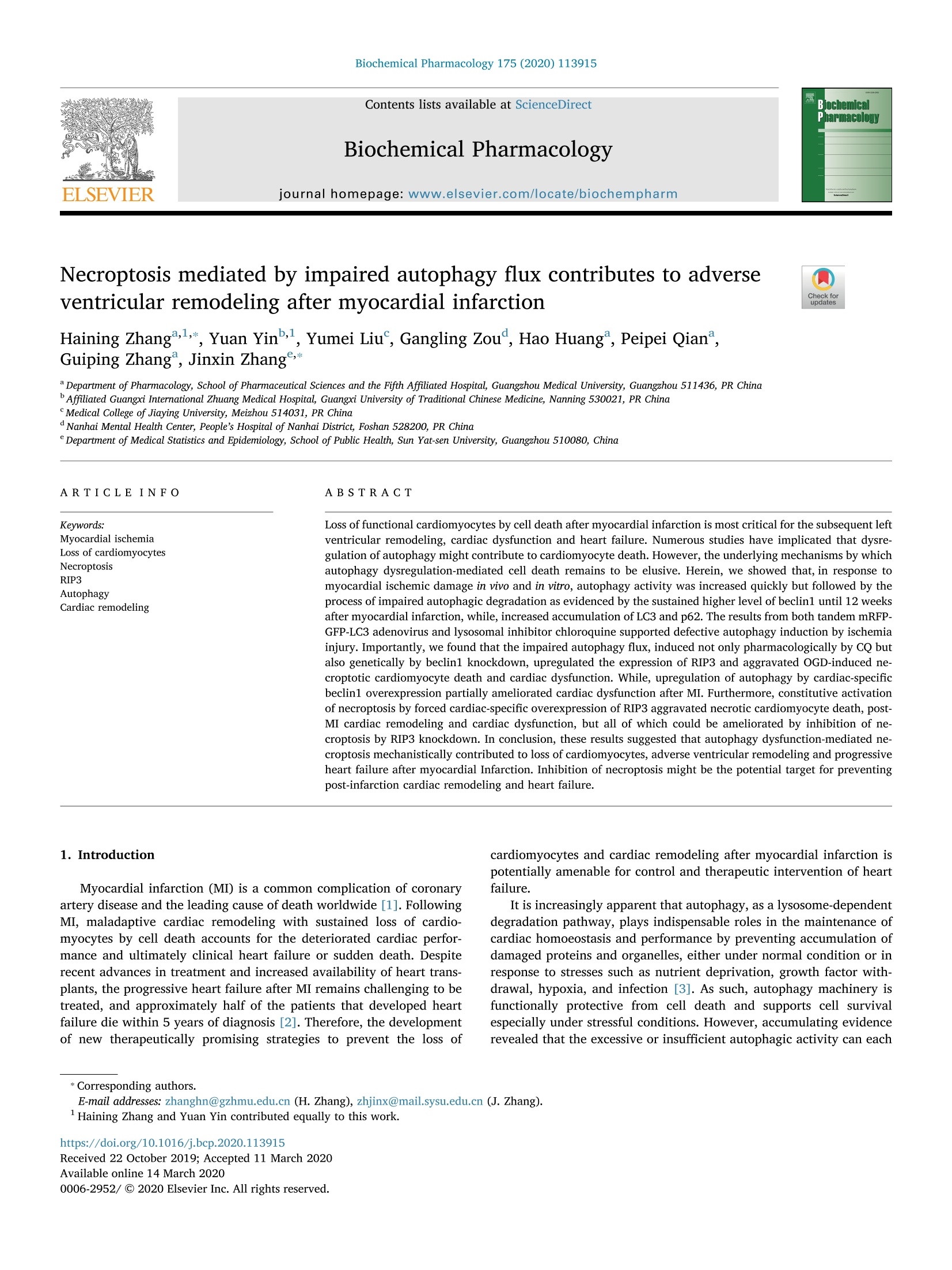
北京隆福佳生物科技有限公司为您提供《心肌细胞中beclin1 和LC3II/I 的表达检测方案(其它培养箱)》,该方案主要用于动物内脏组织中生化检验检测,参考标准《暂无》,《心肌细胞中beclin1 和LC3II/I 的表达检测方案(其它培养箱)》用到的仪器有Invivo2 500低氧工作站(低氧培养箱)。
我要纠错
推荐专场
其它培养箱
更多
相关方案
 咨询
咨询