方案详情文
智能文字提取功能测试中
比利时根特大学基础医学科学系 纳米脂肪移植:基础研究和临床应用Nanofat Grafting Basic Research and Clinical ApplicationsSPECIAL TOPIC纳米脂肪移植:基础研究和临床应用 Plastic and Reconstructive Surgeryber 2013 Nanofat Grafting: Basic Research and Clinical Applications Patrick Tonnard, M.D. Alexis Verpaele, M.D. Geert Peeters, M.D. Moustapha Hamdi, M.D., Ph.D. Maria Cornelissen, Ph.D. Heidi Declercq, Ph.D. Ghent and Brussels, Belgium Background: The indications for fat grafting are increasing steadily. In microfat grafting, thin injection cannulas are used. The authors describe their experi-ence of fat injection with even thinner injection needles up to 27 gauge. The fat used for this purpose is processed into “nanofat.” Clinical applications are described. Preliminary results of a study, set up to determine the cellular con-tents of nanofat, are presented. Methods: Nanofat grafting was performed in 67 cases to correct superficial rhytides, scars, and dark lower eyelids. Three clinical cases are described. In the research study, three fat samples were analyzed. The first sample was a clas-sic lipoaspirate (macrofat). The second sample was microfat, harvested with a multiport small-hole cannula. The third was microfat processed into nano-fat. Processing consisted of emulsification and filtering of the lipoaspirate. Fat samples were analyzed for adipocyte viability. Cells from the stromal vascular fraction and the CD34+ subfraction were quantified. The stem cell quality was investigated by culturing the cells in standard and adipogenic media. Results: No viable adipocytes were observed in the nanofat sample. Adipose-derived stem cells were still richly present in the nanofat sample. Cell cultures showed an equal proliferation and differentiation capacity of the stem cells from the three samples. Clinical applications showed remarkable improve-ments in skin quality 6 months postoperatively. No infections, fat cysts, granu-lomas, or other unwanted side effects were observed. Conclusions: Nanofat injections might become a new concept in the lipofilling area. In clinical situations, nanofat seems to be suitable for skin rejuvenation purposes. (Plast. Reconstr. Surg. 132: 1017, 2013.) he first reports on autologous fat grafting T were published in the early twentieth cen-tury.1 It became more widely implemented after the introduction of liposuction by Illouz in the 1980s.2 Since the standardization of the fat grafting technique by Coleman one decade later, lipofilling has become a very important and valu-able tool in plastic surgery.3,4 Adipose-derived stem cells were first discovered in 2001.5 Since then, much research into these multipotent mesenchy-mal-derived progenitor cells has been performed. Currently,theapplicationsofadipose-derived stem cell therapy extend far beyond the field of plastic surgery.6–11 From Plastic and Aesthetic Surgery, Coupure Centrum for Plastic Surgery Ghent; the Department of Plastic Surgery, University Hospital of Brussels; and the Department of Ba-sic Medical Sciences, Ghent University. ReceivedforpublicationDecember16,2012;accepted February 27, 2013. Copyright © 2013 by the American Society of Plastic Surgeons DOI: 10.1097/PRS.0b013e31829fe1b0 The initial goal of fat grafting was to treat vol-ume losses created by disease, trauma, or aging. Fat was injected with relatively large blunt can-nulas (±2 mm diameter). For delicate areas such as eyelids and lips, smaller injection cannulas becamepopular.Lipofillingwithcannulasas small as 0.7 mm in diameter, also called microfat grafting, has been described.12–15 For these indi-cations, fat is harvested with small-hole cannulas Disclosure: The authors have no financial interest to declare in relation to the content of this article. No external funding was received. Supplemental digital content is available for this article. Direct URL citations appear in the text; simply type the URL address into any Web browser to access this content. Clickable links to the material are provided in the HTML text of this article on the Journal ’s Web site (www. PRSJournal.com). 布鲁塞尔大学医院整形外科 to obtain a lipoaspirate with smaller fat particles. Wepreviouslydescribedmicrofatgraftingin the deep dermal layer of the skin with 23-gauge sharp needles for treatment of fine rhytides in the face.16 To work even more superficially with still finer sharp needles (27 gauge), the harvested fat was mechanically emulsified and filtered until a liquid suspension was obtained. We call this “nanofat.” In this article, we describe the technique and report our experience with nanofat grafting. We presentthreeclearclinicalcases.Inaddition, we provide the results of an experimental study to determine the cellular content of the nanofat. The viability of the adipocytes and, more impor-tantly, the number and activity of the adipose-derived stem cells are investigated. The findings of the nanofat sample analysis are compared to two other lipoaspirates obtained by standard fat harvesting techniques. PATIENTS AND METHODS Macrofat, Microfat, and Nanofat Harvesting Liposuction was performed in a 40-year-old femalepatientduringanabdominoplastypro-cedure.Informedconsenttousethelipoaspi-rate for analysis was obtained from the patient. Fat was harvested from the lower abdomen after infiltration with a modified Klein solution (lido-caine 800 mg/liter and adrenaline 1:1,000,000). The harvested fat was rinsed and filtered through a sterile nylon cloth with 0.5-mm pore size that was mounted over a sterile canister. Two different aspiration cannulas (Fig. 1) were used. Three dif-ferent lipoaspirate samples were analyzed. In detail, a high-negative-pressure liposuction procedure was performed using a standard lipo-suction device. In the first group, a standard 3-mm Mercedes type liposuction cannula with large side holes (2 e 7 mm) (Fig. 1) was used. In the second and third groups, fat was harvested with a multi-port 3-mm cannula with sharp side holes of 1 mm in diameter (Fig. 1); both were obtained from Tulip Medical Products (San Diego, Calif.). After saline rinsing and filtering, no further processing of the lipoaspirates from the first group (called“macrofat”) and second group (called “microfat”) was performed. In the third group, the lipoaspi-ratewasmechanicallyemulsifiedafterrinsing. Emulsification of the fat was achieved by shifting the fat between two 10-cc syringes connected to each other by a female-to-female Luer-Lok con-nector. (See Video, Supplemental Digital Content 1, which illustrates how the microfat lipoaspirate Fig. 1. (Above ) Syringe with microfat in a condition before the emulsifciationprocessandsyringewithahomogeneousfat emulsion after the emulsifciation process. Note the difefrence in color and consistency. (Below ) A standard 3-mm Mercedes type liposuction cannula with large side holes (2 × 7 mm) and a 3-mm multiport cannula, containing several sharp side holes of 1-mm diameter. The sharp side holes augment the harvesting yield. isprocessedintonanofat,http://links.lww.com/PRS/A855.) After 30 passes, the fat changed into an emulsion. At the end of the fragmentation process, the fat became liquid and took on a whit-ish appearance (Fig. 1). After this emulsification process, the fatty liquid was again filtered over the sterile nylon cloth and the effluent was collected in a sterile recipient (see Video, Supplemental Digital Content 1, http://links.lww.com/PRS/A855). This was done to remove the connective tissue remnants that would block the fine needles. This effluent is called “nanofat.” Patients Between May of 2010 and September of 2012, nanofat grafting was performed in 67 cases for a variety of indications. It was used for skin reju-venationpurposesincombinationwithclassic microfat grafting or sharp needle intradermal fat Video1.SupplementalDigitalContent1illustrateshowthemicrofat lipoaspirate is processed into nanofat, http://links.lww.com/PRS/A855. grafting.16 Nanofat grafting was used for the reju-venation of perioral skin [38 cases (58 percent)], glabellar skin [15 cases (23 percent)], or sun-dam-aged skin at the breast cleavage [eight cases (11percent)]. Four scars (6 percent) and two patients with dark lower eyelids (2 percent) were treated with nanofat as well. In all cases, the nanofat was prepared as described above. A 27-gauge needle was mounted on the syringe for superficial intra-dermalandsubdermalinjection.Injectionwas performed until a yellowish discoloration of the skin showed up. [See Video, Supplemental Digi-tal Content 2, which demonstrates the injection of nanofat at the lower eyelid (note the yellowish dis-coloration of the skin after injection), http://links. lww.com/PRS/A856; and see Video, Supplemental Digital Content 3, which shows the injection of nanofat at the area of the breast cleavage (note the superficial fanwise intradermal injections in which the whole skin area can be covered), http://links.lww.com/PRS/A857.] Three clinical examples are described below. Adipocyte Viability The viability of the lipoaspirates, obtained by thethreeharvestingtechniques,wasevaluated using fluorescence microscopy after a live/dead staining. After rinsing the lipoaspirates, 1 ml of phosphate-buffered saline solution supplemented with 2 -l of calcein AM (1 mg/ml) (AnaSpec, Fre-mont, Calif.) and 2 A l of propidium iodide (1 mg/ml) (Sigma-Aldrich, St. Louis, Mo.) was added. Lipoaspirates were incubated for 10 minutes at room temperature, washed twice with phosphate-buffered saline solution, and evaluated by fluores-cencemicroscopy(OlympusinvertedResearch System Microscope, type U-RFL-T, Cell software; Olympus Belgium, Aartselaar, Belgium). Cell Culture Stem Cell Isolation Isolationoftheadipose-derivedstemcells from the different lipoaspirates (macrofat, micro-fat, and nanofat) was as follows. An equal aliquot Video 2. Supplemental Digital Content 2 demonstrates the injec-tion of nanofat at the lower eyelid, http://links.lww.com/PRS/A856. Note the yellowish discoloration of the skin after injection. Video 3. Supplemental Digital Content 3 shows the injection of nanofat at the area of the breast cleavage, http://links.lww. com/PRS/A857. Note the super coial fanwise intradermal injec-tions in which the whole skin area can be covered. of 0.1% collagenase type II (Sigma-Aldrich) in phosphate-buffered saline containing 1% penicil-lin/streptomycin was added to the lipoaspirates. The mixture was incubated for 45 minutes at 37°C on a gyratory shaker (Laboshake; C. Gerhardt, Königswinter, Germany). Fetal bovine serum was added to a final concentration of 10% to stop enzymeactivityfollowedbycentrifugationat 800 rpm for 5 minutes. The overlying fluid and adipose phases were aspirated and discarded. The stromal cell pellet was resuspended in phosphate-bufferedsalineandfilteredthrougha70-u m Falcon cell strainer (Becton Dickinson, Franklin Lakes, N.J.). The amount of cells was counted with a Türk solution (Merck, Whitehouse Station, N.J.). Half of the stromal vascular fraction was cultivated in a T75 Falcon flask in standard medium [Mesen-PRO basal medium supplemented with MesenPro RS growth supplement (Life Technologies, Carls-bad, Calif.) and L -glutamine]. As an additional control experiment, the other half of the stromal vascular fraction was further processed through magnetic-activated cell sorting, with a CD34 kit obtained from Miltenyi Biotec (Auburn, Calif.) accordingtothecompany’sprotocol.Briefly, cells were suspended in magnetic-activated cell-sorting buffer (300 i l) and Fc receptor-blocking reagent (100 p l) was added to avoid unspecific CD34 labeling of cells by means of Fc receptors. CD34microbeads(100 wereaddedtothe cell suspension and incubated for 30 minutes at 4°C. After adding magnetic-activated cell-sorting rinse buffer (5 ml) and centrifuging (5 minutes,800rpm),thecellpelletwasresuspendedin Fig. 2. Fluorescence microscopy of lipoaspirates after calcein AM/propidium iodide staining. (Above ) Macrofat (lipoaspirate harvestedwitha3-mmstandardcannula).(Center )Microfat (lipoaspirate harvested with a 3-mm multiport cannula). (Below ) Nanofat (lipoaspirate harvested with a 3-mm multiport cannula followed by an emulsipciation procedure). Macrofat (above ) and microfat(center )showgoodadipocyteviabilityandadipose tissue structure in contrast with the nanofat (below ), where no adipocytes and no normal adipose tissue structure are visible. 300 u l of magnetic-activated cell-sorting rinse buf-fer. The cell suspension was passed through the magnetic-activated cell-sorting column (Miltenyi Table 1.Results of the Cell Count from the Three Single Fat Samples SVF* CD34+ Fraction* CD34+/SVF Ratio (%) Macrofat (standard cannula) 3,075,000 200,000 6.5 Microfat (multiperforated cannula) 2,360,000 105,000 4.5 Nanofat (multiperforated cannula plus emulsification) 1,975,000 100,000 5.1 SVF, stromal vascular fraction. *No. of cells per 100 ml of lipoaspirate. Biotec) twice. The CD34+ fraction was collected, counted, and cultivated in a T25 Falcon flask in standard medium. Fig. 3. Phase-contrast microscopic images of the stem cell cul-tures derived from the stromal vascular fraction after 7 days.(Above ) Stem cell culture derived from the macrofat. (Center ) Stem cell culture derived from the microfat. (Below ) Stem cell culturederivedfromthenanofat.Noteatypicalfibroblastic morphology of the proliferating cells in the three samples. Stem Cell Differentiation The adherent stromal vascular fraction cells and the CD34+ cells were seeded onto Thermanox 150 u m Fig. 4. Phase contrast microscopic images of stem cells derived from the three lipoaspirates and cultured in adipogenic culture medium for 10 days. Stem cells derived from the stromal vascu-lar fraction from macrofat (above ), microfat (center ), and nanofat (below ). Note the appearance of fat vacuoles in all 3 samples. 100u m Fig. 5. Light microscopic images (oil red O staining) of stem cells derived from the three lipoaspirates and cultured in adipo-genic culture medium for 10 to 28 days. Stem cells derived from the stromal vascular fraction (above and center ) or the CD34+(below ) fraction from macrofat (above ), microfat (center ), and nanofat (below ). coverslips (Nunc, Roskilde, Germany) at a con-centration of 40,000 cells per well in a 24-well plate (Greiner Bio-One, Gloucestershire, United Kingdom)andculturedinstandardmedium. After confluence, standard medium was replaced by adipogenic differentiation medium (Stempro AdipogenicMedium;Invitrogen)andcultured for 14 to 21 days. Adipocyte differentiation was noticed by the intracellular accumulation of lipid droplets. Lipid droplets were evaluated by phase-contrast micros-copy and verified using light microscopy after oil red O staining. RESULTS Adipocyte Viability After calcein AM/propidium iodide staining, the three different lipoaspirate samples were eval-uated on their adipose tissue quality and adipo-cyte viability. In the macrofat (Fig. 2, above ) and microfat (Fig. 2, center ), adipose tissue with a nor-mal histologic structure could be visualized. The adipocytes were viable (green), and only a very few dead cells could be observed. This was in high contrast with the nanofat (Fig. 2, below ), where the adipose tissue structure was completely disturbed and replaced by an oily emulsion. No viable adipo-cytes were noticed in the nanofat. Stem Cell Isolation and Culture After isolation of the stromal vascular frac-tion and the CD34+ subpopulation from the three lipoaspirates, a cell count was performed. The number of viable stem cells derived from the stro-mal vascular fraction ranged from 1.9 to 3.0 s 106cells/100 ml lipoaspirate, independent of the pro-cessing method of the adipose tissue. The num-ber of CD34+ cells in this stromal vascular fraction ranged from 0.1 to 0.2 s 106 cells/100 ml lipoaspi-rate, resulting in a 4.5 to 6.5 percent CD34+–stro-mal vascular fraction ratio (Table 1). The stromal vascular fraction and CD34+ cells wereculturedinstandardmedium.Adherent cells in both stromal vascular fraction and CD34+fractionsformedmonolayersandpresenteda fibroblasticmorphology.Nodifferencesincell cultures were noticed between the three different lipoaspirate samples (Fig. 3). Stem Cell Differentiation Todemonstratethestemcellnatureof the stromal vascular fraction and CD34+ frac-tion, cells were plated in control or adipogenic mediumfordifferentiationintomatureadi-pocytes. After 10 days of culture in adipogenic medium, phase contrast microscopic evaluation showed the presence of spherical cells contain-inglipidvacuoles,indicatingdifferentiation into the adipogenic lineage. No differences in quality and quantity of adipocytes between the different lipoaspirate samples were noticed(Fig. 4). Stem cells cultured in standard medium remained fibroblast-like. Light microcopy after oil red O staining confirmed the presence of Clinical Results Between May of 2010 and September of 2012, nanofat grafting was performed in 67 cases. Intra-dermal injection was performed with a 27-gauge needle. The endpoint of injection was until a yel-lowish discoloration of the skin appeared. This discolorationusuallydisappearedafewhours after the injection. The clinical results gradually improved over time and were maximal from 4 to 6months postoperatively. There were no important complications seen in this series. No infections, fat cysts, granulomas, or other unwanted side effects were observed. In injections of larger areas such as the décolletage or the face, there was a tempo-rary erythema of the injected area that lasted for 1.5 to 2 days. CASE REPORTS Case 1 A 41-year-old woman complained about fan-shaped vertical rhytides and sun-damaged skin in the décolleté area (Fig. 6). The rhytides were injected intradermally in a longitudinal fashion with 6 cc of microfat using a 23-gauge needle. Injection was per-formed until a slight overcorrection was seen. Next, the whole triangular surface of the décolleté area was injected at a super-ficial subdermal level with 12 cc of nanofat using a 27-gauge needle in repeated fan-shaped patterns. The first few weeks after injection, the treated area appeared overcorrected but gradually normalized toward the result seen at 3 months (Fig. 6). Case 2 A 33-year-old woman sought improvement of the aesthetics of her lower eyelids. She presented with a moderate bulging of the lower eyelid fat compartments with a marked eyelid-cheek junction. The skin of the lower eyelid was heavily pigmented with an extension of this pigmentation along the nasojugal groove(Fig. 7, left ). The patient mentioned that the discoloration had been present since childhood. A lower eyelid blepharoplasty with fat redraping over the lower and lateral orbital rim was proposed Fig. 7. Case 2. (Left ) A 33-year-old woman with lower eyelid blepharochalasis and dark coloration of the lower eyelid skin extending into the nasojugal groove. (Right ) Seven months after fat redraping, blepharoplasty, and intradermal injection of 1.6cc nanofat in the lower eyelid and pigmented nasojugal groove. Fig. 8. Case 3. (Left ) A 61-year-old woman with perioral rhytides and who refused to undergo any laser resurfacing procedure.(Right ) Result 7 months after injection of 4 cc of intradermal microfat graft into the rhytides, accompanied by intradermal injec-tion of 6 cc of nanofat in the perioral skin. Note the better skin quality in the treated area. together with intradermal infiltration of nanofat to improve the pigmentation of the skin. The operation was performed under local anesthesia combined with intramuscular midazolam seda-tion. A classical infraciliary incision was carried out for the fat redraping and blepharoplasty procedure. After completion of the blepharoplasty, the eyelid skin and the nasojugal groove were injected intradermally with 1.6 cc of nanofat per side. At the end of the injections, the whole lower eyelid was colored yellowish like a giant xanthelasma. The whitish discoloration disappeared after 1 month. The lower eyelid skin remained erythematous for 3months, followed by a gradual lightening of the skin (Fig. 7, right ). Case 3 A 61-year-old woman consulted for perioral rejuvenation, mainly the correction of the vertical rhytides of the upper and lower lips. She refused to have any laser resurfacing procedure(Fig. 8). Microfat grafting at the rhytides of the lip together with nanofat grafting of the whole perioral region and cheeks were performed. Then, 4 cc of microfat was injected with a 23-gauge needle into the rhytides (sharp needle intradermal fat grafting technique), and 6 cc of nanofat was used for intradermal injec-tion into the skin of the upper and lower lips and in both cheek areas. DISCUSSION For microfat grafting, usually performed in thefacialarea,bluntinjectioncannulasrang-ing from 0.7 to 0.9 mm are used with very good results. To provide a smooth injection through these fine cannulas, the fat particles need to be sufficiently small. If the fat particles are too large, passage through the injection cannula would be difficult. A disrupted injection will follow, which may result in an unequal lipofilling with irregular fat deposits. To provide a lipoaspirate with smaller parti-cles for microfat grafting procedures at the lower eyelid,Trepsatusedamultiperforatedharvest-ing cannula of 2 mm with 1-mm side holes and 19-gauge injection cannulas. A multiperforated liposuction cannula 3 mm in diameter with 2-mm side holes was used to harvest fat for injection of other parts of the face.12 Nguyen et al. described the use of a similar multiperforated harvesting cannula with side holes of 1 mm.14 Coleman and Mazzola reported the use of injection cannulas up to 22 gauge.13 Nguyen et al. applied blunt injec-tion 25-gauge cannulas in a mouse model and mentioned the use of blunt 21- or 23-gauge can-nulas for fat grafting in clinical cases.14 Toensureasmoothfatinjectionthrough27-gauge sharp needles, the aspirated fat has to be processed mechanically to provide a liquid fat emul-sion, which we call nanofat. A yield of 1 ml of nano-fat per 10 ml of lipoaspirate can be expected using a nanofat processing procedure as described above. Our initial goal of local injections with nano-fat using 27-gauge needles was to use it as filler for superficial rhytides. Because of the reduced num-ber of viable adipocytes in the emulsified fat, the filling capacity of nanofat is obviously very limited. After noting a clear skin rejuvenation effect in our clinical cases, we started to use nanofat to improve skin quality. Otherstudiesusedlipofillingasaninstru-ment for skin regenerative purposes such as treat-mentofradiotherapyulcersorscars.17–19One study reported a clear and statistically significant improvement in dermal elasticity after injection of facial scars in 14 patients.18 The mechanism for this regenerative effect on damaged skin remains unknown.Improvedelasticityispresumablya consequenceofincreasedcollagenandelastin synthesis and remodeling. These effects are most likely triggered by stem cells rather than by grafted adipocytes. Moreover, the nanofat sample analysis revealed that adipocytes were destroyed during the emulsification process. Toisolatestemcells,thestromalvascular fractionhastobeseparatedfromtheadipo-cytes. The stromal vascular fraction contains dif-ferent types of cells, such as endothelial cells, monocytes, macrophages, granulocytes, and lymphocytes. The stromal vascular fraction also includes a substantial amount of mesenchymal stem cells (adipose-derived stem cells). These multipotent stem cells have the ability to adhere to plastic culture plates and to form fibroblast-like colonies. Likewise, stem cells can be isolated by culturing the adherent stromal vascular frac-tion cells. It is remarkable that these multipo-tent stem cells are richly present in fat tissue, in contrast to bone marrow or other sources of multipotent mesenchymal stem cells.20 Adi-pose-derived stem cells have an extensive prolif-erative capacity and the ability to differentiate into the mesoderm, ectoderm, and endoderm lineages.21–24 Ithasbeendemonstratedthatcellswitha CD34+phenotyperepresentacellpopulation with a great stem cell proliferative capacity.21,25–32In this study, stem cells were isolated in two ways. First, cells from the stromal vascular fraction were selected on their adherence to the plastic plate. These adherent cells were further cultured. Sec-ond,asacontrolexperiment,theCD34+sub-population was isolated from the stromal vascular fraction and cultured as well. The lipoaspirate viability evaluation shows that the nanofat sample has lost the normal fat tissue structure and that adipocytes are eliminated dur-ing nanofat processing. However, a large number of good quality mesenchymal stem cells are still present in the nanofat sample. Classic fat grafting is mostly used to build up large volumes, especially in breast reconstructive cases. In those cases, it is crucial to preserve as many viable adipocytes as possible. Nanofat does not has the capacity to build up a significant fat volume. Consequently, nanofat is evidently not suitable for these indications. In fact, because of the lack of adipocytes, the volumetric effect of nanofat is obviously very limited. Therefore, the indicationsfornanofatinjectionaredifferent when compared with microfat grafting. Usually, nanofat grafting is combined with other modali-tiesofmicrofatgraftingsuchassharpneedle intradermal fat grafting to obtain a soft-tissue fill-ing effect, where the nanofat is layered more fan-wise in an intradermal level to enhance the skin quality. In our experience, as opposed to microfat, the effect of nanofat usually appears with a delay of 4 weeks to 3 months. In the case where dark circles were treated, the beneficiary effect was pre-ceded by a rather prolonged erythematous phase, which most likely is attributable to the soft-tissue rearrangement. In fact, it may be questioned whether a nano-fat transfer actually is a “fat grafting” procedure, as adipocytes did not survive the emulsification process. The major effect of nanofat injection is probablyastemcellactivity.Likewise,nanofat injection might rather be considered as an in vivo tissue-engineeringprocess.Itmightbelogical to discard the dead adipocyte fraction from the nanofat and to inject the purified stromal vascu-lar fraction only. However, isolating the stromal vascular fraction out of the nanofat before injec-tion in routine clinical cases would be time con-suming, complicated, and expensive. Besides, it requires specific laboratory equipment and expe-rience. Moreover, it is known that apoptotic cells release cytokines and attract macrophages that induce growth factors and play an important role in regeneration of the damaged tissue.33 Thus, co-injection of fragmented adipocytes might have a stimulating effect on stem cell differentiation and tissue regeneration. This is the first report of a new fat grafting technique that we have been using since May of 2010. Clinical assessment was performed by our observations and preoperative and postoperative photographs. No other objective measurements orobjectiveskinqualitytestshavebeenper-formed. In addition to the three clinical cases, we present the results of our laboratory pilot study comparing three fat samples. More fat samples have to be analyzed to determine the content of the nanofat in a statistically significant way. Fur-thermore,histologicevidenceofthenanofat effect is not present yet. Further studies will have to determine whether the clinical observations can be correlated with preoperative and postop-erative histologic analysis. These studies are in progress. CONCLUSIONS Nanofat injection may become another con-cept in the lipofilling area. In this study, it seems thatstemcellsareresponsiblefortheclinical resultsseenafternanofatinjections.Clinically, nanofatappearssuitableforskinrejuvenation procedures. Further studies have to be performed tofindstatisticallysignificantevidenceforthe nanofat effect. Geert Peeters, M.D. Department of Plastic Surgery University Hospital of Brussels Laarbeeklaan 101 1090 Brussels, Belgium gpeetersg@gmail.com REFERENCES 1. Neuber G. Fetttransplantation. Verh Dstch Ges Chir . 1893;22:66.2. Illouz YG. Body contouring by lipolysis: A 5-year experience with over 3000 cases. Plast Reconstr Surg . 1983;72:591–597. 3. Coleman SR. Long-term survival of fat transplants: Controlled demonstrations. Aesthetic Plast Surg . 1995;19:421–425. 4. Coleman SR. Structural fat grafts: The ideal filler? Clin Plast Surg . 2001;28:111–119. 5. Zuk PA, Zhu M, Mizuno H, et al. Multilineage cells from human adipose tissue: Implications for cell-based therapies. Tissue Eng . 2001;7:211–228. 6. Okura H, Saga A, Soeda M, et al. Intracoronary artery trans-plantation of cardiomyoblast-like cells from human adipose tissue-derivedmulti-lineageprogenitorcellsimproveleft ventricular dysfunction and survival in a swine model of chronic myocardial infarction. Biochem Biophys Res Commun .2012;425:859–865. 7. Fawzy El-Sayed KM, Dörfer C, Fändrich F, et al. Adult mesen-chymal stem cells explored in the dental field. Adv Biochem Eng Biotechnol . E-published ahead of print September 1, 2012. 88.. O O h h J J S S ,, P P a a r r k k I I S S ,, K K i i m m K K N N ,, Y Y o o o o n n d d o o H H ,, K K i i m m S S H H ,, H H a a Y Y .. Transplantation of an adipose stem cell cluster in a spinal cord injury. Neuroreport 2012;23:277–282. 9. Puissant B, Barreau C, Bourin P, et al. Immunomodulatory effectofhumanadiposetissue-derivedadultstemcells:Comparison with bone marrow mesenchymal stem cells. Br J Haematol . 2005;129:118–129. 10. Yoo KH, Jang IK, Lee MW, et al. Comparison of immuno-modulatory properties of mesenchymal stem cells derived from adult human tissues. Cell Immunol . 2009;259:150–156. 11. Zhou B, Yuan J, Zhou Y, et al. Administering human adipose-derived mesenchymal stem cells to prevent and treat experi-mental arthritis. Clin Immunol . 2011;141:328–337. 12. TrepsatF.Midfacereshapingwithmicro-fatgrafting(in French). Ann Chir Plast Esthet . 2009;54:435–443. 13. ColemanSR,MazzolaRF.FatInjection:FromFillingto Regeneration . St. Louis: Quality Medical; 2009. 14. Nguyen PS, Desouches C, Gay AM, Hautier A, Magalon G. Development of micro-injection as an innovative autologous fat graft technique: The use of adipose tissue as dermal filler. J Plast Reconstr Aesthet Surg . 2012;65:1692–1699. 15. Dasiou-PlakidaD.Fatinjectionsforfacialrejuvenation:17yearsexperiencein1720patients.JCosmetDermatol .2003;2:119–125. 16. Zeltzer AA, Tonnard PL, Verpaele AM. Sharp-needle intra-dermal fat grafting (SNIF). Aesthet Surg J . 2012;32:554–561. 17. Rigotti G, Marchi A, Galiè M, et al. Clinical treatment of radiotherapytissuedamagebylipoaspiratetransplant:A healing process mediated by adipose-derived adult stem cells. Plast Reconstr Surg . 2007;119:1409–1422; discussion 1423. 18. Sardesai MG, Moore CC. Quantitative and qualitative dermal change with microfat grafting of facial scars. Otolaryngol Head Neck Surg . 2007;137:868–872. 19. Akita S, Yoshimoto H, Ohtsuru A, Hirano A, Yamashita S. Autologousadipose-derivedregenerativecellsareeffec-tive for chronic intractable radiation injuries. Radiat Prot Dosimetry 2012;151:656–660. 20. Zhu X, Du J, Liu G. The comparison of multilineage differ-entiation of bone marrow and adipose-derived mesenchymal stem cells. Clin Lab . 2012;58:897–903. 21. Traktuev DO, Merfeld-Clauss S, Li J, et al. A population of multipotent CD34-positive adipose stromal cells share peri-cyte and mesenchymal surface markers, reside in a periendo-thelial location, and stabilize endothelial networks. Circ Res .2008;102:77–85. 22. Brzoska M, Geiger H, Gauer S, Baer P. Epithelial differen-tiation of human adipose tissue-derived adult stem cells. Biochem Biophys Res Commun . 2005;330:142–150. 23. Cao Y, Sun Z, Liao L, Meng Y, Han Q, Zhao RC. Human adipose tissue-derived stem cells differentiate into endothelialcellsinvitroandimprovepostnatalneo-vascularizationinvivo.BiochemBiophysResCommun .2005;332:370–379. 24. Declercq HA, De Caluwé T, Krysko O, Bachert C, Cornelissen MJ. Bone grafts engineered from human adipose-derived stem cells in dynamic 3D-environments. Biomaterials 2013;34:1004–1017. 25. Boquest AC, Shahdadfar A, Frønsdal K, et al. Isolation and transcription profiling of purified uncultured human stro-mal stem cells: Alteration of gene expression after in vitro cell culture. Mol Biol Cell 2005;16:1131–1141. 26. BoquestAC,ShahdadfarA,BrinchmannJE,CollasP. Isolation of stromal stem cells from human adipose tissue. Methods Mol Biol . 2006;325:35–46. 27. Bailey AM, Kapur S, Katz AJ. Characterization of adipose-derivedstemcells:Anupdate.CurrStemCellResTher .2010;5:95–102. 28. Suga H, Matsumoto D, Eto H, et al. Functional implications of CD34 expression in human adipose-derived stem/progen-itor cells. Stem Cells Dev . 2009;18:1201–1210. 29. Gronthos S, Franklin DM, Leddy HA, Robey PG, Storms RW, Gimble JM. Surface protein characterization of human adipose tissue-derived stromal cells. J Cell Physiol .2001;189:54–63. 30. FolgieroV,MiglianoE,TedescoM,etal.Purification andcharacterizationofadipose-derivedstemcellsfrom patients with lipoaspirate transplant. Cell Transplant .2010;19:1225–1235. 31. Deans RJ, Moseley AB. Mesenchymal stem cells: Biology and potential clinical uses. Exp Hematol . 2000;28:875–884. 32. LinCS,NingH,LinG,LueTF.IsCD34trulyanega-tivemarkerformesenchymalstromalcells?Cytotherapy 2012;14:1159–1163. 33. Mahdavian Delavary B, van der Veer WM, van Egmond M, Niessen FB, Beelen RH. Macrophages in skin injury and repair. Immunobiology 2011;216:753–762.
关闭-
1/10
-
2/10
还剩8页未读,是否继续阅读?
继续免费阅读全文产品配置单
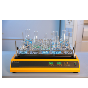
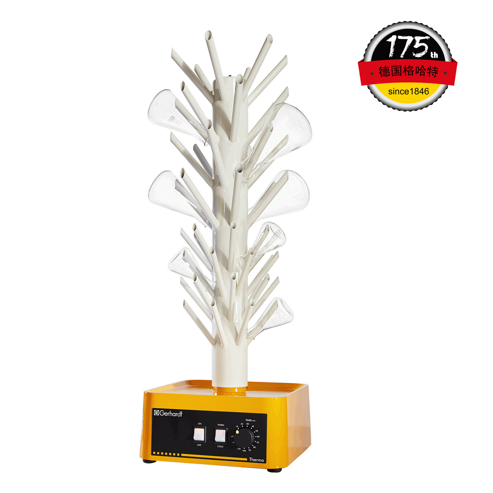
中国格哈特为您提供《纳米脂肪细胞样品的振荡》,该方案主要用于其他中振荡检测,参考标准《暂无》,《纳米脂肪细胞样品的振荡》用到的仪器有格哈特强力高重现振荡器LS500/RO500、格哈特快速干燥仪STL56、德国移液器MM。
我要纠错
推荐专场

快速干燥仪
更多
相关方案
 咨询
咨询