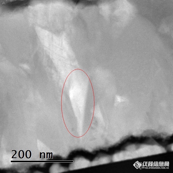
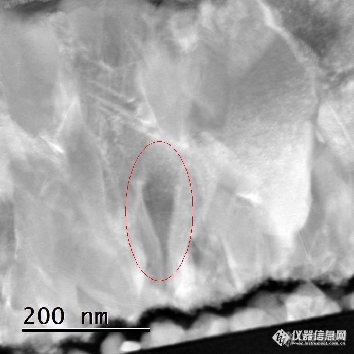
Sorry not be able to type Chinese on this computer! Attached are two ADF images, image 1 is taken at higher collection angle than image 2, so i would expect diffraction contrast in image 2, and mostly Z contrast in image 1. You can notice that there are areas (arrowed in the images) that appear dark in image 1 but bright in image 2. Initially, I thought the bright area in image 1 indicates a heavier material, but actually both brighter and darker areas in image 1 has the same composition suggested by EDX. So I am confused why I see the contrast in image 1 for same material? if it is still a diffraction contrast, then why it is reversed compared to that in image 2? Anybody has some idea about this? Any suggestions would be appreciated! Thank you! Image 1: Image 2: