

 仪器对比
仪器对比


 关注
关注


技术参数:
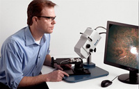
美国Phoenix Research实验室 Micron III提供全功能小动物眼睛in vivo 图像。视网膜图像达到空前的分辨率,包括亮场图像,荧光血管造影成像和荧光探针成像。眼睛前部成像选项包括一个高分辨率狭缝成像器和一个独立的小梁网成像部件,都能够产生荧光成像。对于视网膜功能研究,系统还可以提供多焦点和PERG研究
眼睛成像新标准
Micron III is making a fundamental contribution to eye research at professional research laboratories around the world
视网膜成像
亮场图像: 大鼠、小鼠的白内障和暗色素
荧光血管造影成像:精度可达4um
荧光探针成像: GFP, YFP, CFP, mCherry
眼睛前部成像
角膜, 晶状体的狭缝成像和小梁网成像
视觉电生理
通过视网膜的mfERG(多焦视网膜电图)和PERG (图形视网膜电图)
精确传输任意刺激模式
可实现mfERG 和 PERG
需要和 diagnosys LLC公司的电子装置和软件搭配使用
用户评价:
“Best rodent imaging system I’ve ever used.
Produces exceptionally high quality digital fundus images of rodents and small animals.The resolution and contrast are much better than any other instruments we have tried. It is robust and easy for the students to use.We have had good success with bright field, as well as fluorescence
imaging of gfp expression in the retina and fluorescein angiography.The ability to capture video, select and output individual frames later is a big help when focusing on the retina in the small rodent eyes.”
John Flannery, PhD
Professor of Vision Science and Molecular and Cell Biology
Associate Director, Helen Wills Neuroscience Institute
University of California, Berkeley
“I have always sought good fundus images.
It used to take all day for an experienced researcher to get just one. Now with our Micron from Phoenix, even a new user can learn to capture publishable rat and mouse fundus images in less than 30 minutes."
Robert L. Peiffer, Jr, DVM, PhD
Senior Investigator
Merck & Co, Inc,
具体图片展示查看请登录www.bio-sun.com.cn


相关产品

