
方案详情文
智能文字提取功能测试中
HORIBAScientificHORIBAExplore the futureAutomotive Test Systems 1 Process & Environmental1Medical 1 Semiconductor1Scientific Raman Spectroscopy Applied to Geology and Earth Science Dr. Renata Lewandowska, Application Scientist HORIBA Jobin Yvon SAS, 231 rue de Lille, Villeneuve d'Ascq, France Raman Spectroscopy is a scattering technique probing the vibrational modes of molecules and crystals. Raman signaturesprovide information about the chemical nature and the crystalline structure of samples. This characterisation method is fast,non-destructive and does not require any particular sample preparation. Raman spectroscopy can be used for the identification ofchemical species; its high spectral resolution allows closelyrelated chemical substances to be distinguished. Further-more, this method is sensitive to atomic structure: differentcrystalline phases can be easily differentiated, includingdisordered/amorphous materials. Rutile and anatase are two polymorphs of titanium dioxide (Ti0,):even if their chemical composition is the same, their crystalstructure is different which results in significantly different Ramanspectra. Inthis secondexample, variouscarbonates were analyzed. Theirchemical or structural differences canbe easily highlighted thanks to the highspectral resolution of the method. Areference matrix of multiple species wasmapped out using the unique DuoScanMmacro-mapping option; the false colorimage on the right reveals the locationof the various species on this macro-sizesample based on their low frequencyRaman signatures. Courtesy of Mark Van Zuilen, Centre for Geobiology of the University of Bergen The figure below shows typical spectral differences existingbetween crystalline and amorphous (disordered) structures of thesame chemical species. The highly crystalline structures exhibitssharp and well-defined peaks while the disordered/amorphousmaterials generate broader features. Confocal Raman microscopy allows sub-micron features and objects analysisand leads to high spatial discrimination within heterogeneous samples, not onlyat the surface but also in depth, in the case of laminar samples or inclusions. The analysis of inclusions can be quite challenging due to their small size and the matrixmaterial lying on the top of them. Confocal Raman microscopy allows signal emitted from the matrix and adjacent partsof the liquid/gaseous phase inclusion to be minimised. The figure below (left) shows typical Raman spectra from the inner(gaseous) and outer (liquid) part of the inclusion, the figure on the right side shows the video image and mapping results. In the example (right), the inclusion contains bothgaseous and liquid phases of CO,. The Raman spectraof these two phases are very similar with two characte-ristic pairs of peaks. However, a very small spectral shiftneeds to be efficiently resolved to detect their presenceand identify each of them. 6000-5000-4000-Gas CO3000 Liquid CO,2000-10011250 1300 1350 400Raman Shift (cm) Raman Imaging of minerals. Hyperspectral Raman imaging provides informa-tion on the chemical and structural nature, aswell as spatial distribution of elements within thecrystal. Depending on the grain size and level ofheterogeneity, optimal mapping step and laserspot sizes can be adjusted from sub-micron tohundreds of microns to prevent the analyst frommissing mineral constituents while optimizingmapping speed. The figure (right) has been obtained from a meteoritecross section. This chemical image shows a very broaddistribution of grain size, from sub-micron to severaltens of microns. Raman peak positions are very sensitive to environ-mental conditions. Peaks shift can be observed underthe influence of pressure or temperature changes.These fluctuations can be calibrated (using the photolu-minescence of ruby, for example) and used for structuralor phase transition analysis. Raman peaks shift when the bonding length varies, structural modifications induced by temperature and/. or pressure variations to be followed in real time. Raman Spectroscopy is a scattering technique probing the vibrational modes of molecules and crystals. Raman signatures provide information about the chemical nature and the crystalline structure of samples. This characterisation method is fast, non-destructive and does not require any particular sample preparation.
关闭-
1/1

产品配置单
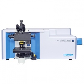
上海巨纳科技有限公司为您提供《金红石,锐钛矿中结晶结构,化学性质检测方案 》,该方案主要用于金红石,锐钛矿中结晶结构,化学性质检测,参考标准《暂无》,《金红石,锐钛矿中结晶结构,化学性质检测方案 》用到的仪器有HORIBA HR Evolution高分辨拉曼光谱仪。
我要纠错
推荐专场

相关方案
-
方案 稀土溶液中La、Ce、Pr、Nd、Sm、Eu、Gd、Tb、Dy、Ho、Er、Tm、Yb、Lu、Y检测方案(ICP-AES)
稀土/稀有金属 | La、Ce、Pr、Nd、Sm、Eu、Gd、Tb、Dy、Ho、Er、Tm、Yb、Lu、Y

-
方案 稀土溶液中La、Ce、Pr、Nd、Sm、Eu、Gd、Tb、Dy、Ho、Er、Tm、Yb、Lu、Y检测方案(ICP-AES)
稀土/稀有金属 | La、Ce、Pr、Nd、Sm、Eu、Gd、Tb、Dy、Ho、Er、Tm、Yb、Lu、Y
- 查看全部方案
 咨询
咨询