方案详情文
智能文字提取功能测试中
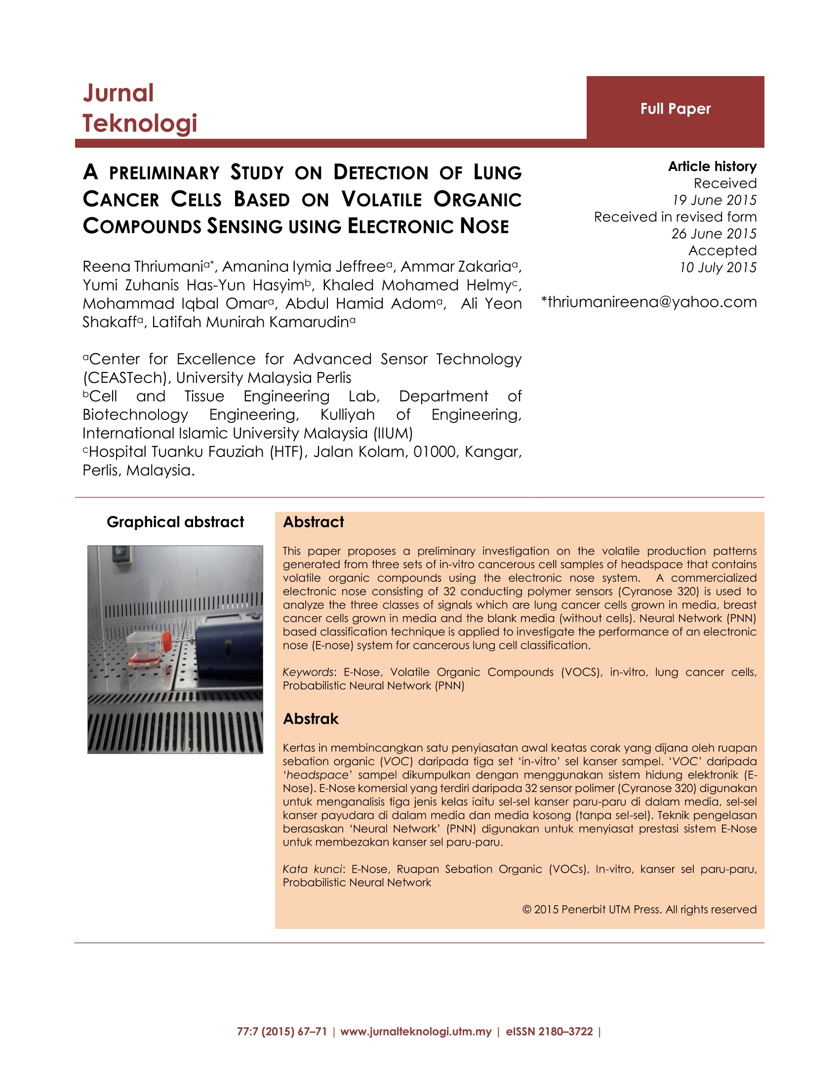
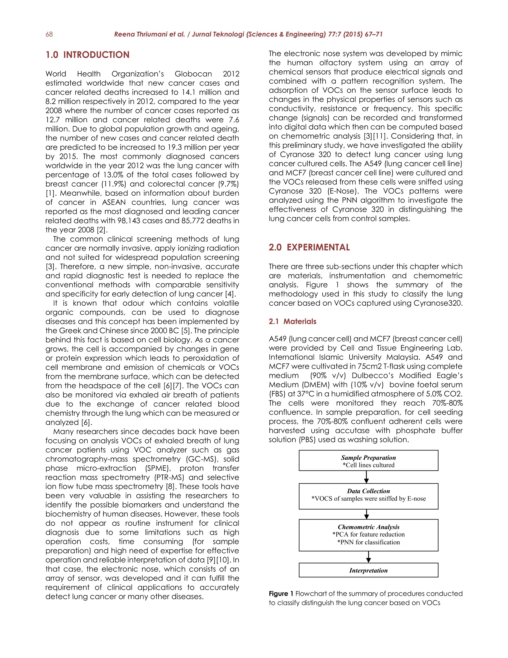
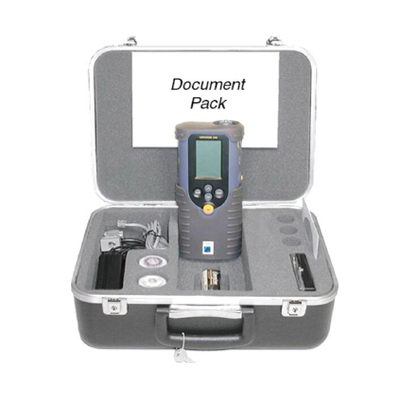
JurnalTeknologiFull Paper Reena Thriumani et al. / Jurnal Teknologi (Sciences & Engineering) 77:7 (2015) 67-7168 77:7 (2015) 67-71|www.jurnalteknologi.utm.my|elSSN 2180-3722| A PRELIMINARY STUDY ON DETECTION OF LUNGCANCERCELLSS BASED ON VOLATILE ORGANICCOMPOUNDS SENSING USING ELECTRONIC NOSE Reena Thriumania*, Amanina lymia Jeffreea, Ammar Zakaria,Yumi Zuhanis Has-Yun Hasyimb, Khaled Mohamed Helmyc,Mohammad labal Omara, Abdul Hamid Adoma, Ali YeonShakaffa, Latifah Munirah Kamarudina ·Center for Excellence for Advanced Sensor Technology(CEASTech),University Malaysia PerlisbCell andTTissue Engineering Lab,DepartmentOTBiotechnology Engineering, KulliyahOTfEngineering,International Islamic University Malaysia (IIUM)cHospital Tuanku Fauziah (HTF), Jalan Kolam, 01000, Kangar,Perlis, Malaysia. Article history Received19 June 2015Received in revised form26 June 2015Accepted10 July 2015 Graphical abstract Abstract This paper pproposes a preliminary investigation on the volatile production patternsgenerated from three sets of in-vitro cancerous cell samples of headspace that containsvolatile organic compounds using the electronic nose system.A commercializedelectronic nose consisting of 32 conducting polymer sensors (Cyranose 320) is used toanalyze the three classes of signals which are lung cancer cells grown in media, breastcancer cells grown in media and the blank media (without cells). Neural Network (PNN)based classification technique is applied to investigate the performance of an electronicnose (E-nose) system for cancerous lung cell classification. Keywords: E-Nose, Volatile Organic Compounds (VOCS), in-vitro, lung cancer cells,Probabilistic Neural Network (PNN) Abstrak Kertas in membincangkan satu penyiasatan awal keatas corak yang dijana oleh ruapansebation organic (VOC) daripada tiga set ‘in-vitro’sel kanser sampel.‘VOC’daripada'headspace'sampel dikumpulkan dengan menggunakan sistem hidung elektronik (E-Nose). E-Nose komersial yang terdiri daripada 32 sensor polimer (Cyranose 320) digunakanuntuk menganalisis tiga jenis kelas iaitu sel-sel kanser paru-paru di dalam media,sel-selkanser payudara di dalam media dan media kosong (tanpa sel-sel). Teknik pengelasanberasaskan ‘Neural Network' (PNN) digunakan untuk menyiasat prestasi sistem E-Noseuntuk membezakan kanser sel paru-paru. Kata kunci: E-Nose, Ruapan Sebation Organic (VOCs), In-vitro, kanser :selparu-paru,Probabilistic Neural Network C 2015 Penerbit UTM Press. All rights reserved 1.0 INTRODUCTION World HealthOrganization's Globocan 2012estimated worldwide that new cancer cases andcancer related deaths increased to 14.1 million and8.2 million respectively in 2012, compared to the year2008 where the number of cancer cases reported as12.7 million and cancer related deaths were 7.6million. Due to global population growth and ageing,the number of new cases and cancer related deathare predicted to be increased to 19.3 million per yearby 2015. The most commonly diagnosed cancersworldwide in the year 2012 was the lung cancer withpercentage of 13.0% of the total cases followed bybreast cancer (11.9%) and colorectal cancer (9.7%)[1]. Meanwhile, based on information about burdenof cancer in ASEAN countries, lung cancer wasreported as the most diagnosed and leading cancerrelated deaths with 98,143 cases and 85,772 deaths inthe year 2008[2]. The common clinical screening methods of lungcancer are normally invasive, apply ionizing radiationand not suited for widespread population screening[3]. Therefore, a new simple, non-invasive, accurateand rapid diagnostic test is needed to replace theconventional methods with comparable sensitivityand specificity for early detection of lung cancer [4]. It is known that odour which contains volatileorganic compounds, can be used to diagnosediseases and this concept has been implemented bythe Greek and Chinese since 2000 BC [5]. The principlebehind this fact is based on cell biology. As a cancergrows, the cell is accompanied by changes in geneor protein expression which leads to peroxidation ofcell membrane and emission of chemicals or VOCsfrom the membrane surface, which can be detectedfrom the headspace of the cell [6][7]. The VOCs canalso be monitored via exhaled air breath of patientsdue to the exchange of cancer related bloodchemistry through the lung which can be measured oranalyzed [6]. Many researchers since decades back have beenfocusing on analysis VOCs of exhaled breath of lungcancer patients using VOC analyzer such as gaschromatography-mass spectrometry (GC-MS), solidphase micro-extraction(SPME), protontransferreaction mass spectrometry (PTR-MS) and selectiveion flow tube mass spectrometry [8]. These tools havebeen very valuable in assisting the researchers toidentify the possible biomarkers and understand thebiochemistry of human diseases. However, these toolsdo not appear as routine instrument for clinicaldiagnosis due to some limitations such as highoperation costs,p,time consuming(for samplepreparation) and high need of expertise for effectiveoperation and reliable interpretation of data [9][10]. Inthat case, the electronic nose,which consists of anarray of sensor, was developed and it can fulfill therequirement of clinical applications to accuratelydetect lung cancer or many other diseases. The electronic nose system was developed by mimicthe human olfactory system using an array ofchemical sensors that produce electrical signals andcombined with a pattern recognition system. Theadsorption of VOCs on the sensor surface leads tochanges in the physical properties of sensors such asconductivity, resistance or frequency. This specificchange (signals) can be recorded and transformedinto digital data which then can be computed basedon chemometric analysis [3][11]. Considering that, inthis preliminary study, we have investigated the abilityof Cyranose 320 to detect lung cancer using lungcancer cultured cells. The A549 (lung cancer cell line)and MCF7 (breast cancer cell line) were cultured andthe VOCs released from these cells were sniffed usingCyranose 320 (E-Nose). The VOCs patterns wereanalyzed using the PNN algorithm to investigate theeffectiveness of Cyranose 320 in distinguishing thelung cancer cells from control samples. 2.0 EXPERIMENTAL There are three sub-sections under this chapter whichare materials,,instrumentation and chemometricanalysis. Figure1showsthesummary ofthemethodology used in this study to classify the lungcancer based on VOCs captured using Cyranose320. 2.1 Materials A549 (lung cancer cell) and MCF7 (breast cancercell)were provided by Cell and Tissue Engineering Lab,International Islamic University Malaysia. A549 andMCF7 were cultivated in 75cm2 T-flask using completemedium (90%v/v) Dulbecco’s Modified Eagle'sMedium (DMEM) with (10% v/v) bovine foetal serum(FBS) at 37°C in a humidified atmosphere of 5.0% CO2.The cellss were monitored they reachi70%-80%confluence. In sample preparation, for cell seedingprocess, the 70%-80% confluent adherent cells wereharvested using accutase with phosphate buffersolution (PBS) used as washing solution. Figure 1 Flowchart of the summary of procedures conductedto classify distinguish the lung cancer based on VOCs A standardized concentration of each cell line (1x10⁵cells/ ml) was seeded into 25cm2 surface treated T-flasks for the E-nose experiment. The samples wereprepared in triplicates and incubated for 72 hours in ahumidified atmosphere of 5% CO2 at 37°℃. The blankmedium (without cells) samples were also prepared intriplicates and incubated together with A549 andMCF7 samples as a control data. 2.2 Instrumentation In this study, the commercially available E-nose,known as Cyrano Science'Cyranose 320 was used.This portable and handheld E-nose contain an arrayof 32 individual polymer sensors blended with carbonblack composite with additional pumps and valves todraw sample of the sensing array. As shown in figure 2,whenthe) sensorsinteractt withthe evolatilecompounds, they swell and lead to changes of thecarbon pathway's conductivity and causeetheresistance value to increase, which can be monitoredas the sensor signal and used to characterize aspecific smell. The Cyranose 320 settings for bothdetermination methods and raw data and classtraining can be stored for additional analysis inWindows based PC using PCnose software [11][12]. 2.3 Sampling Procedure The headspaces of VOCs of the cancerous cell andblank medium in the 25cm2 T-flask were sniffed withCyranose 320. The snout of Cyranose 320 was insertedinto the closed T-25cm2 flasks for 15 minutes. Eachmeasurement was repeated for 3 times. To inspect theVOCs emitted by the cancerous cells at differentconfluencestates,the sampling process wasconducted once every 24th hour (1st day) until 72thhour (3rd day). 2.4 Chemometric Analysis Commercially available softwares; Statistical Packagefor Social Science (SPSS 17) and Matlab (Matwork,Natrick, MA) were used to process the raw data andidentify the effectiveness to improve the effectivenessof E-nose system performance in detecting andidentifying the lung cancer cell.l.Table 1 show theparameters used in theedata analysis andclassification. a) Dimensionality Reduction The 10 end points of the steady state value (Rss) thatcan be expressed as the stationary values reachedduring the measurement were extracted from theeach response. The high dimensionality of features inthe raw data obtained can cause an increase in timeand space required for data processing. Therefore,the unsupervised Principal Component Analysis (PCA)techniques were used to improve the accuracy ofclassification by reducing its dimension while retainingthe important features in different basis [13]. Based on Kaiser's rule, the principal components witheigenvalues greater than 1.0 are selected and usedas input of the classifier in this study. Table 1 shows thenumber of principal component selected for eachdata [14]. Figure 2 Snout of Cyranose 320 Inserted Into T-25cm2 FlaskContaining Cell Culture b) Classification In this work, a well applied algorithm in patternrecognition, probabilistic neuralnetwork (PNN)classifier is used to classify lung cancer from controlsamples.5.PNN neuralnetwork algorithmWasdeveloped by SSpechtin1989 with a simpleconstruction and wide application [15]. PNN whichdefined as an implementation of Kernel discriminantanalysis contains operations which are organized intomulti-layered feed forward network with four layerswhich are input layer, hidden layer, summation layerand output layer [16] as shown in figure 3. AlthoughPNN algorithm required large memory for training, it iswidely used as classifier in many applications since itrequired less training time, able to learn quickly ascompared to other than several neural networksmodel and able to meet to the optimal classifier as thesize of the representative training set increases andtraining samples can be added or removed withoutextensive retraining [16][17]. Thus, PNN is describedand considered as a capable algorithm to be used aslung cancer classifier in this paper. The spread parameter of PNN algorithm, a, is crucialto obtain accurate classification. This is because,o oftoo small value lead to a solution that does notgeneralize from the input vectors while o of too largevalue can result in large output value of radial basisneurons for all inputs used in the design of the network.In that case, determinations of o value with the rangeof 0.1 to 0.9 were conducted for each data set toobtain an appropriate o with respect to classificationaccuracy[18]. Table 1 shows the number of principalcomponent selected for each data. The 10 end points of the steady state value (Rss) thatcan be expressed as the stationary values reachedduring the measurement were extracted from theeach response. The high dimensionality of features inthe raw data obtained can cause an increase in timeand space required for data processing. Therefore, the unsupervised Principal Component Analysis (PCA)techniques were used to improve the accuracy ofclassification by reducing its dimension while retainingthe important features in different basis [13]. Figure 3 The diagram of PNN built with 4 layers 3.0 RESULTS AND DISCUSSION In this preliminary study, we used Cryanose 320 E-noseto detect lung cancer cells from breast cancer cellsand blank medium with the aid of PNN classifier. Thedata collected once every 24 hours for 3 days andgrouped into 3 classes which are A549 (lung cancercell line), MCF7 (breast cancer cell line) and blankmedium. The PCA wasusedto reduce thedimensionality of data and speed up the evaluationbefore PNN classification was conducted. All of theexperimental results were averaged over several runsof randomly generated 60/40, training/test splits of thedata. The spread factor of PNN, which range startingfrom 0.1 to 0.9 is conducted on each 4 dataset. Table 2 demonstrated the result of accuracy,sensitivity and specificity of a PNN classifier ondetecting VOCs of lung cancer cell from breastcancer cell and blank medium. All the dataset arechecked through spread factor and the results showthat outputs successfully rated over 99% of all spreadvalues. However, for spreading factor values 0.7, theoutput matched highest percentage for all 4 datasets.The percentage ofaccuracy, sensitivity' andspecificity on day 1, day 2 and day 3 matched 100%respectively when used spread value 0.7. Table 1 Properties and parameters of dataset for PNNalgorithm Parameters Dataset Day 1 Day 2 Day 3 3 days Total Number of Features 32 32 32 32 Total Samples 450 450 450 1350 After PCA (Features) 5 4 4 5 Training Samples 5x270 4x270 4x270 5x810 Testing Samples 5x180 4x180 4x180 5x540 Classes 3 3 3 3 The accuracy of data from a combination of all 3 daysshows 99.98±0.15%, while the sensitivity and specificityrecords 100% and 99.98±0.23% respectively. For spread value 0.3, the percentage of accuracy,sensitivity and specificity were similar with the outputsfrom spread value, 0.7; however the percentage ofspecificity of data on a combination of all data werelesser 0.01% than spread value 0.7.y. In nutshell, it can be said that the testing of lungcancer cell detection using an electronic nose systemwith the aid of PNN classifier was done successfully.These studies show that this method able to rapidlyand effectively discriminates the cancer cell typesand also from the control which is served as baselineresponse. The PNN classifier with spread value 0.3 and0.7 can be used for the best performance as it isproved to be a fast learning algorithm and does notneed an iterative training process. Table 2 The accuracy, sensitivity and specificity of PNNclassification Considering that, the spread value, a,0.3 and 0.7 canbe used for all the datasets to achieve efficient lungcancer classification system. The PNN analysis appliedin this study reached 100% classification and thesephenomena might occur because of the dataset wasrelatively small and the variability associated with theclassification is still high. Therefore, for more accuratequantification of the general performance of PNNusing e-nose data, further experimentation with largerdatasets is necessary [191. 4.0 CONCLUSION Thiss study proposed a lung cancer cellllVOCsdetection system which consists of a commerciallyavailable electronic nose (Cyranose 320) and patternrecognition algorithm (using PNN). The results provedthat this proposed system is able to detect lungcancer VOCs by achieving over 99% of classificationaccuracy. Although the current pattern classificationtechnique produces pleasing results, but further studymay improve the classification accuracy and speedof the operations by using different types of pre-processing and classification methods. Future work willbe extended to a larger number of samples where thenormal lung cancer cell lines and other types of lungcancer cell lines beside A549 will be investigated. Acknowledgement This work is supported by Exploratory Research GrantScheme by Ministry of Education Malaysia (ERGS 9010-00018). The author would like to thank the Animal Celland Tissue Culture Lab, Department of BiotechnologyEngineering, International Islamic University Malaysiaand Hospital Tuanku Fauziah, Kangar, Perlis for thegreat collaboration. References ( [1] Ferlay.J, Soerjomataram.J, , Ervik.M, D ikshit.R , E Eser.S, Mathers.C, R ebelo.M, Parkin.DM, Forman.D, a n d Br a y.F. 2013. Cancer I ncidence a nd Mortality Worldwide: IARC Cancer Base GLOBOCAN 2012 Cancer Incidence and Mortality Worldwide: IARC Cancer Base.(11)11. ) ( [2] M. K imman, R. N orman, S . Jan, D . K ingston, and M. Woodward. 2 012. The Burden of Cancer in MemberCountries of the Association of Southeast A sian N ations (ASEAN). Asian Pacific J. C ancer Prev. 13(2): 411-420. ) [3] C. Brunner, W. Szymczak, V. Hollriegl, S. Mortl, H. Oelmez,aBergner, R. M. Huber, C.Hoeschen, and U.Oeh. 2014.Discrimination Of Cancerous And Non-Cancerous CellLines By Headspace-Analysis With PTR-MS. Anal. Bioanal.Chem.(397)6:2315-2324. ( [4] U. Kallur i ,M. Naiker , and M. A Myers. 2014. Cell CultureMetabolomics I n T he D iagnosis O f Lung Cancer-TheInfluence Of Cell C ulture Conditions. J . Breath Res. (8)2: 1 - 10. ) [5] R. Fend, A.H. J. Kolk, C. Bessant, P. Buijtels, P. R. Klatser, andA. C. Woodman,2006. Prospects For Clinical ApplicationOfElectronic-NoseTechnology To EarlyDetectionn OfMycobacterium Tuberculosis In Culture And Sputum. JJ.Clin. Microbiol. (44) 6:2039-2045. [6] G. Peng, M. Hakim, Y. Y. Broza,S. Billan, R. Abdah-Bortnyak,a Kuten, U. Tisch, and H. Haick, 2010. Detection Of Lung,Breast, Colorectal, And Prostate Cancers From ExhaledBreath Using A Single Array Of Nanosensors. Br. J. Cancer(103)4:542-551. [7] D. A. P. Daniel, K. Thangavel, and R. S. C. Boss.2012. AReview Of Early Detection Of Cancers Using BreathAnalysis. Int. Conf. Pattern Recognition, Informatics Med.Eng. Mar. 433-438. ( [8] R. Thriumani, A. Zakaria, M. 1 . Omar, A. H . A dom, A. Y. M . Sharaff, L . M. Kamaruddin, N . Yusuf, and K. M. Helmy. 2014.Review On Exhaled Volatil e Organi c Compounds FromLung Cancer And A d vances Of E-Nose As No n -Invasive Detection Method. IEEE 5th Control and System GraduateResearch Colloquium.213-218. ) ( [9] Manolis.A. 1983. The Diagnostic Po tential Of Breath Analysis. Clin. Chem. 29:5-15. ) ( [10] A. D. Wilson and M. Baietto. 2011.Advances In Electronic- Nose Technologies Developed For Biomedical ApplicationsSensors (Basel.) (11)1:1105-1176. ) ( [11] T.Mohamed, A. Sabeel,S.E. Eluwa,and S. Mohamed.2000. Detection of Volatile Compounds in Urine.3-6. ) ( [12] R. Dutta, E. L. Hines, J . W. G ardner, a n d P. Bo i lot. 2002. Bacteria Classification Using Cyranose 320 Electronic Nose.Biomed.Eng. Online.1: 4. ) [131 R. Palaniappan and K. V. R. Ravi. 2006. Improving VisualEvokeddPotential FFeatureClassificationForPersonRecognition Using PCA AndNormalization. PPatternRecognit. Lett. 27(7): 726-733. [14] Jolliffe.l.T. 2002. Principal Component Analysis. SpringerSeries in Statistics. [15] C. Ji. D, F. Song. B, and H. Yi. H. 2009. Equipment FaultDiagnosis based on Probabilistic Neural Networks andSimulation Analysis. Fire Control Command Control.34:82-85. ( [16] M. M ishra, A. R. Jena, a nd R. Das, 2013. A P r obabilistic Neural Network A p proach for Classification o f V e hicle. 2(7):367-371. ) ( [171 N. Bhattacharyya and A. Jana 2 008. Incremental PNN Classifier for a Versatile Electronic Nose 242-247. ) ( [18] R. Antony, M. S. G. Nandagopal, S. Rangabhashiyam,andN. Selvaraju.2014. Probabilistic N eural Network Prediction Of L iquid- Liquid Two Phas e Flows I n A C ircularMicrochannel. J. Sci. Ind.Res.73:525-529. ) ( [19] H. Zhang, M . O . B a laban, an d J. C . P rincipe. 20 0 3.Improving P attern R ecognition Of Electronic N ose D a ta With Time-Delay Neural N etworks. Sensors Actuators BChem.96(1-2): 385-389. ) 电子鼻系统是利用一系列产生电信号的化学传感器模拟人类嗅觉系统,并与模式识别系统相结合而开发的。VOC在传感器表面的吸附导致传感器物理性能的变化,如电导率、电阻或频率。这种特定的变化(信号)可以被记录下来并转换成数字数据,然后根据化学计量学分析进行计算[3][11]。鉴于此,在这项初步研究中,我们研究了Cyranose 320利用肺癌培养细胞检测肺癌的能力。培养A549(肺癌细胞系)和MCF7(乳腺癌细胞系),并用Cyranose 320(电子鼻)嗅探这些细胞释放的VOC。利用PNN算法对VOCS模式进行了分析,研究了Cyranose 320对肺癌细胞与对照细胞的鉴别效果。本章分为三个小节,分别是材料、仪器和化学计量学分析。
关闭-
1/5

-
2/5

还剩3页未读,是否继续阅读?
继续免费阅读全文产品配置单
图拉扬科技有限公司为您提供《血细胞中VOCs检测方案(感官智能分析)》,该方案主要用于全血/血清/血浆中VOCs检测,参考标准《暂无》,《血细胞中VOCs检测方案(感官智能分析)》用到的仪器有Cyranose 320 电子鼻。
我要纠错
相关方案


 咨询
咨询


