脑动脉瘤模型中流动特性,流场,速度矢量场,速度场检测方案(粒子图像测速)
检测样品 全血/血清/血浆
检测项目 流动特性,流场,速度矢量场,速度场

 金牌会员
752 篇解决方案
金牌会员
752 篇解决方案
方案详情文
智能文字提取功能测试中
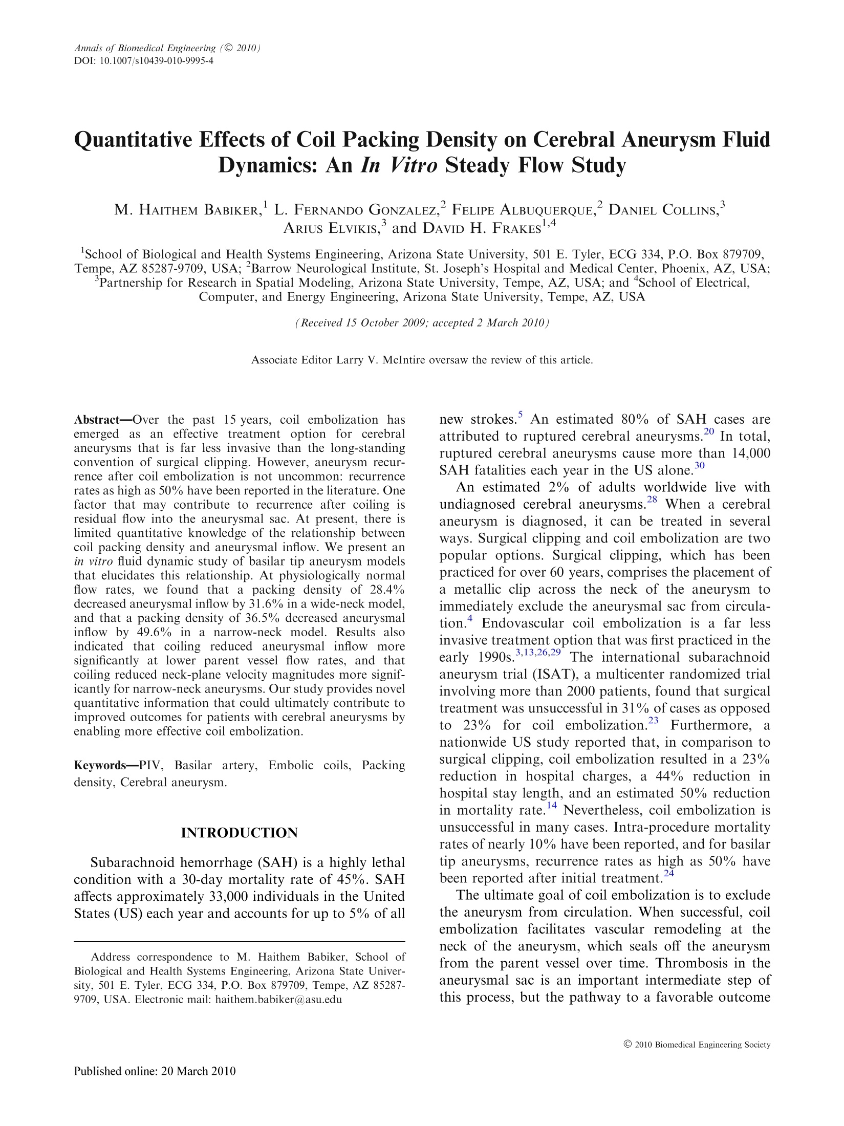
Annals of Biomedical Engineering (@ 2010)DOI: 10.1007/s10439-010-9995-4 BABIKER et al. C 2010 Biomedical Engineering SocietyPublished online: 20 March 2010 Quantitative Effects of Coil Packing Density on Cerebral Aneurysm FluidDynamics: An In Vitro Steady Flow Study M. HAITHEM BABIKER, L. FERNANDO GONZALEZ,’FELIPE ALBUQUERQUE,’DANIEL COLLINS,’ARIUs ELVIKIS.and DAVID H. FRAKES1,4 School of Biological and Health Systems Engineering, Arizona State University, 501 E. Tyler, ECG 334, P.O. Box 879709,Tempe, AZ 85287-9709,USA;Barrow Neurological Institute, St. Joseph’s Hospital and Medical Center, Phoenix, AZ, USA;Partnership for Research in Spatial Modeling, Arizona State University, Tempe, AZ, USA; and School of Electrical,Computer, and Energy Engineering, Arizona State University, Tempe, AZ, USA (Received 15 October 2009; accepted 2 March 2010) Associate Editor Larry V. McIntire oversaw the review of this article. Abstract-Over the pastst15 years, coil embolization hasemerged ass2an effective treatment option for cerebralaneurysms that is far less invasive than the long-standingconvention of surgical clipping. However, aneurysm recur-rence after coil embolization is not uncommon: recurrencerates as high as 50% have been reported in the literature. Onefactor that may contribute to recurrence after coiling isresidual flow into the aneurysmal sac. At present, there islimited quantitative knowledge of the relationship betweencoil packing density and aneurysmal inflow. We present anin vitro fluid dynamic study of basilar tip aneurysm modelsthat elucidates this relationship. At physiologically normalflow rates, we found that a packing density of 28.4%decreased aneurysmal inflow by 31.6% in a wide-neck model,and that apacking density of 36.5% decreased aneurysmalinflow by 49.6% in a narrow-neck model. Results alsoindicated thatcoiling reduced aneurysmall inflow moresignificantly at lower parent vessel flow rates, and thatcoiling reduced neck-plane velocity magnitudes more signif-icantly for narrow-neck aneurysms. Our study provides novelquantitative information that could ultimately contribute toimproved outcomes for patients with cerebral aneurysms byenabling more effective coil embolization. Keywords-PIV, Basilar artery, Embolic coils, Packingdensity, Cerebral aneurysm. INTRODUCTION Subarachnoid hemorrhage (SAH) is a highly lethalcondition with a 30-day mortality rate of 45%. SAHaffects approximately 33,000 individuals in the UnitedStates (US) each year and accounts for up to 5% of all ( Address correspondence to M. H aithem B a biker, S c hool o f Biological and Health Systems E ngineering, Arizona S tate Univer- sity, 50 1 E . Tyler, ECG 334, P.O. Box 879709, Tempe, AZ 85287-9709, USA. E l ectronic mail: h aithem.babiker@asu.edu ) new strokes. An estimated 80% of SAH cases areattributed to ruptured cerebral aneurysms.20 In total,ruptured cerebral aneurysms cause more than 14,000SAH fatalities each year in the US alone. An estimated 2% of adults worldwide live withundiagnosed cerebral aneurysms.28When a cerebralaneurysm is diagnosed, it can be treated in severalways. Surgical clipping and coil embolization are twopopular options. Surgical clipping, which has beenpracticed for over 60 years, comprises the placement ofa metallic clip across the neck of the aneurysm toimmediately exclude the aneurysmal sac from circula-tion.4 Endovascular coil embolization is a far lessinvasive treatment option that was first practiced in theearly 1990s.3.13,26,29The international subarachnoidaneurysm trial (ISAT), a multicenter randomized trialinvolving more than 2000 patients, found that surgicaltreatment was unsuccessful in 31% of cases as opposedto23%for coili embolization.23Furthermore.anationwide US study reported that, in comparison tosurgical clipping, coil embolization resulted in a 23%reduction in hospital charges, a 44% reduction inhospital stay length, and an estimated 50% reductionin mortality rate.14 Nevertheless, coil embolization isunsuccessful in many cases. Intra-procedure mortalityrates of nearly 10% have been reported, and for basilartip aneurysms, recurrence rates as high as 50% havebeen reported after initial treatment.24 The ultimate goal of coil embolization is to excludethe aneurysm from circulation. When successful, coilembolization facilitates vascular remodeling at theneck of the aneurysm, which seals off the aneurysmfrom the parent vessel over time. Thrombosis in theaneurysmal sac is an important intermediate step ofthis process, but the pathway to a favorable outcome begins with the immediate goal of coil embolization: toeliminate aneurysmal inflow by occluding the aneu-rysm. When embolization is incomplete, there isresidual flow from the parent vessel into the aneurysm,which may be one factor that contributes to recur-rence. Accordingly, post-treatment aneurysmal inflowis a highly relevant clinical parameter. Aneurysmal inflow is evaluated during treatmentbased on visual inspection of dynamic angiograms.This evaluation serves as the short-term measure ofembolic success or failure in the procedure room.When little or no contrast agent can be visualized..entering the aneurysm, embolization is considered tobe complete. This is a qualitative exercise, however,and embolizations that are classified as complete basedon angiographic inspection may correspond to a broadrange of aneurysmal inflows. Once a procedure iscomplete, follow-up examinations may include mag-netic resonance and/or conventional angiography, butboth modalities are qualitative in this context and lackthe capability to quantify aneurysmal inflow. Quantitative knowledge of the relationship betweencoil packing density and aneurysmal inflow couldmitigate evaluation uncertainty and facilitate morecomplete embolization. Clinical studies have correlatedhighppacking densityywwith (decreased! aneurysmalrecurrence, but have never quantified the relationshipbetween packing density and aneurysmal inflow.27.33Numerous studies have investigated the effects of coilembolization on aneurysmal fluid dynamics in silicousing computationalfluid ddynamicsS((CFD).6,8.22However, authors of those studies have reported thatcoiled aneurysm fluid dynamics are extremely difficultto simulate well because of highly complex post-deployment coil geometries and fluid structure inter-actions.8,22 Experts in the field have also noted the shortcom-ings of previous experimental work, and have recog-nized1 a need for different methods.34sSpecifically,previous experimental studies have been both limitedand primarily qualitative.2,7,12,15,25,31,32Flow visuali-zation was used qualitatively in several studies toobserve post-embolization reductions in aneurysmalinflow.12,15 Other ex vivo and in vivo studies havefocused on intra-aneurysmal pressure measurements tocharacterize changes in fluid dynamics after coildeployment.2,25Sorteberg et al. used pressure andtemperature measurements at a single point inside theaneurysm1to quantify local flow variationsi nidi-rectly.31,32Jou et al. also quantified fluid dynamicchanges indirectly based on the visualization of con-trast agent flow with fluoroscopy.Canton et al. usedtwo-dimensional (2D) particle image velocimetry (PIV)as a qualitative tool to visualize redirected flow at theaneurysmal neck after coiling. However, none of these studies measured post-embolization flows directly, nordid they quantify post-embolization fluid dynamics inrelation to coil packing density. We present an in vitro steady flow study of the fluiddynamics in coiled cerebral aneurysms that quantifiesthe relationship between coil packing density andaneurysmal inflow using three-dimensional (3D) stereoPIV. The study investigates a type of aneurysm that isparticularly challenging to embolize, the basilar tipaneurysm, through parametric idealized models. Thiswork is the initial phase of an ongoing effort toestablish a foundation for understanding coiled aneu-rysm fluid dynamics through experimentation withparametric idealized and anatomical models understeady and pulsatile flow conditions. Our ultimate goalis to facilitate improved outcomes for patients withcerebral aneurysms. To our knowledge, this studyrepresents the first time that the relationship betweencoil packing density and aneurysmal inflow has beenquantified, or that 3D PIV has been used to image flowin aneurysm models treated with endovascular coils. METHODS Idealized computational models comprising basilarartery bifurcations and basilar tip aneurysms weredesigned in Solidworks (Solidworks, Concord, MA,USA). The models were designed under physiciansupervision and were based on in vivo angiographicimages of basilar tip aneurysm anatomies. One suchimage is shown in Fig. 1a. In each model, the main axisof each outlet vessel was oriented to the main axis ofthe parent vesselelat a 60° angle. Four millimeterdiameters were specified for the parent vessel andoutlet vessels, and a 4 mm diameter spherical aneurysmwas attached at the parent vessel bifurcation. Differentaneurysmal neck-plane sizes were achieved by varyingthe overlap between the spherical aneurysms and theparent vessel bifurcations. Overlaps of 0.5 and 1 mmwere specified for narrow-neck and wide-neck models, FIGURE 1. In vivo angiographic image of basilar tip aneu-rysm (a), wide-neck computational model with neck-planeindicated in black (b), and wide-neck physical model duringexperimentation (c). respectively. These correspond to aneurysmal neck-plane diameters of 2.64 mm (66.0% of the aneurysmaldiameter) and 3.46 mm (86.5% of the aneurysmaldiameter). The neck-plane represents the boundary thatfluid must cross to enter the aneurysm. It is defined hereas the smallest circular cross section of the aneurysmalsphere that shares boundaries with the parent vessel. Forclarity, weuse the term“neck-plane”to refer only to thiscircular cross section, and not to the entire geometricplane that it resides within. In Fig. 1b, a wide-neckcomputational model is shown and the projection of theneck-plane onto the model surface is indicated by theblack line. In each model, an entrance length of 8 cm(20 vessel diameters) was included upstream of thebifurcation to ensure that flow was fully developedbefore entering the bifurcation region. Each computational model was used to construct aphysical core model from pot metal with computernumerical controlled (CNC) cutting (Shelley Medical,Toronto, ON, Canada). Transparent physical modelswere molded around the pot metal cores with Sylgard184 silicon elastomere (Dow Corning, Midland, MI,USA), and the cores were then melted out. A wide-necktransparentlost-core physical model is shown in Fig. 1c. The transparent models were connected to a flowloop with flexible polyvinyl chloride tubing. Equalvascular resistances were imposed at each model out-let. A blood analog working fluid composed of water,glycerol,and aqueous sodium iodide was configured toachieve a refractive index of 1.43, matching that ofSylgard 184, and a dynamic viscosity of 3.40 cP at anoperating temperature of 25 °C. The working fluid wasseeded with 8 um fluorescent Rhodamine-B particles(Thermo Scientific, Waltham,MA, USA) that experi-ence peak excitation and emission at 542 and 618 nm,respectively. Flow through the loop was driven bya Compuflow 1000 piston pump (Shelley Medical,Toronto,ON, Canada). Data were acquired with aFlowmaster 3D stereo PIV system (Lavision, Ypsi-lanti, MI,USA). Flow within the models was illumi-nated by a 0.5 mm thick laser sheet from a 532 nm SoloPIV III dual cavity pulsed YAG laser (New WaveResearch, Fremont, CA, USA). Two Imager Intensecross-correlation CCD cameras were used to acquireimages with matrix sizes of 1376 by 1040 and pixeldimensions of 9.88 um square. Low-pass optical filterswith a 572 nm cutoff (Omega Optical, Brattleboro,VT, USA) were installed on both cameras. Those filtersallowed the fluorescent seed particles to be imagedeffectively despiteeintense laser reflections fromdeployed coils. Axium bare platinum detachable coils (EV3, Plym-outh, MN, USA) were used to embolize aneurysmswithin the transparent models. To facilitate deploy-ment, microcatheters were integrated into the flow loop both upstream and downstream of the model interface.A balloon-assisted technique was used during coildeployment to prevent coil herniation into the parentvessel, as shown in Fig. 2a. Coilss were deployedaccording to standard clinical practice. ThisWasaccomplished through the initial deployment of a longcoil to create a basket for further coil deployments.Smaller coils were then sequentially deployed to achieveincreasing packing densities until no further coilscould be deployed without herniation. A clinical “coilpusher” tool was also used after deployment, whennecessary, to ensure that the coil mass was orientedinternal to the aneurysm and that the aneurysmal neck-plane region could be visualized clearly. Data wereacquired after each deployment. An example of a coilpacking sequence is shown in Figs. 2b-2d. Coil packingdensity was calculated analytically and is expressed inour results as the percentage of aneurysmal sac volumeoccupied by the deployed coils. The coil volumes usedin calculation were based upon manufacturer’s specifi-cations. Steady flow rates of 3 and 5 mL/s were inves-tigated for each different packing density in bothmodels. These flow rates were chosen to span a physi-ologic range of normal and diseased conditions.s.1l66Three planes within the fluid domain were examined foreach combination of experimental parameters: thecenter plane defined by the parent and outlet vessel FIGURE 2. Demonstration of balloon-assisted coil deploy-ment technique (a) and progressive stages of a coil deploy-ment sequence in a narrow-neck basilar tip aneurysm model:0% packing density (b), 19.74% packing density (c), and28.09% packing density (d). main axes, and the two planes displaced orthogonallyfrom the center plane by 0.5 mm. At each of theseplanes, 130 image pairs were acquired at a rate of 5 Hz,with a time separation of 20 us between images in apair. Velocity vectors were then calculated throughcross-correlation within DaVis software (Lavision,Ypsilanti, MI, USA). Interrogation windows of pro-gressively smaller dimensions were used down to 32 by32 pixels with a 25% overlap between neighboringwindows. Results from each of the 130 image pairs werethen averaged to determine a single flow velocity vectorfield for each plane. RMS errors associated with theaveraging process were carefully monitored to ensurethat no significant transient flows were present. The volumes of acquired velocity data were thenanalyzed to quantify cross-neck flow, defined as thecumulative flow volume crossing the neck-plane of theaneurysm per second, and RMS velocity magnitude atthe neck-plane of the aneurysm, calculated as: where n is the number of data points within theaneurysmal neck-plane andV; indicates the flowvelocity magnitude at point i. It is noteworthy that theacquired flow velocity volumes did not span the entireaneurysmal neck-plane in either model configuration,but did span 68.1 and 53.6% of the neck-plane in thenarrow and wide-neck models, respectively. Further-more, each acquired volume was symmetric about themain axes of the parent vessel and outlet vessels, andincluded the region of the aneurysmal neck-planetoward which the highest magnitude flow velocitieswere directed from the parent vessel. Accordingly, weare confident that these flow velocity volumes arehighly indicative of the overall fluid dynamics at thecorresponding neck-planes of the aneurysm models. For 3D streamtracing and visualization, a modifiedcontrol grid interpolation (MCGI) was used to recon-struct isotropically resolved 3D velocities throughoutthe fluid domain spanned by each set of three acquiredvelocity data planes. MCGI is an established algo-rithm for fluid flow velocity data reconstruction thatperforms with high accuracy while minimizing diver-gence errors. Displacement fields linking similar fluidstructures in neighboring velocity magnitude imageswere determined by minimizing: where E is the optical flow-based error associated withthe displacement field defined by control point row and column displacements and B, I is an image com-prising flow velocity magnitudes, the vector n=(n, n2) denotes image matrix coordinates, R is theregion of support, and 0 and o represent basis func-tions that implement a bilinear interpolation to pop-ulate a complete displacement field based on u and p.Sets of displacement fields were then used to facilitatethe directional interpolation of individual velocitycomponents, whereby an isotropically resolved volumeof 3D flow velocity vectors was reconstructed for eachacquired data set. This framework reduces computa-tional requirements in comparison to pure optical flow,at a small expense in terms of displacement field flex-ibility. More information can be found in the litera-ture.9-11 RESULTS Consistent flow across the neck of the aneurysm wasobserved in the uncoiled models, as shown in Fig. 3where vector plots corresponding to different parentvessel flow rates are overlaid on a wide-neck model. Inthat figure, we have chosen to invert the conventionalcolor map so as to display greater contrast in regionsof the vector field characterized by smaller flowvelocity magnitudes. Figure 3 also displays the largevortices that formed near the center of the aneurysmalsac in the uncoiled models. which are a commonfinding in the literature.12,19,21 After coil deployment,consistent reductions in cross-neck flow were observed.For both models, and for all flow rates examined,increased packing density led to decreased cross-neckflow. As shown in Fig. 4, every incremental increase inpacking density was accompanied by a decrease incross-neck flow. A final packing density of 36.5% in anarrow-neck model led to cross-neck flow decreases of49.6 and 30.5% for parent vessel flow rates of 3 and5 mL/s, respectively. A final packing density of 28.4%in a wide-neck model led to cross-neck flow decreasesof 31.6and 27.3% for parent vessel flow rates of3 and5 mL/s, respectively. These results can also be appre-ciated qualitatively in Fig. 5, where several features ofthe flow field corresponding to the more denselypacked aneurysm are consistent with decreased flowacross the neck of the aneurysm. Specifically,the cross-neck components of the flow vectors at the neck-planein Fig. 5b are visibly shorter than those in Fig. 5a(indicating more significant occlusion in Fig. 5b), andthe high-velocity jet oriented along the main axis of theparent vessel terminates further upstream in Fig. 5b ascompared to Fig. 5a. Another visual representation ofthe cross-neck flow reduction realized through coilingis provided in Fig. 6, where the respective 3D stream-traces correspond to pre- and post-treatment fluid Velocity Magnitude (m/s) Velocity Magnitude (m/s) FIGURE 3. Velocity magnitude vectors within the center plane of flow velocity data acquired from an uncoiled wide-neck basilartip aneurysm model at parent vessel flow rates of 3 (a) and 5 mL/s (b). FIGURE 4. (Cross-neck flow rate (mL/s) vs. coil packingdensity (%). dynamic conditions in a narrow-neck model. In thatfigure, both sets of streamtraces originate from iden-tical locations within the model center-plane, spreaduniformly across the inlet of the bifurcation region. Similar trends were observed with respect to RMSvelocity magnitude at the neck-plane. As shown inFig. 7, all increases in packing density also led todecreases in RMS velocity magnitude at the neck-plane, with two exceptions. A final packing density of 36.5% in a narrow-neck model led to RMS velocitymagnitude decreases of 34.7 and 23.1% for parentvessel flow rates of 3 and 5 mL/s, respectively. A finalpacking density of 28.4% in a wide-neck model led toRMS velocity magnitude decreases of 7.7 and 16.1%for parent vessel flow rates of 3 and 5 mL/s, respec-tively. These results can also be appreciated qualita-tively in Fig. 5, where the color-coding indicatesdecreased flow velocity magnitudes at the neck-planefor the more densely packed case. In order to char-acterize inter-acquisition variations in our results,six repeat data acquisitions were performed for anuncoiled narrow-neck model at a steady parent vesselflow rate of 3 mL/s. Cross-neck flow rates variedfrom the mean by an average of 4.89% with a stan-dard deviation of 3.31% of the mean. RMS velocitymagnitudes at the neck-plane varied from the meanby an average of 3.77% with a standard deviation of2.43% of the mean. DISCUSSION AND CONCLUSIONS The most direct conclusion from our results is thatcoil embolization significantly reduces both aneurys-mal inflow and RMS velocity magnitude at the neck ofthe aneurysm. However, our results also indicate thateven very dense coil packings, by clinical standards, donot embolize completely. In the wide-neck model FIGURE 5.Velocity magnitude vectors within the center plane of flow velocity data acquired from a narrow-neck basilar tipaneurysm model at packing densities of 19.74% (a) and 32.27% (b) for a parent vessel flow rate of 3 mL/s. configuration for example, as much as 72.7% ofthecross-neck flow present before coiling persisted at thehighest packing density investigated. This suggests thatcomplete embolization, as defined by current clinicalevaluationnmethods., mayaallow more significantaneurysmal inflow than would be clinically expected. Two other important conclusions relate specificanatomical and physiologic parameters to post-treat-ment fluid dynamics. First, our results indicate thatcoiling reduces aneurysmal inflow more significantly atlower parent vessel flow rates. This finding is consistentwith a previous study that evaluated post-treatmentaneurysmal inflow.Second, our results indicate thatcoiling reduces RMS velocity magnitude at the neck-plane more significantly, and more consistently, fornarrow-neck aneurysms. We propose that coil embo-lization may be more effective in establishing a flowstagnation point at the neck-plane for these types ofaneurysms, which leads to more significant reductionsin flow velocity magnitude at the neck. Results also showed that the ratio of cross-neck flowreduction to incremental increase in packing density isgreater athigher packing densities. Simply put,reductions in aneurysmal inflow per volume unit of coiladded were greater for the smaller coils deployed lastin the coiling sequence, as compared to the largest coilsdeployed first. This indicates that deployment of thelast coil in a sequence can be very important forachieving acceptably low aneurysmal inflow rates, even though that coil may change packing density by asmall amount. These novel quantitative results supportclinical findings that high packing density contributesto decreased aneurysmal recurrence through morecomplete embolization.27,33 There are several ways that quantitative knowledgeof the relationship between coil packing density andaneurysmal inflow could facilitate improved outcomesfor patients treated with coil embolization. First,neurosurgeons could target an optimal, patient-specificpacking density prior to intervention that would limitaneurysmal inflow to an acceptable level. This wouldencourage the deployment of a sufficient coil volume, ifpossible, and could reduce the number of proceduresresulting in incomplete embolization. A target density....would also provide a landmark for neurosurgeonsindicating when to stop packing an aneurysm withcoils, which would decrease both prolonged anesthesiarisks and the risk of aneurysmal rupture due to over-packing. Finally, if a target density could not beachieved in practice, then clinicians would be aware ofincreased recurrence risk, and could monitor the pa-tient more closely or pursue alternative treatment. Several limitations of this study are noteworthy.First, because every coil deployment is different, fluiddynamics may vary significantly between two differentdeployments that achieve the same packing density.Unfortunately, the high cost and limited availability ofcoils prohibit extensive repeat experimentation. We felt FIGURE6.Velocity magnitude color-coded 3D streamtraceswithin reconstructed flow velocity volumes from a narrow-neck basilar tip aneurysm model at 0% packing density (un-treated case: bluelgreen) and at 32.27% packing density(treated case: yellow/red) for a parent vessel flow rate of 3 mL/s. Both sets of streamtraces originate from identical locationswithin the model center-plane, spread uniformly across theinlet of the bifurcation region. FIGURE 7. Neck-plane RMS velocity magnitude (m/s) vs. coilpacking density (%). that instead of repeating experiments for a few packingdensities in one model, our available coil resourceswould be better used in this initial study by exploring abroad range of packing densities in different models.However, one component of future work will be to quantify variations in fluid dynamic results amongdifferent deployments that achieve the same packingdensity. Another limitation of this study is that, within theaneurysmal sac, coils occlude the particles that mustbe imaged to perform PIV. Accordingly, PIV cannotbe used to measure velocities in the aneurysmal sacdirectly after dense coiling. Instead, we chose to mea-sure velocities at the neck-plane of the aneurysm,whichrelatee(directlvytoointra-aneurysmalfluiddynamics since all flow entering the aneurysmal sacmust cross the neck-plane. To avoid any problems withocclusion at the neck-plane, a clinical “coil pusher”tool was used to ensure that coils were sufficientlydisplaced from the neck-plane to allow for accuratevelocity measurements to be taken there with PIV. This study is also limited in that we explored onlyidealized models and varied a small number of physi-ologic and geometric parameters in those models.Physiologically, we focused on models with unregu-lated compliance under steady flow conditions. Geo-metrically, we focused on varying aneurysmal neck sizeand coil packing density, but there are many othergeometric parameters that affect flow conditions incoiled aneurysms. For example, previous computa-tional studies have reported that the angle between themain axes of the parent vessel and aneurysm canstrongly influence fluid dynamics in basilar tip aneu-rysms.18 Clearly, it would be impractical to vary thisparameter in a large number of in vitro models, whichis one disadvantage of bench-top work in comparisonto simulation. Furthermore, our study was focused onthe instantaneous changes in aneurysmal inflow thatinduce long-term exclusion of the aneurysmal sac fromcirculation. The mid- and long-term effects of clottingand other biological processes on aneurysmal fluiddynamics are factors that were not investigated, butneed to be considered. Nevertheless, we feel that ourdata elucidate the effects of varying two importantgeometric parameters over a physiologic range ofsteady flow rates spanning normal and diseased con-ditions. Future work will explore the effects of varyingother parameters, and will include both anatomicalmodels and pulsatile flow conditions. While general trends in risks associated with over-packing and prolonged anesthesia are well-known,specific quantitative risks associated with these factorsare less well-known. For this reason, we have consid-ered the impact that our study may have on reducingthese risks, but have not tried to quantify that impactas of yet. This quantification is a component of the riskassessment that is one goal of future work. This study is the initial phase of an ongoing effort toestablish a foundation for understanding coiled aneu-rysm fluid dynamics through in vitro experimentation with PIV. We have quantified the relationship betweencoil packing density and clinically relevant features ofcoiled aneurysm fluid dynamics, including cross-neckflow, for parametric idealized basilar tip aneurysmmodels. Our study provides novel quantitative infor-mation that could contribute to improved outcomesfor patients with cerebral aneurysms by enabling moreeffective coil embolization. ACKNOWLEDGMENTS The authors thank EV3 (Plymouth, MN,USA), andspecifically Victoria Schuman, Ph.D., for their guid-ance and for the generous donation of embolic coils,balloons, and catheters that made this study possible.The authors also thank Hristo Nikolov,M.S. (ImagingResearch Laboratories, Robarts Research Institute,University of Western Ontario, London, Canada), forhis valuable contributions to our work. ( REFERENCES ) ( Allen, B. T. , C. B. Anderson , B . G. Rubin, R. W.Thompson,M. W. Flye, P. Y oung-Beyer, P. Fri s e l la, andG. A. Sicard. T he i n fluence of anesthetic t echnique onperioperative c omplications a f ter carotid e n darterectomy.J. Vasc. Surg. 19:834-842,1994. ) ( Boecher-Schwarz, H.G., K. Ringel, L. Kopacz, A. H e imann,andO. Kempski. Ex vivo study of the physical effect of co i lson pressure and flow dynamics in experimental aneurysms. Am. J. Neuroradiol . 21: 1 532-1536, 2 000. ) ( Brilstra, E., G. Rinkel, Y. V. D. Graaf, W. V. Rooij, andA. A lgra. T r eatment of intracrania l aneurysms by embo-lization with coils: a systematic review. Stroke. 30:470-476, 1999. ) ( "Brisman, J ., J. Song, and D .Newell. Cerebral a n eurysms. N . E ngl. J. Med. 355:928 - 939, 2 006. ) ( Broderick, J. P., T. B rott, T. Tomsick, R . Miller, andG. Huster. Intracerebral hemorrhage more t han t wice ascommon a s s u barachnoid h emorrhage. J. N eurosurg.78:188 - 191,1993. ) ( °Byun, H., and K. Rhee. CFD m o deling of blood flowfollowing coil embolizatio n of aneurysms . Med. Eng.Phys. 26:755-761,2004. ) ( 'Canton, G ., D . L e v y, and J . L a sheras. C hanges i n t h eintraaneurysmal p r essure d u e to hydrocoil em b olization.Am. J. Neuroradiol. 26:904-907,2005. ) ( Cha, K . , E. Balaras, B . Lieber, C. Sadasivan, andA. Wakhloo. M o deling the interaction of coil s with the local blood f low a fter c oil embolization o f i n tracranial aneurysms. J . Biomech. E ng.1 2 9:873-879, 2 0 07. ) ( Frakes, D ., L . D asi, K. Pekkan, H . K itajima, K. Sund-areswaran, A. Yoganathan, and M. Smith. A new methodfor registration-base d medical image interpolation . IEEETrans. Med. Imaging. 2 7:370-377,2008. ) ( F rakes, D . , K. Pekkan, L . Dasi, H. Kitajima, D . Zelicourt,H. L. Leo, J. Carberry , K. Sundereswaran, H. Simon,and ) ( A. P . Yoganathan.Modified control grid interpolation f orthe v olumetric r e construction of fluid f l ows. E x p. Fl u ids.45:987-997,2008. ) ( Frakes, D. H., C. P . Conrad, T . M. H ealy, J . W. Monaco, M . Fogel, S . S harma, M. S m ith, and A . P . Y o ganathan.Application of a n ad a ptive control gri d int e rpolationtechnique to morphological vascular reconstruction. IEEE T rans. Biomed. E ng. 5 0:197-206, 2003. ) ( Gobin, Y. , J. C o unord, P. Fl a ud, an d J. Du f faux. In vitrostudy of haemodynamics in a g i ant saccular aneurysm model:influence of flow dynamics in the parent vessel an d effects ofcoil embolisation. Neuroradiology 36:530-53 6 , 19 9 4. ) ( Hayakawa, M., Y . Mu r ayama, G. R . Duc k wiler, Y. P .Gobin, G. G u glielmi, a n d F. Vi n uela. Na t ural history ofthe neck r emnant o f a c e rebral aneurysm tr e ated with theGuglielmi detachable coil s ystem. J. N eurosurg. 94:561- 568, 2 000. ) ( 14 H t igashida, R., B . J. Lahue, M. T. T orbey, L. N. Hopking, E . L eip, and D . F . Hanley. Treatment o f u n ruptured intracranial aneurysms: a n ationwide assessment o f effec-tiveness. Am. J. Neuroradiol. 28:146-151,2007. ) ( Imbesi, S. G., and C.W. Kerber. Analysis of slipstream flowin a w ide-necked b a silar a r tery a n eurysm: evaluation ofpotential treatment regimens. A m . J. Neuroradiol. 22 : 721-724, 2 001. ) ( Jou, L., A . Mohamed, D . L e e, and M. M a wad. 3 D Rota-tional digita l subtractio n angiography may underestimateintracranial aneurysms: findings f r om tw o bas i lar an e u-rysms. Am. J. Neuroradiol.28:1690-1692,2007. ) ( Jou, L . , D. Saloner, and R. Higashida. De t ermining intra-aneurysmal flow for coiled cerebral aneurysms with digital f luoroscopy. Biomed. E ng. Appl. Basis Commun. 1 6 :43-48, 2004. ) ( 1 L 8 iou, T ., Y. Li,and W. Juan. N umerical and experimentalstudies o n pulsatile flow in aneurysms arising laterally froma c urved parent vessel a t various a n gles. J . i Biomech. 40:1268-1275,2007. ) ( Mantha, A. R . , G. Benndorf, A . H ernandez, a nd R . W . M etcalfe. S tability of pulsatile blood flow at the ostium ofcerebral aneurysms. J. Biomech.42:1081-1087,2009. ) ( 20- Mayberg, M. , H. Ba t jer, R . Dacey, M. Diringer, E. C. Haley, L . L . S ternau, J. Torner, H. P. Admans, and W. F einberg. Guidelines for the management of aneurys-mal s ubarachnoid hemorrhage: a statement for healthcareprofessionals from a special w riting group o f the S t roke Council. American Heart Association. Circulation 90:2592- 2605, 1 994. ) ( Meckel, S., A . E . S t adler, F. Santini, E. W. R adu, D . A. R ifenacht, M . Markl, an d S. G. Wet z el. In v ivo visualiza-tion and a nalysis of 3-D hemodynamics in c e rebral aneu-rysms w ith f low-sensitize d 4-D MR i maging at 3 T . N euroradiology 5 0:473-484, 2 008. ) ( 2Mitsos, A ., N . K akalis, Y. Ventikos, and J. Byrne. Hae- modynamic simulation of aneurysm c o iling in an anatom-ically accurate computational f luid dynamics m o del:technical note. Neuroradiology 50:341-347, 2 008. ) ( Molyneux, A., R. Kerr, L . Yu, M . C l arke, M. Sneade, J . Yarnold, and P. Sandercock. I nternational subarachnoid aneurysm t r ial ( ISAT) of ne u rosurgical clipping versusendovascular coiling i n 2143 p atients with ruptured i ntra-cranial aneurysms: a r andomised comparison of effects onsurvival, dependency, s eizures, r ebleeding, s ubgroups,an d aneurysm occlusion. Lancet 366:809 - 817, 2 005. ) ( ourt , 24Murayama, Y . , Y . Nien, G. D uckwiler, Y. R. Jahan, J. F r azee, N. Martin, and F . V inuela. Guglielmi ) ( detachablee coil embolization ( o f cerebral l aneurysms: 11 years’ e xperience. J. N eurosurg. 98:959-966,20 0 3. ) 2N5ovak, P., R. Glikstein,. and G. Mohr. Pulsation-pressure relationship in experimental aneurysms: obser-vation of aneurysmal hysteresis. Neurol. Res. 18:377-382,1996. ( 2Ogilvy, C . S. Neurosurgical clipping versus e ndovascularcoiling of patients with ruptured i ntracranial aneurysms.Stroke 34:2540 - 2542,2003. ) ( 2 } 7 Raymond, J., F . Guilbert, A.A. WVeill, S. Georganos,L. Juravsky, A . Lambert,J. Lamoureux, M. Chagnon, andD. Roy. L ong-term a n giographic recurrences a f ter selective endovascular treatment of aneurysms with detachable coils. Stroke 34:1398-1403, 2 003. ) ( 28Rinkel, G ., M . Djibuti, A . A lgra, a nd J. V . G ijn. Prevalence and risk o f rupture of intracranial aneurysms a s y stematic review. Stroke 29:251-256, 1 998. ) ( Roy, D . , G. Milot,and J. Raymond. Endovascular treat-ment of unruptured aneurysms. Stroke 32:1998-2004,2001. ) 3Schievink, W. Intracranial aneurysms. N. Engl. J. Med.336:28-40,1997. ( 31 S c orteberg, A., W. S orteberg, B . Aagaard, A. R a ppe, andC. S trother. Hemodynamic versus hy d rodynamic effe c ts ofGuglielmi detachable coils o n intra-aneurysmal p r essureand flow at varying pulse rate and systemic pressure. Am.J.Neuroradiol. 25:1049-1057,2004. ) ( .32 S c orteberg, A., W. S orteberg, A . R appe, a n d C. Strother. E ffect of Guglielmi detachable c oils o n i ntraaneurysmalflow: experimental study i n canines. Am. J . N euroradiol.23:288-294, 2 002. ) akhloo, A., M. Gounis, J. Sandhu, N. AkA. Schenck, and I. Linfante. Complex-shaped platinumcoilsforbrainaaneurysms:higher packing density,improved biomechanical stability, and midterm angio-graphic outcome. Am. J. Neuroradiol. 28:1395-1400,2007."Wakhloo, A., B. Lieber, G. Canton, D. Levy, J. Lasheras,and C. Geindreau. Changes of intra-aneurysmal pressureduring coiling. Am. J. Neuroradiol. 27:471-474,2006.
关闭-
1/9
-
2/9
还剩7页未读,是否继续阅读?
继续免费阅读全文产品配置单

北京欧兰科技发展有限公司为您提供《脑动脉瘤模型中流动特性,流场,速度矢量场,速度场检测方案(粒子图像测速)》,该方案主要用于全血/血清/血浆中流动特性,流场,速度矢量场,速度场检测,参考标准《暂无》,《脑动脉瘤模型中流动特性,流场,速度矢量场,速度场检测方案(粒子图像测速)》用到的仪器有德国LaVision PIV/PLIF粒子成像测速场仪、Imager sCMOS PIV相机。
我要纠错
推荐专场


CCD相机/影像CCD
更多
相关方案
 咨询
咨询