

方案详情文
智能文字提取功能测试中
Int J Legal Med (2017) 131:497-500DOI 10.1007/s00414-016-1414-4CASE REPORT Int J Legal Med (2017) 131:497-500498 Imaging mass spectrometry of elements in forensiccases by LA-ICP-MS Estelle Lauer·Max Villa· Morgane Jotterand·Raquel Vilarino·Marc Bollmann·Katarzyna Michaud· Silke Grabherr·Marc Augsburger.Aureli,2en Thomas2 Abstract Laser ablation inductively coupled plasma massspectrometry (LA-ICP-MS) was performed to map elementsin thin formalin-fixed paraffin-embedded tissue sections oftwo forensic cases with firearm and electrocution injuries,respectively. In both cases, histological examination of thewounded tissue regions revealed the presence of exogenousaggregates that may be interpreted as metallic depositions.The use of imaging LA-ICP-MS allowed us to unambiguouslydetermine the elemental composition of the observed aggre-gates assisting the pathologist in case assessments. To the bestofour knowledge, we demonstrate for the first time the use ofimaging LA-ICP-MS as a complementary tool for forensicpathologists and toxicologists in order to map the presenceof metals and other elements in thin tissue sections of post-mortem cases. Keywords Imaging mass spectrometry·LA-ICP·MetalTissue section·Histology· Trace elements Introduction Imaging mass spectrometry (IMS) is an innovative approachfor the molecular and elemental mapping in tissues whilemaintaining a high correlation between the resulting ionicimages and the histology of the sections [1-3]. IMS is steadily 区Aurelien Thomasaurelien.thomas@chuv.ch 1 University Centre of Legal Medicine,Lausanne-Geneva, Switzerland 2 Faculty of Biology and Medicine, Lausanne University Hospital,University of Lausanne, Lausanne, Switzerland gaining in popularity in the field of forensic sciences [4, 5].For instance, thetechnology has been recently used to performchemical imaging of latent fingerprinting, providing not onlyphysical identification of individuals but also an associatedchemical information [6, 7]. Although a plethora ofdesorption/ionization techniques can be used for IMS experi-ments, including matrix-assisted laser desorption/ionization(MALDI), desorption electrospray ionization (DESI), andsecondary ion mass spectrometry (SIMS), laser ablationinductively coupled plasma (LA-ICP) is the method of choicefor imaging of metals and in a broader extent trace elementsoffering high sensitivity, wide dynamic range of concentra-tion, limited matrix effect, and multiplex capability [8-10].In the IMS experiment, an ablation laser is used to raster thetissue section using a grid array of fixed lateral resolution. Atevery coordinate, a mass spectrum of the selected elements isacquired. Although user fixes the lateral resolution, a physicallimitation occurs with the capability of focusing the laserbeam. With respect to the ablation laser actually commercial-ized, a lateral resolution of 5 um is achievable [11, 12]. In thisarticle, we present the use of laser ablation inductivelycoupled plasma mass spectrometry (LA-ICP-MS) for theimaging of elements in formalin-fixed paraffin-embedded(FFPE) tissue sections of two forensic post-mortem cases. Incombination of conventional histological staining, the tech-nology allowed us to reveal the presence of metals in the tissuesections, providing an approach of interest in the toolbox ofthe forensic pathologists and toxicologists. Case history ( Case 1 ) A 47-year-old train driver was found dead in a locomotivewith patterns of electrocution. After reconstitution, it was established that the driver was electrocuted when he tried togather a key which was fallen behind the dashboard of thelocomotive. A full autopsy was performed including histologicalexamination and toxicological analyses. No ethanol was detect-ed in blood, and only caffeine was found in blood and urine. Noinner lesion indicating preexisting illnesses of an acute innercause of death was seen. However, areas of burns were observedon the left temporal area of the scalp and on the right hand, withpartial amputation of fingers, which might be in relation toelectrocution.During histological examination, small metallicfragments were observed in the epidermal tissue of the rightwrist, which could come from the metallic structure of the loco-motive. In order to identify these fragments, elementary analysisby ICP-MS was performed. Case 2 A 42-year-old man was found dead with 12 wounds inducedby bullets or fragments of bullets. A full autopsy was per-formed including histological examination and toxicologicalanalyses. No ethanol was detected in blood, and only caffeinewas observed in blood and urine. At least 6 trajectories weredetermined, and the cause of death was consecutive of thegunshot injuries in head, neck, and thorax area. No bulletswere detected in the body. Only small metallic fragments wereobserved in wounds on the hands. During histological exam-ination, small metallic fragments were observed in the epider-mal tissue, which were suspected to be fragments ofthe bullet.In order to identify these fragments, elementary analysis byICP-MS was performed. Material and methods Sample collection Tissue samples for histological and ICP-MS investigationswere collected during the autopsy from the lesion of the rightwrist according to standard procedures. Samples were resizedwith a scalpel to obtain a fragment of about 2×1×0.3 cmprior formalin fixation (4%). Materials Xylol and ethanol were purchased from Sigma-Aldrich(St. Louis, MO). Preparation of FFPE tissue sections FFPE tissue sections were sectioned at a thickness of 5 umusing a Leica RM2235 microtome (Leica Biosystems,Muttenz, Switzerland) and mounted on standard microscopicglass slides. Paraffin removal was carried out using washing in xylene (100 %, three times for 5 min) and graded ethanolwashes (100, 96, and 70 %, twice each for 5 min). The slideswere then allowed to dry at room temperature for 1 h. Imaging LA-ICP-MS Imaging LA-ICP-MS measurements were carried out on aquadrupole instrument (Agilent 7700 ICP series, Darmstadt,Germany) hyphenated to a laser ablation system (NWR-213,New Wave, Fremont, USA). Material on tissue section wasablated by a Nd:YAG/213-nm laser focused on 18 um diameterprior to be transported towards the ICP ion source by a heliumcarrier gas of 800 mL/min. The LA-ICP MS parameters wereoptimized to provide the best signal-to-noise ratio while keep-ing the maximum reproducibility between analyses. In thisreSway, the energy output was set at 23 %corresponding to a laserfluency of 0.35 J/cm . The lateral resolution was fixed at 20 um in the raster modewith a scan speed and a repetition frequency of 20 Hz and20 um/s, respectively. 27Alt, 43Cat, 47Tit, 53Cr*, 55Mn*, 56Fe*,60Ni*, 63Cu*,66zn*. 95Mo*, 107Ag*, 118Snt, 197Aut, and 208Pb* weremonitored for a duty cycle of 1 s. Raw data were acquiredusing the MassHunter software (Agilent, Darmstadt,Germany). Data files (.d) of IMS experiment were successivelyconverted to mzML and imzML formats using msconvert andimzMLConverter tools, respectively. Ion images were recon-structed and visualized using the MSIReader v0.06 software[13]. For the first case, two distinct regions of the tissue section,with or without the presence of brownish depositions, wereanalyzed serially (Fig. 1). Results and discussion Two different forensic cases are presented to illustrate theinterest of imaging LA-ICP-MS as a complementary histo-pathological tool for investigations of metals and other ele-ments in thin tissue sections. For both cases, imaging MSexperiments were performed on FFPE tissue sections fromwounded epidermal tissues harvested during the autopsies. For the first case, tissue sections were prepared from a skinsample of the right wrist. Hematoxylin & eosin (H&E) stain-ing revealed electric lesions with detachment of the epidermisat one end and black particles potentially associated with themetallic conductor (Fig. 1a). At higher magnification, an elon-gation of the cell nuclei with palisade arrangement wasobserved in the stratum basale, which are typical microscopicsigns of electrocution wounds. Based on this observation,imaging LA-ICP-MS was performed to potentially determinethe metal composition ofthese aggregates. Interestingly, agood correlation was observed between the brownish areasobserved with H&E staining and some ion images obtained Fig. 1 H&E (magnification ×2) and ion images of epidermal tissuesections from a skin sample of the right wrist. Two different regionswere analyzed by LA-ICP-MS as demonstrated by the dashed squares Pb m/z 208 Fe m/z 56 Au m/z 197 on the H&E image. Brownish depositions are labeled by the blackarrows. For each element, ion images were normalized on the sameintensity scale between the two regions Fig. 2 a Optical and ion imagesof a left wound epidermal tissuesection. Ion image of Pb iscorrelated with location of theblack corpses. b Graph shows theintensity of Pb in pixels extractedfrom two regions of the tissuesection. Region A and Region Bcorrespond to 60 pixels selectedin the region of the black corpseslabeled by the left black arrowand in the region between the twoblack arrows (a), respectively Fe m/z 56 Cu m/z 63 b Region A Region B by LA-ICP-MS, notably for Fe and Pb elements. Indeed, asignificant enhancement of the MS signal-to-noise ratio is ob-served by LA-ICP-MS for these specific elements confirmingthat a non-physiological deposition occurred (Fig. 1, bottompanel). In addition, an enhancement of the S/N ratio was con-comitantly observed for Al, Cr, Ti, Cu, Sn, Ni, and Zn withinthese regions of interest (data not shown). Unlike these metalswe were not able to determine a potential deposition of Au(Fig. 1), Ag, and Mo (data not shown). Taken together, theseresults are in agreement with the type ofelectrocution involvingan electrical panel. With respect to the histological analyses,IMS brought a complementary level of information byconfirming a metallic deposition occurred at the epidermalregion due to the electrocution. For the second case, histology samples were taken fromskin injuries of the hands. Metallic fragments were visiblemacroscopically. On histology [3], there are numerous skininjuries and abrasions, with numerous gray and brownish for-eign bodies on the skin and within the subcutaneous tissue(Fig. 2a). There are also focal associated subcutaneous hem-orrhages. Outside the injuries, the microscopic structure of theskin is normal. With respect to the panel of 14 elements si-multaneously measured by imaging LA-ICP-MS, Pb elementwas found to be selectively highly abundant in the foreignmatter, allowing the unambiguous distinction of these regions(Fig. 2a). As demonstrated in Fig. 2b, an around 1000 timesenhancement of the Pb signal was measured in these regionswith respect to an arbitrary area selected outside in the tissuesection. In contrast, no meaningful enhancement was poten-tially measured for the other elements of the panel, as forinstance with Fe and Cu (Fig. 2a). Such an elevated abun-dance of Pb is in agreement with the case history of firearmdeath and was thus interpreted as fragments of the projectiles.Since we aim at demonstrating the presence or absence ofelements in a tissue section, it is important to note that weperformed herein a qualitative comparison of raw signal in-tensities measured for each m/z feature between different re-gions of interest. Absolute quantification can be performed byIMS and will be of interest to determine the absolute concen-tration of elements at each pixel and then to allow the directcomparison of different tissue sections [2, 11, 14]. Conclusions With the aim of illustrating the potential of imaging LA-ICP-MS, fatal cases of electrocution and gunshot are presented.Histopathological investigations revealed the presence ofexogenous aggregates that may be interpreted as metallicdepositions with respect to the case histories. Within the sametimeframe than other tissue investigations, imaging LA-ICP-MS allowed the unambiguously determination of elementsfrom thin tissue sections, representing a complementary tool for forensic pathologists and toxicologists. Considering theremarkable evolution and actual capabilities of the technology,we can foresee that IMS, and notably imaging LA-ICP-MS aspresented in this case report, will bring new opportunities forforensic investigations in the future Compliance with ethical standards Conflict of interest The authors declare that they have no conflict ofinterest. Ethical approvalAll procedures performed in studies involving hu-man participants were in accordance with the ethical standards of theinstitutional and/or national research committee and with the 1964Helsinki Declaration and its later amendments or comparable ethicalstandards. For this type of study, formal consent is not required. Allsamples of deceased subjects were collected on request and in agreementwith judicial authorities during autopsies made in our institute. References 1. Norris JL, Caprioli RM (2013) Analysis of tissue specimens bymatrix-assisted laser desorption/ionization imaging mass spectrometryin biological and clinical research. Chem Rev 113(4):2309-2342 2. Thomas A, Chaurand P (2014) Advances in tissue section prepara-tion for MALDI imaging MS. Bioanalysis 6(7):967-982 3. Jungmann JH, Heeren RM (2012) Emerging technologies in massspectrometry imaging. J Proteomics 75(16):5077-92 4. Porta T, Varesio E, Kraemer T, Hopfgartner G (2011)Molecularimaging by mass spectrometry: application to forensics. SpectroscEur 23(5):6-13 5. Hoffmann WD, Jackson GP (2015) Forensic mass spectrometry.Annu Rev Anal Chem 8:419-40 6. Ifa DR,Manicke NE, Dill AL, Cooks RG (2008) Latent fingerprintchemical imaging by mass spectrometry. Science 321(5890):805 7. Bradshaw R, Rao W, Wolstenholme R, Clench MR, Bleay S,Francese S (2012)Separation of overlapping fingermarks by matrixassisted laser desorption ionisation mass spectrometry imaging.Forensic Sci Int 222(1-3):318-326 8. Sabine BJ (2013) Imaging of metals in biological tissue by laserablation inductively coupled plasma mass spectrometry (LA-ICP-MS):state of the art and future developments. J Mass Spectrom: JMS 48(2):255-268 9. Becker JS, Zoriy M, Matusch A. Wu B, Salber D, Palm C (2010)Bioimaging of metals by laser ablation inductively coupled plasmamass spectrometry (LA-ICP-MS). Mass Spectrom Rev 29(1):156-175 10. Chaurand P (2012) Imaging mass spectrometry of thin tissue sec-tions: a decade of collective efforts. J Proteome 75:4883-4892 11..Becker JS, Matusch A, Wu B (2014) Bioimaging mass spectrometryof trace elementsrecent advance and applications ofLA-ICP-MS: areview. Anal Chim Acta 835C:1-18 12. Rompp A, Spengler B (2013)Mass spectrometry imaging with highresolution in mass and space. Histochem Cell Biol 139(6):759-83 13. Robichaud G, Garrard KP, Barry JA, Muddiman DC (2013)MSiReader: an open-source interface to view and analyze highresolving power MS imaging files on Matlab platform. J Am SocMass Spectrom 24(5):718-21 14. Hamm G, Bonnel D, Michel C et al (2012) Toward quantitativeimaging mass spectrometry. Curr Trends Mass Spectrom No July,1-7 Springer 应用激光消融电感耦合等离子体质谱(LA-ICP-MS)对两例火器伤和电刑伤法医薄层福尔马林固定石蜡包埋组织切片进行元素图谱绘制。在这两例中,损伤组织区域的组织学检查显示存在外源性聚集物,可以解释为金属沉积。影像学LA-ICP-MS的使用使我们能够明确地确定观察到的聚集物的元素组成,帮助病理学家进行病理评估。就我们所知,我们首次展示了将LA-ICP-MS成像技术作为法医病理学家和毒理学家的辅助工具,以绘制死后病理薄组织切片中金属和其他元素的存在情况。
关闭-
1/4
-
2/4
还剩2页未读,是否继续阅读?
继续免费阅读全文产品配置单
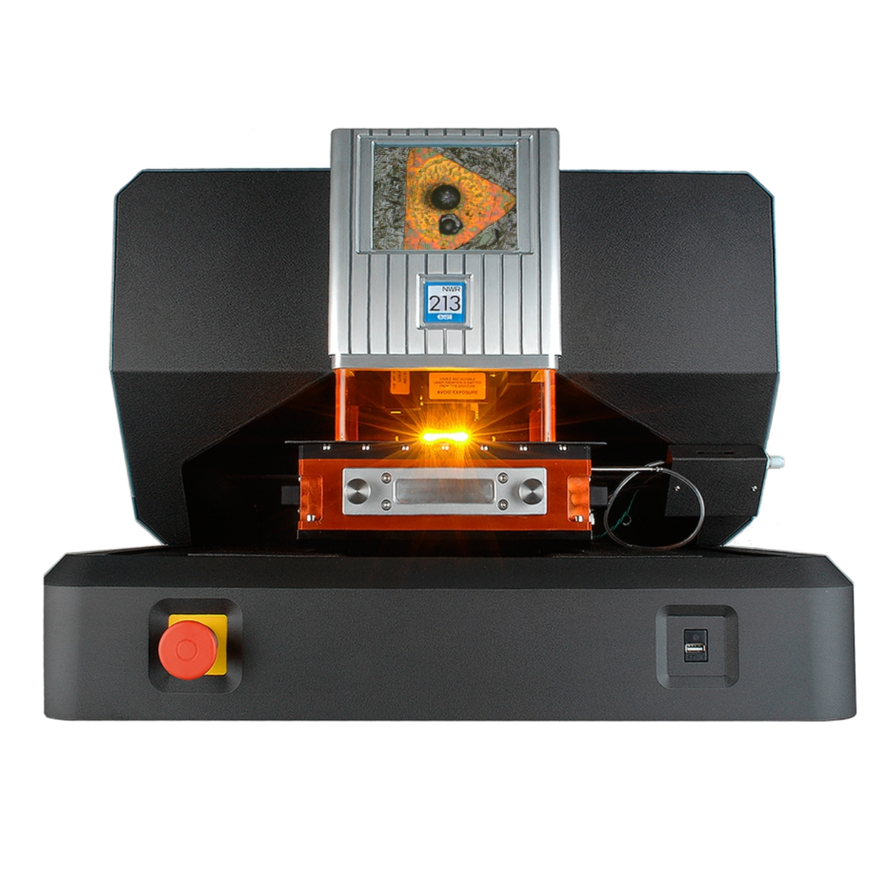
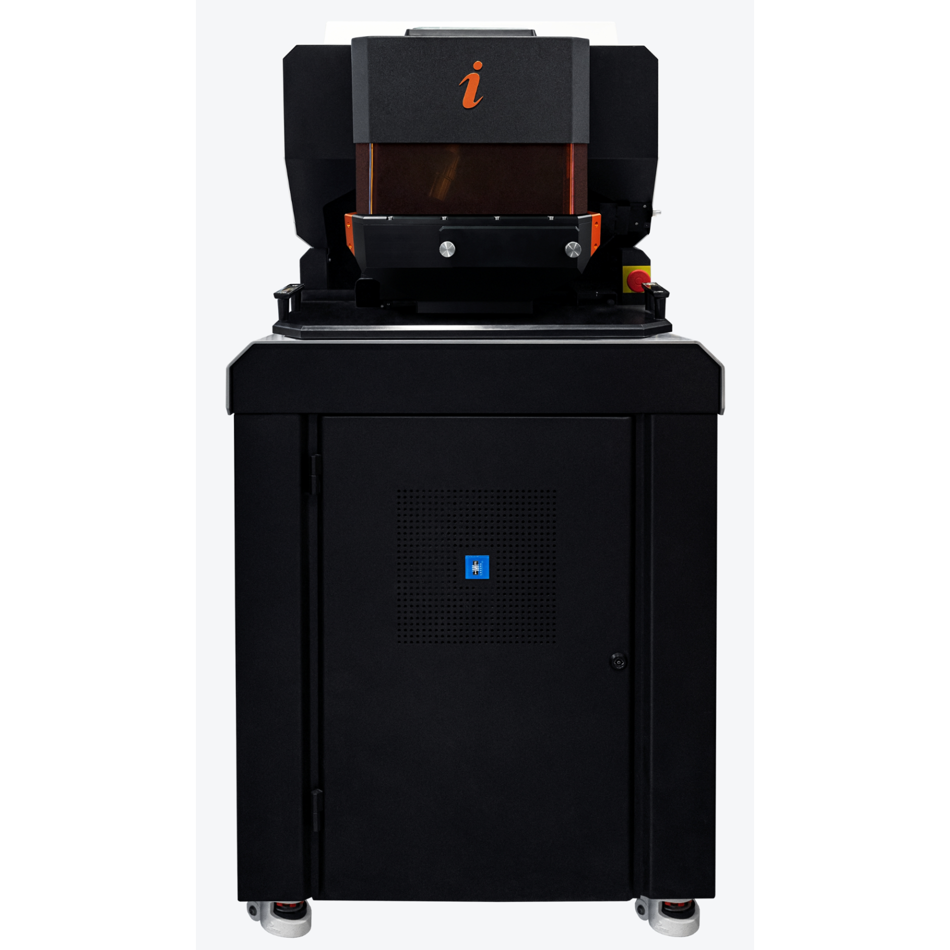
上海凯来仪器有限公司为您提供《法医学案件中元素成像质谱检测方案(激光剥蚀进样)》,该方案主要用于司法鉴定中元素成像质谱检测,参考标准《暂无》,《法医学案件中元素成像质谱检测方案(激光剥蚀进样)》用到的仪器有ESL213 灵活的激光剥蚀系统。
我要纠错
推荐专场
相关方案
 咨询
咨询