

方案详情文
智能文字提取功能测试中
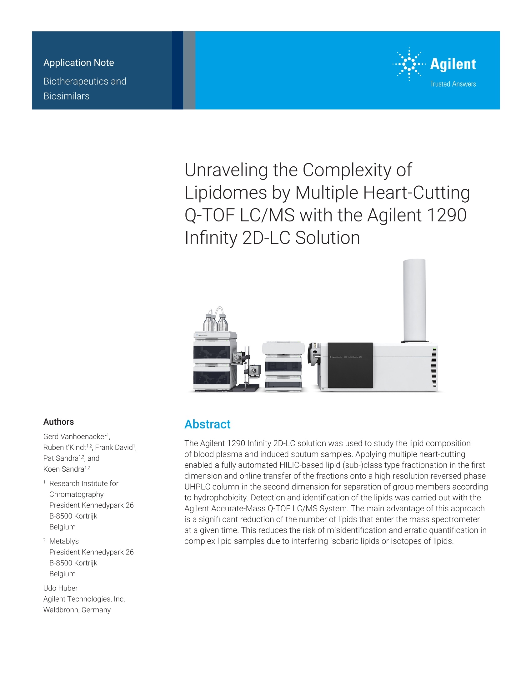
Authors Gerd Vanhoenacker1.Ruben t'Kindt12, Frank David1,Pat Sandra12, andKoen Sandra1.2 1 Research Institute for ChromatographyPresident Kennedypark 26B-8500 Kortrijk Belgium 2 Metablys President Kennedypark 26 B-8500 Kortrijk Belgium Udo Huber Agilent Technologies,Inc. Waldbronn,Germany Unraveling the Complexity ofLipidomes byy MIul t iple Heart-Cutting 1201Q-TOF LC/MS with the Agilent 1290Infinity 2D-LC Solution Abstract The Agilent 1290 Infinity 2D-LC solution was used to study the lipid compositionof blood plasma and induced sputum samples. Applying multiple heart-cuttingenabled a fully automated HILIC-based lipid (sub-)class type fractionation in the firstdimension and online transfer of the fractions onto a high-resolution reversed-phaseUHPLC column in the second dimension for separation of group members accordingto hydrophobicity. Detection and identification of the lipids was carried out with theAgilent Accurate-Mass Q-TOF LC/MS System. The main advantage of this approachis a signifi cant reduction of the number of lipids that enter the mass spectrometerat a given time. This reduces the risk of misidentification and erratic quantification incomplex lipid samples due to interfering isobaric lipids or isotopes of lipids. Introduction The lipidome can be described asthe complete lipid content in a givensample. Lipids exist in different classeswith different physicochemical andbiological properties. Each class containsmany members that differ in the lengthof the fatty acid side-chain and thenumber of double bonds, among others.Lipid extracts generally are complexmixtures with a significant structuraldiversity and a large dynamic range. As aresult, the measurement of the total lipidcontent is an analytical challenge andrequires a high performance separationstep followed by high-resolution massspectrometry. Several approaches are available forthis type of analysis,and state-of-the-artUHPLC on high performance LC columnsenables the high-resolution separationrequired for detailed profiling of lipids incomplex biological matrixes. We haverecently reported on lipidomic analysesin blood plasma? and induced sputum.3These studies have shown that a betterchromatographic separation is beneficialfor MS data analysis and facilitates lipididentification and quantification. Several options are available forimproving the chromatographicseparation. Multidimensionalchromatographic solutions are anefficient way to drastically increasepeak capacity. In two-dimensionalLC (2D-LC), comprehensive LCxLC isknown to substantially increase thechromatographic resolution as longas the two dimensions are orthogonaland the separation obtained in thefirst dimension is maintained upontransfer to the second dimension.4In the comprehensive LCxLC mode,each fraction collected from the firstdimension column is transferred toa second dimension separation for analysis. A less complex approachin 2D-LC is heart-cutting (LC-LC),where only one or a few fractions aretransferred to the second dimension.The advantage of this multidimensionalmode is that the second dimensionanalysis time is not limited by the 2D-LCmodulation time. Therefore, a betterseparation of the transferred fractionscan be achieved. The application of 2D-LC for complexlipid samples is not new, andseveral column combinations havebeen reported. The most obviouscombinations are a normal phase orHILIC separation in the first dimensionand a reversed-phase (RPLC)mechanism in the second dimension.Such combinations have proven toresult in good orthogonality for lipidsamples. The first dimension providesa separation based on the polarhead-group of the lipids, resulting in alipid (sub-)class separation (for example,phospholipids, ceramides, acylglycerols).Here, neutral lipids will elute first beforemore polar and charged lipids. RPLC,conversely, separates the lipids basedon hydrophobicity, that is, the carbonchain length and unsaturation (numberof double bonds) of the fatty acid sidechains. The application of RPLC in thesecond dimension is also beneficial forcoupling with the mass spectrometer. The fractionation of lipids into differentclasses and the chromatographicanalysis of the members of each classare attractive. The combination ofHILIC and RPLC has been performedoffline5.6 or online using a specialinterface for solvent evaporation.78 Inthis Application Note, a fully automatedmultiple heart-cutting Q-TOF LC/MSanalysis of complex lipid extracts withthe Agilent 1290 Infinity 2D-LC solution isdemonstrated. Experimental Samples and sample preparation The lipid extraction protocol has beendescribed in detail in reference 2.Briefly, 300 pL of methanol was addedtop 50 pL of plasma or 120 pL of lungsputum. After vortex mixing, 1 mL oftert-butyl methyl ether (MTBE) wasadded, and the samples were incubatedfor one hour at room temperature. Theaddition of 260 pL of water induced aphase separation, generating a lowerhydrophilic and an upper lipophilic layerwith a protein layer at the bottom. A1 mL amount of the upper phase wasremoved and dried in a centrifugalvacuum evaporator. The dried phase wasreconstituted in 100 uL (plasma) or 40 uL(lung sputum) of MTBE/isopropanol50/50 (v/v), respectively. Instrumentation An Agilent 1290 Infinity 2D-LC solutionconnected to an Agilent 6530 Q-TOFmass spectrometer was used for theexperiments. Agilent 1290 Infinity Binary Pump(first dimension) (G4220A) Agilent 1290 Infinity Binary Pump(second dimension) (G4220A) Agilent 1290 Infinity Autosampler(G4226A) Agilent 1290 Infinity Thermostat(G1330A) Agilent 1290 Infinity ThermostattedColumn Compartment (G1316C) Agilent 1290 Infinity valve drive (2x)(G1170A) Agilent 2D-LC hardware solutionkit (G4236A), including loop kitfor 2D-LC (option #001), capillarykit for 2D-LC (option #003), andmultiple heart-cutting upgrade kit(option #007) Agilent 6530 Accurate-Mass Q-TOFLC/MS (G6530A) The Jet Weaver mixer was removedin the first dimension pump to reducethe delay volume. To obtain sufficientbackpressure on the first dimensionseparation, a calibration capillary(G1312-67500) was installed betweenthepump and the autosampler. Method Software Agilent OpenLAB CDS ChemStationEdition (revision C.01.07) withAgilent 1290 Infinity 2D-LC Software(revision A.01.02) Agilent MassHunter for instrumentcontrol (revision B.05.01) Agilent MassHunter for data analysis(revision B.06.00) Results and discussion When lipid samples are analyzed withthe described HILIC method, they elutein five to six groups in order of polarity,from neutral lipids to more polar andcharged lipids. The separation of theindividual lipids within each class islimited, and typically broad peaks arenoted in real samples. This is shownfor a plasma sample in Figure 1. Thelipid classes selected in this ApplicationNote together with the peak codes andMHC fraction are summarized in Table 1.Applying the same sample to RPLC willgive a different elution pattern where theseparation is based on hydrophobicityand the selectivity for the individual lipidswithin a class is significant (Figure 2).It is evident that the separation modesprovide good orthogonality in thismultidimensional LC setup. Table 1. Lipids, peak codes in figures, andHILIC fractions. Lipid Class (Abbreviation) Code MHC Fraction Free fatty acids (FFA) A 1 Monoacylglycerols (MG) B 1 Diacylglycerols (DG) C 1 Triacylglycerols (TG) D 1 Cholesterol (Chol) E 1 Cholesterol esters (CE) F 1 Ceramides (CER) G 1 Glycoceramides (Hex-CER) H 1 Glyconceramides (Hex -CER) 2 Phosphatidylinositols (PI) J 2 Phosphatidylethanolamines (PE) K 3 Glycosphingolipids (GSL) L 3 Phosphatidylserines (PS) M 4 Phosphatidylcholines (PC) N 4 Sphingomyelins (SM) O 5 Lysophospholipids (LysoPL) P 5 Lysophospholipids (LysoPL) P 5 Figure 1. Total ion chromatogram (positive and negative ionization) for the analysis of a plasmasample with the first dimension HILIC mode. ×106 Figure 2. Total ion chromatogram (positive and negative ionization) for the analysis of a plasmasample with the second dimension RPLC mode (flow: 0.5 mL/min, gradient: 0 to 5 minutes: 60 to80% B, 5 to 17 minutes: 80 to 100%B). Using the Agilent 1290 Infinity multipleheart-cutting 2D-LC solution, a fullyautomated transfer of the HILIC fractionsto RPLC and the subsequent analysiscan be realized. The collected fractionsare stored in loops installed on the2-position/4-port duo-valve and a singlemultiple heart-cutting valve, and thesefractions are analyzed sequentially.Filling and emptying of the loops isperformed in a countercurrent fashion toreduce carryover. The order, timing, andlocation of storage and analysis of thefractions can be fully controlled by the2D-LC add-on software for the AgilentOpenLAB CDS ChemStation Edition. Ascheme of the valve configuration isshown in Figure 3. The MS signals for the collectedfractions of the plasma and thesputum sample can be seen in Figure 4(for positive ionization) and Figure 5(for negativeionization). Figure 3.Configuration of the 2-position/4-port duo-valve for the multiple heart-cutting method.The six loops are installed on the multiple heart-cutting valve. Figure 4. Total ion chromatogram (positive ionization) for the analysis of a plasma sample withthe first dimension HILIC mode and BPC (+) of the multiple heart-cutting analyses of plasma andinduced sputum. The advantage of performing agroup-type separation before separationof individual lipids is demonstratedin Figure 6. In the one-dimensionalRPLC separation, there is (partial)overlap of a nearly isobaric PE andPC (m/z difference is 0.0364 Da),complicating data analysis. With the1290 Infinity multiple heart-cutting 2D-LCsolution, PCs are efficiently separatedfrom PEs in the first dimension, andboth isobaric compounds can beanalyzed in separate fractions. This isa clear demonstration of the benefitof the multiple heart-cutting approachfor identification and quantification ofindividual lipids in complex mixtures. Figure 5. Total ion chromatogram (negative ionization) for the analysis of a plasma sample withthe first dimension HILIC mode and BPC (-) of the multiple heart-cutting analyses of plasma andinduced sputum. PC(p-16:0/18:1) PC(p-18:0/16:1) A m/z744.5901 m/z744.5901 PE(18:0/18:2) m/z 744.5538 PE(18:0/18:2) Figure 6. Extracted ion chromatogram (positive ionization) for selected PEs and PCs in a plasmasample. (A) shows the one-dimensional RPLC separation, (B) traces show the results for themultiple heart-cutting analyses where PEs and PCs are separated in the first dimension HILICseparation. To further illustrate the performanceof multiple heart-cutting 2D-LC andthe usefulness of this technique inlipidome studies, some specific structureelucidations are presented in Figure 7though Figure 12. The nomenclatureproposed by the International LipidClassification and NomenclatureCommittee (ILCNC) is used. Fraction1 ×106 A BPC(+) Fraction 1 2.0 1.5 Formula t (min) m/z Identity 1 C55H101.N 11.45 871.7629 TG(52:4)NH, 2 11.61 873.7786 TG(52:3)NH, 3 C47H7NO, 11.71 689.6111 CE(20:4)NH, 4 CssH1059N 11.78 875.7942 TG(52:2)NH, 5 C45H7NO, 11.84 665.6111 CE(18:2)NH, 6 CH1070N 12 877.8099 TG(52:1)NH, 7 CSHNO, 12.1 667.6267 CE(18:1)NH, Figure 7. Fraction 1, positive ionization, triglycerides, and cholesterol esters in plasma.TG and CE represent the most apolar lipids. Both groups are detected as ammoniaadducts in the positive ESl mode. Feature Feature (Glycophingolipids) Formula t (min) m/z Identity 1 C32H63NO。 9.85 509.480794 CER(NS)(32) 2 C34H6NO, 10.11 537.512094 CER(NS)(34) 3 C36H,NO, 10.33 565.543394 CER(NS)(36) 4 C38H7NO 10.53 593.574694 CER(NS)(38) 5 C40H7NO。 10.69 621.605994 CER(NS)(40) 6 C42H83NO, 10.83 649.637294 CER(NS)(42) Figure 8. Fraction 1, negative ionization, sphingolipids in sputum. Ceramides and smaller glycosphingolipids are preferentially detected in negative ESlmode. Elution order is based on the attached sugar moiety and the fatty acid side chain. Acquisition time (min) Feature Formula t (min) m/z Identity 1 C52H,7NO18 59.46 1023.671 HexHexHexCER(NS)(34) 2 C3H03P 59.68 836.5415 PI(18:1/16:0) 3 C7H303P 59.75 886.5571 PI(18:0/20:4) 4 C7H8593P 59.85 888.5728 PI(18:0/20:3) 5 C45H85013P 59.94 864.5728 PI(18:1/18:0) Figure 9. Fraction2,negative ionization, phosphatidylinositols insputum. Phosphatidylinositols encompass approximately 5% of detectedglycerophospholipids in sputum. HexHexHexCer(NS)(34) is present in bothFractions 1 and 2. Acquisition time (min) Feature Formula t (min) m/z Identity 1 C57H104N021 49.22 1152.713 NeuAcHexHexCER(d18:1/16:0) 2 C0HN.02 49.35 1226.75 HexNAcHexHexHexCER(d18:1/16:0) 3 CHNO.P 49.78 689.4996 PE(32:1) 4 CHNO.P 49.85 715.5152 PE(16:0/18:2) 5 C43HNO,P 49.9 747.5203 PE(p16:0/22:6) 6 C4HNO,P 49.95 723.5203 PE(p16:0/20:4) 7 C39H76NO,P 50.13 701.5359 PE(p16:0/18:1) 8 C43H7 NO,P 50.16 751.5516 PE(p18:0/20:4) 9 CH8NO,P 50.31 729.5672 PE(p18:0/18:1) 10 CHNOP 50.22 745.5622 PE(18:0/18:1) Figure 10. Fraction 3, negative ionization, phosphatidylethanolamines,andglycosphingolipids in sputum. Plasmalogen phosphatidylethanolamines (PEp) are highlyabundant in lung sputum. Complex glycosphingolipids containing N-acetylneuraminicacid (Neu5Ac) elute in this fraction. x105 Feature Formula t (min) m/z 1 C39H78NO,0P 39.66 751.5363 2 CHNOP 39.72 851.5676 3 CHNOP 39.73 777.552 4 CHNOP 39.76 827.5676 5 CHNOP 39.78 803.5676 6 CSHNO,P 39.86 829.5833 7 C41HNOP 39.91 8 C43H84NO,P 39.96 805.5833 PC(16:0/18:1)formate 9 C7H86NOP 39.99 855.5989 PC(18:0/20:4)formate 10 CSHNO,P 40.02 831.5989 PC(36:2)formate 11 CHNO.P 40.17 833.6146 PC(18:0/18:1)formate Figure 11. Fraction 4, negative ionization,phosphatidylcholines (PC) insputum and plasma. Phosphatidylcholines are highly abundant in bothsamples. Both samples show different PC patterns. PCs are detected asformate adducts in negative ESl mode. Feature Formula t (min) m/z Identity 1a C7H,NO,P 26.75 565.33797 PC(0:0/18:2)formate 1b C27H52NO,P 26.89 565.33797 PC(18:2/0:0)formate 2a C27H5NO,P 27.19 567.35362 PC(0:0/18:1)formate 2b C27H54NO,P 27.33 567.35362 PC(18:1/0:0)formate 3a CHsNO,P 27.68 569.36927 PC(0:0/18:0)formate 3b C7H56NO,P 27.82 569.36927 PC(18:0/0:0)formate 4 CH,,N,O.P 29.33 720.5418 SM(d18:1/14:0)formate 5 C40H81N20。P 29.63 748.5731 SM(d18:1/16:0)formate 6 C2H850P 29.9 776.6044 SM(d18:1/18:0)formate 7 C48H95N0P 30.32 858.6826 SM(d18:1/24:1)formate 8 CHNO.P 30.18 856.667 SM(d18:1/24:2)formate Figure 12. Fraction 5, negative ionization, lysophosphatidylcholines andsphingomyelins in sputum. Sn-2 lysophospholipids elute before sn-1lysophospholipids in RPLC. Both lipid subclasses are detected as formateadducts in negative ESl mode. Conclusion The Agilent 1290 Infinity 2D-LCsolution was used for fully automatedmultiple-heart-cutting analyses of theblood plasma and induced sputumlipidome. The lipids were fractionatedin HILIC mode, and the fractions werethen transferred to and analyzed withRPLC. Detection with the Agilent 6530Accurate-Mass Q-TOF LC/MS Systemfacilitated identification of the individuallipids in each fraction. Selected dataclearly demonstrate the power ofthe system in lipidomics and mightbe extended to other metabolomicapplications. References 1...Sandra, K.; Sandra, P. LipidomicsFrom an Analytical Perspective,Curr. Opin. Chem. Biol.2013, 17,847-853. 2. Sandra, K.; et al.ComprehensiveBlood Plasma Lipidomics by LiquidChromatography/QuadrupoleTime-of-Flight Mass Spectrometry,J. Chromatogr. A2010, 1217,4087-4099. 3. Telenga,E.;et al. UntargetedLipidomic Analysis in ChronicObstructive Pulmonary Disease,Am. J. Respir. Crit. Care Med. 2014,190,155-164. 4 Francois, l.; Sandra, K.; Sandra, P.Comprehensive LiquidChromatography: FundamentalAspects and PracticalConsiderations -a Review,Anal. Chim. Acta.2009,64,14-31. 5. Lisa, M.;Cifkova, E.; Holcapek, MLipidomic Profiling of BiologicalTissues Using Off-LineTwo-Dimensional High-PerformanceLiquid Chromatography-MassSpectrometry,J. Chromatogr. A2011,1218,5146-5156. 6. Cifkova,E.; Holcapek, M.;Lisa, M. Nontargeted LipidomicCharacterization of Porcine OrgansUsing Hydrophilic InteractionLiquid Chromatography andOffline Two-Dimensional LiquidChromatography-Electrospraylonization Mass Spectrometry, Lipids2013,48,915-928 7, Nie, H.; et al. Lipid Profiling ofRat Peritoneal Surface Layers byNormal-andReversed-Phase 2DLC QTOF-MS, J. Lipid Res. 2010, 51,2833-2844. 8. Li, M.; et al. Lipid Profiling of HumanPlasma from Peritoneal DialysisPatients Using an Improved 2D(NP/RP) LC-QTOF MS Method,Anal. Bioanal.Chem. 2013,405,6629-6638. 9.Fahy,E.;et al. Lipid Classification,Structures and Tools, Biochim.Biophys. Acta. 2011, 1811, 637-647. www.agilent.com/chem For Research Use Only. Not for use in diagnostic procedures. 使用Agilent 1290 Infinity 2D-LC解决方案研究脂质成分血浆和诱导痰样本。 进行多次心脏切割首先实现了基于HILIC的全自动脂质(亚)类类型的分馏馏分的尺寸和在线转移到高分辨率反相中第二维的UHPLC色谱柱用于分离组成员疏水性。 脂质的检测和鉴定使用安捷伦精确质量Q-TOF LC / MS系统可以显着减少进入质谱仪的脂质数量在给定的时间。 这样可以减少错误识别和错误量化的风险由于干扰同量异位脂质或脂质同位素而产生的复杂脂质样品。
关闭-
1/14
-
2/14
还剩12页未读,是否继续阅读?
继续免费阅读全文产品配置单
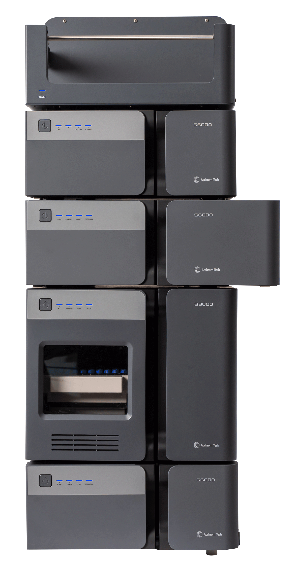
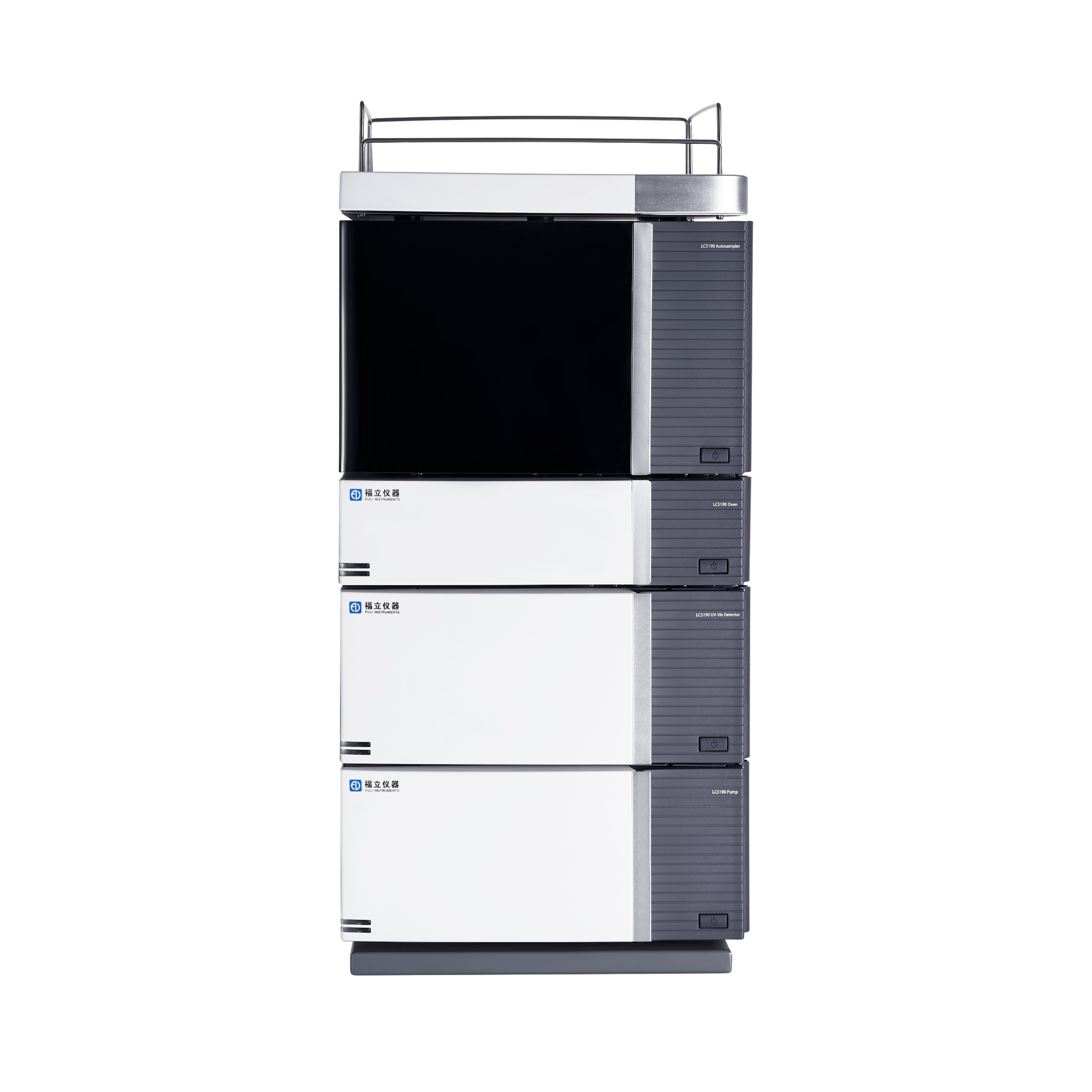
安捷伦科技(中国)有限公司为您提供《血浆和诱导痰中脂质检测方案(液相色谱仪)》,该方案主要用于全血/血清/血浆中生化检验检测,参考标准《暂无》,《血浆和诱导痰中脂质检测方案(液相色谱仪)》用到的仪器有安捷伦 1290 Infinity 二元液相色谱系统(1290 LC)。
我要纠错
推荐专场
相关方案
 咨询
咨询