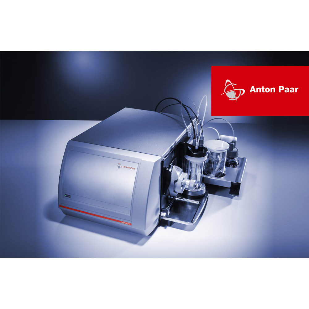
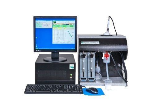
狂犬病毒感染的细胞中外泌体可能参与病毒感染过程检测方案(Zeta电位仪)
检测样品 其他
检测项目 外泌体可能参与病毒感染过程


方案详情文
智能文字提取功能测试中
Virologica Sinica (2019) 34:59-65https://doi.org/10.1007/s12250-019-00087-3www.virosin.orgwww.springer.com/12250RESEARCH ARTICLE Virologica Sinica60 Exosomes Released from Rabies Virus-Infected Cells May be Involvedin the Infection Process Jingyu Wang·Fan Wul · Chuntian Liul·Wenwen Dai · Yawei Teng· Weiheng Su1r2. Wei Kong2·Feng Gao12. Linjun Cai12. Ali Hou1o.Chunlai Jiang1o Received: 2 September 2018 / Accepted: 10 December 2018/ Published online: 6 February 2019 @ Wuhan Institute of Virology, CAS 2019 Abstract Exosomes are cell-derived vesicles that are secreted by many eukaryotic cells. It has recently attracted attention as vehiclesof intercellular communication. Virus-infected cells release exosomes, which contain viral proteins, RNA, and pathogenicmolecules. However, the role of exosomes in virus infection process remains unclear and needs to be further investigated.In this study, we aimed to evaluate the effects of exosomes on rabies virus infection. OptiPrepM density gradientcentrifugation was used to isolate exosomes from rabies virus-infected cell culture supernatants. A rabies virus G proteinenzyme-linked immunosorbent assay and acetylcholinesterase activity assays were performed to verify the centrifugationfractions. Exosomes were then characterized using transmission electron microscopy and Western blotting. Our resultsshowed that rabies virus infection increased the release of exosomes. Treatment with GW4869 and si-Rab27a, twoexosomal secretion inhibitors, inhibited exosome release. Furthermore, the inhibitors reduced the levels of extracellular andintracellular viral RNA. These data indicated that exosomes may participate in the viral infection process. Moreover, ourresults establish a basis for future research into the roles of exosomes in rabies virus infection and as potential targets fordeveloping new antiviral strategies. Keywords Exosomes· Rabies virus· Isolation· Virus infection Introduction Exosomes are nanovesicles ranging in size from 30 to150 nm that are released from all cells, allowing them to bedetected in various body fluids (Simons and Raposo 2009;Thery et al. 2002; Yanez-Mo et al. 2015). Exosomes wereoriginally discovered as secreted waste products in studiesof the transformation of reticulocytes into mature red bloodcells. However, exosomes have recently been described asintraluminal vesicles (ILVs), which form multivesicular ( 区Ali Houhoual@jlu.edu.cn ) 区 Chunlai Jiangjiangcl@jlu.edu.cn 1 National Engineering Laboratory for AIDS Vaccine, Schoolof Life Science, Jilin University, Changchun 130012, China 2 Key Laboratory for Molecular Enzymology and Engineeringof the Ministry of Education, School of Life Science, JilinUniversity, Changchun 130012, China bodies (MVBs) that subsequently fuse with the cytoplasmicmembrane to be released into the extracellular space(Harding et al. 2013; Thery et al. 2002). Exosomes notonly containnwaste discarded by cellsbut also playimportant roles in disease and pathology, particularlyinfectious diseases (Anderson et al. 2016; Chahar et al.2015; Liu et al.2017;Meckes and Raab-Traub 2011;Nagashima et al.2014). Rabies virus has a negative single-stranded RNA gen-ome, and in some animals who are seropositive for rabies,the viral RNA. genome is detectable using polymerasechain reaction (PCR) on saliva samples, but no virus par-ticles can be directly detected (Warrel and Warrell 2004).Additionally, it is still unclear how rabies virus spreadsamong host cells (Schnell et al.2010). Exosomes can haveopposing roles in viral infections. First, infected cells shedexosomes containing viral proteins and the whole virusgenome, promoting the spread of infection to other cells(Nour and Modis 2014: Raab-Traub and Dittmer 2017;Simons and Raposo 2009). This mechanism of virus spreading can potentially help the virus evade host immunedefenses, such as neutralizing antibodies. However, exo-somes can also transfer antiviral molecules induced by hostimmune responses to inhibit virus replication (Raab-Trauband Dittmer 2017; Schwab et al. 2015). For example,humaniimmunodeficiencyvirus (HIV)-1 Nef proteinpresented in exosomes shed from virus-infected cells caninduce apoptosis of CD4+T cells (Luo et al. 2015). Thus,the pathogenicity of HIV-1 is achieved by eliminating theaction of CD4T cells (Fevrier et al. 2011). Moreover, Nefprotein can be transmitted from infected macrophages touninfected B cells through the vesicle transport pathway(Lenassi et al. 2010). Exosomes from HIV-infected cellscontain transactivating response (TAR) element RNA andcan stimulate the expression of pro-inflammatory cytokinesin primary macrophages (Sampey et al. 2016). Viral pro-teins and whole-virus genomes were found to be present inexosomes secreted by hepatitis Cvirus (HCV)-infectedcells, enabling the establishment of infections and resis-tance to neutralizing antibodies (Liu et al. 2014; Ramakr-ishnaiah et al. 2013). Exosomes derived from humanT-lymphotropicvirustype 1(HTLV-1)-infected cellsincorporated viral proteins and mRNA and played impor-tant roles in HTLV-1-induced pathogenesis (Jaworski et al.2014). Enterovirus 71 (EV71)-infected cells secrete exo-some-packaged viral genomic RNA and miR-146a, facili-tating viral pathogenesis (Fu et al. 2017). Exosomessecreted from Epstein-Barr virus-infected B cells were alsofound to harbor virus-associated microRNA (miRNA), andEpstein-Barr virus-infectednasopharyngeal carcinoma(NPC) cells secrete exosomes with viral miRNAs that canbe transported into uninfected cells (Baglio et al. 2016;Meckes et al. 2010; Verweij et al. 2013). In hepatitis Bvirus (HBV)-infected cells, interferon-induced moleculesare transferred intercellularly vial exosomestoserveantiviral proteins (Li et al. 2013). Although there are many studies focus on the roles ofexosomes in virus infection process, the exact function anddetailed mechanism of exosomes during the process needfurther investigation. How the rabies virus spreads amonghost cells and the role of exosomes in rabies virus infectionprocess still remain unclear. In this study, we explored theroles of exosomes in rabies virus infection using Opti-PrepTM density gradient centrifugation to isolate exosomesfrom rabies virus-infected Vero cell culture supernatantsand demonstrated that rabies virus infection enhanced theproduction of exosomes, which may facilitate virus infec-tion. Exosomes may facilitate infection and the spread ofviruses. Our results provide important insights into themechanisms by which exosomes facilitate the spread ofviruses in host cells. Materials and Methods Cell Culture and Virus Vero cells were cultured as monolayers in Dulbecco'smodified Eagle’s medium supplemented with 10% exo-some-free fetal calf serum (depleted of bovine exosomesby ultracentrifugation for 16 h at 100,000 xg), 100 IU/mLpenicillin, and 100 mg/mL streptomycin at 37 ℃ in anatmosphere containing 5% CO2. Vero cells were infectedwith rabies virus (PM1503 strain) at a multiplicity ofinfection (MOI) of 0.01, and the infected cells were cul-tured for 4 days before exosome isolation. Transmission Electron Microscopy (TEM) Transmission electron microscopy (TEM) was used toobserve the morphological characteristics of the exosomes.The Vero cells (approximately 2 ×10°) were fixed with4% glutaraldehyde and osmic acid at 4C followed bygradient elution using ethanol. Cells were then embeddedfor 3 h, sectioned using an ultramicrotome (LEICA EMUC7), and stained with uranyl acetate and lead citrate. Thepurified exosomes were negatively stained using 2% (w/v)phosphotungstic acid and were immunolabeled with anti-CD63 antibodies. The imagingwas performed atit anacceleration voltage of 80 kV using a transmission electronmicroscope (HITACHI H-7650). Isolation of Exosomes Using OptiPrepM DensityGradient Centrifugation Exosomes were isolated using differential velocity cen-trifugation followed by OptiPrepTM density gradient cen-trifugation. Supernatantsharvested fromVero cells(approximately 4 ×10") were centrifuged at 4℃ and300 ×g foirr10 min. 2000 ×g for20 min. and10,000 ×g for 30 min (Théry et al. 2006). Pellets con-taining cells, dead cells, and cell debris from each of thefirst three centrifugations were discarded, and the super-natants were used in the next step. After centrifugation at100,000 ×g for 2 h at 4℃, the supernatants were dis-carded, and the pellets (containing exosomes and rabiesvirus particles) were resuspended in 500 uL phosphate-buffered saline (PBS). Subsequently, an OptiPrepM den-sity gradient was used to purify the exosomes from rabiesvirus, as previously described (Cantin et al. 2008; Konaduet al. 2016). A discontinuous 6%-24% density gradientwas prepared by diluting solutions of 24%, 22%,20%,18%, 16%, 14%, 12%,10%, 8%, and 6% (w/v) iodixanolfrom a stock solution of OptiPrepM (60%[w/v] aqueousiodixanol [Axis-Shield PoC AS, Oslo, Norway]) with 0.25 mol/L sucrose/TE buffer (pH 7.4). The unpurifiedexosomes (containing rabies virus particles) in a volume of500 pL were overlaid on the top of the gradient and cen-trifuged usingaBeckman Coulter SW40Tiirotor at100,000 ×g for 18 h at 4℃. Identification of OptiPrepTM Density GradientCentrifugation Fractions After centrifugation, each gradient fraction was collectedmanually. The exosomes and rabies virus particles in thefractions were identified by acetylcholinesterase (AChE)assay and rabies virus G protein enzyme-linkedimmunosorbent assay (ELISA), respectively. These twoassays were performed according to the product instruc-tions. AChE is an exosome-specific marker that has beeninvestigated in previous studies (Cantin et al. 2008;Gastpar et al. 2005; Konadu et al.2016; Rieu et al. 2000). Nanoparticle Tracking Analysis (NTA) The sizes and concentrations of the purified exosomefractions were measured using a ZetaView NTA (ParticleMetrix GmbH, Meerbusch, Germany), which is a combi-nation of light scattering and Brownian motion technology.The assays were performed according to the user guide ofthe NTA. Briefly, the exosome sample was diluted in 1 mLPBS and loaded into the cell. The instrument measuredeach sample at 11 different positions throughout the cell.For each measurement, the instrument pre-acquisitionparameters were set to a temperature of 25 °C, a sensitivityof 85, a frame rate of 30 frames per second (fps), a shutterspeed of 100, and a laser pulse duration equal to that ofshutter duration. Western Blot Analysis of Isolated Exosomes Exosome fractions (8%-12%) were pooled, diluted with3 mL PBS, and ultracentrifuged at 100,000 ×g for 2 h.After discarding the supernatant, the pellets were resus-pended in radioimmunoprecipitation assay (RIPA) bufferand heated for 10 min at 97°C. The denatured proteinswere separated using sodium dodecyl sulfate-polyacry-lamide gel electrophoresis on 12% gels and transferredonto nitrocellulose membranes using a Bio-Rad dry blot-ting system (Bio-Rad, Hercules, CA, USA). The mem-branes were blocked with 3% nonfat powdered milk inPBS for1 h at 20-25℃ and then incubated with primaryanti-cluster of differentiation 633 (CD63)antibodies(1:1000, System Biosciences, Palo Alto USA), anti-CD81antibodies (1:1000, Abcam, Cambridge,UK), anti-TSG101antibodies (1:1000, Abcam), and anti-calnexin antibodies(1:1000, Abcam) overnight at 4C. The membranes were washed three times with PBS plus 0.1% Tween (PBST) andthen incubated with alkaline phosphatase-conjugated sec-ondary antibodies (1:2000) for 1 h. Exosome Secretion Inhibition Assay Vero cells (approximately 4×10) were treated withGW4869, or transfected with small interfering RNA(siRNA) against Rab27a (si-Rab27a), or controls. After48 h, exosomes were isolated by using differential velocitycentrifugation followed by OptiPrepTMdensity gradientcentrifugation. RNA Extraction and Reverse Transcription (RT)-PCR Analysis The MiniBEST viral RNA extraction kit (TaKaRa, Shiga,Japan) was used to extract rabies viral RNA. A One-StepSYBR PrimeScript RT-PCR kit (TaKaRa) and rabies virus-specific primers for the N gene (forward primer: 5'-CAA-GATGTGTGCYAAYTGGAG-3' and reverse primer: 5'-AGCCCTGGTTCGAACATTCT-3) were used for ampli-fication and subsequent quantification with a CFX96 sys-tem (Bio-Rad). Statistical Analysis Data represent at least three independent experiments werepresented as mean ± SEM and analyzed using GraphPadPrism software (GraphPad Software Inc., La Jolla, CA).The Student’s t test or one-way analysis of variance wasused for statistical analysis to compare the differencesamong treatment groups. P < 0.05was considered toindicate statistical significance. Results Observation of Exosomes in Ultrathin CellSections Exosomes are essentially ILVs generated by inward bud-ding of endosomal MVBs. The ILVs of MVBs may trafficto the plasma membrane, where they are released into theextracellular space by fusion with the plasma membrane(Harding et al. 2013; Simons and Raposo 2009). Accord-ingly, we investigated the release of exosomes from rabiesvirus-infected Vero cells using ultrathin sections fixed with2% glutaric dialdehyde. As shown in Fig. 1A, MVBsinvaginated inward within the cell. Additionally, ILVswere fused with the plasma membrane and released theexosomes into the extracellular environment (Fig. 1B). Fig. 1 Transmission electronmicroscopy (TEM) images ofexosomes in rabies virus-infected Vero cell ultrathinsections. A The arrow indicatesILVs that formed from MVBsinvaginated inward. B FormedILVs were fused with plasmamembrane. ILVs: intraluminalvesicles; MVB: multivesicularbody; PM: plasma membrane.Scale bars = 500 nm. Fig. 2 OptiPrepTMdensity gradient centrifugationtechnique tcseparate exosomes from rabies virus-infected cell supernatants.Fractions from 6%-24% OptiPrepTM density gradient were collectedand analyzed using AChE activity assays (A) and rabies virus G Isolation and Identification of Exosomes morphology. As shown in Fig. 3D, the exosome fractions(8%-12%) had positive signals of exosome-specific markers(CD63, CD81, and TSG101) and were negative for calnexin,a marker of the endoplasmic reticulum. One major challenge in this study was that exosomes andrabies viruses have similar buoyant densities. Therefore, asimple and reliable method was needed for isolating andpurifying exosomes from rabies virus-infected cell culturesupernatants. In this study, we used a 6%-24%OptiPrepIMdensity gradient centrifugation method to isolate exosomesfrom rabies viral particles. The centrifugation fractionswere identified using AChE assays and rabies virus Gprotein ELISA. As shown in Fig. 2, the exosomes andrabies viral particles were present in the 8%-12% and18%-20% fractions, respectively. Taken together, these exosome-specific markers were foundonly in the exosome fractions (8%-12%), and the isolatedexosomes had a round morphology. These data indicated thatOptiPrepIM density gradient centrifugation could be used toisolate exosomes from rabies virus-infected cell culture super-natants. The data of the rabies virus G protein ELISA impliedthat the exosome fractions did not contain virus particles. Exosome Pathway may Participate in RabiesVirus Infection Next, we further characterized the isolated exosomesusing TEM and western blotting. As shown in Fig. 3A-C, theexosomes were negatively stained with phosphotungstic acidand immunogold labeled with anti-CD63 antibodies. TheTEM data showed that the exosomeshadaround We then investigated the effect of virus infection on exo-some secretion. The exosomes were isolated from the samevolumes of virus-infected cell culture supernatants and protein ELISA (B). All dcata are means of three independentexperiments; AChE, acetylcholinesterase; ELISA, enzymelinkedimmunosorbent assay. Fig. 3 Characterization of exosomes from rabies virus-infected cellculture supernatants. Transmission electron microscopy (TEM)analysis shows purified exosomes negatively stained with (A) 2%phosphotungstic acid and (B, C) immunogold labeled with anti-CD63antibodies. Scale bars = 100 nm. D Western blot analysis of purified control cell culture supernatants. As shown in Fig. 4A, thenumber of exosomes isolated from virus-infected cell cul-ture supernatants (approximately 5.2 × 10/mL) was sig-nificantly (P<0.05) higher than that in the control group(approximately 1.3 ×10 /mL). Next, the roles ofexosomes in the viral infection processwere investigated. GW4869 and si-Rab27a, two exosomalsecretion inhibitors, were used to reduce the release of exo-somes. As shown in Fig. 4B, both GW4869 and si-Rab27aeffectively inhibited exosome release. Then, Vero cells werepretreated with 1, 5, or 10 umol/L GW4869 for 12 h beforethe addition of rabies virus. Then, 48 h after viral infection,the extracellular and intracellular rabies viral RNA wasquantified using RT-PCR. As shown in Fig. 4C, 4D, treat-ment with GW4869 significantly reduced the levels ofextracellular and intracellular rabies virus RNA. When cellswere treated with 100 nmol/L si-Rab27a, we also found thatthe levels of intracellular and extracellular rabies viral RNAwere significantly reduced by the inhibitor (Fig. 4E,4F). These results demonstrated that viral infection increasedexosome secretion. Moreover, exosomal secretion inhibi-tors reduced the levels of extracellular and intracellularviral RNA. These data indicate that exosomes may par-ticipate in the process of rabies viral infection. Discussion Exosomes are membrane-bound vesicles secreted from mostcell types and found in various biological fluids (Alenquerand Amorim 2015; Simons and Raposo 2009). In this study, exosomes using exosome-specific markers CD63, CD81, and TSG101and nonexosomal marker calnexin (CL: cell lysate; RV: rabies virusfraction; EXO: exosome fraction). Results are representative of threeindependent experiments. we used TEM to observe exosome release from rabies virus-infected cells. MVBs were clearly shown to invaginateinward, forming ILVs that fused with the plasma membraneto release the exosomes into the extracellular environment. Several methods are currently used for the isolation andpurification of exosomes from cell culture supernatantsincluding differential centrifugation, sucrose density gradientcentrifugation, ultrafiltration, and antibody-coated immuno-magnetic using commercialized kits (Momen-Heravi et al.2013). However, these commonly used techniques for iso-lating exosomes result in contamination with viral particlesbecause exosomes and virus particles have similar sizes anddensities. An OptiPrepIM density gradient centrifugationprotocol for isolating exosomes from HIV particles wasestablished in previous studies (Cantin et al. 2008; Konaduet al.2016). In the present study, considering the character-istics of large high-density rabies virus particles versus rela-tively small low-density exosomes, we modified the 6%-18%OptiPrepM density to 6%-24%. Our data demonstrated thatthe modified gradient centrifugation could be used to isolateexosomes from the supernatants of rabies virus-infected cells. Several recent studies have confirmed that exosomes playimportant roles in physiological and pathological environ-ments by carrying proteins, mRNAs, miRNAs, and otherhost functional genetic elements to neighboring cells,thereby facilitating the establishment of productive infec-tions and modulating cellular responses. In this study, wefound that the rabies viral infection increased the release ofexosomes from host cells. Moreover, GW4869 and si-Rab27a'a treatments inhibited exosomeproduction andreduced levels of extracellular and intracellular viral RNA. Fig. 4 Quantification of exosomes released from Vero cells andeffects of exosomal secretion inhibitors on rabies viral infectionprocess. (A) Numbers of exosomes in uninfected Vero cell culturesupernatants and rabies virus-infected Vero cell culture supernatantswere measured using nanoparticle tracking analysis (NTA). (B) Ex-osomal secretion inhibitors GW4869 and si-Rab27a blocked exosomerelease. Exosome number was measured using NTA. (C,D) GW4869reduced levels of extracellular and intracellular viral RNA. Vero cellswere pretreated with 1, 5, and 10 umol/L GW4869; DMSO; or culturemedium (control) for 12 h before addition of rabies virus.Then, 48 hafter infection, extracellular and intracellular viral RNA wasextracted,and RT-PCR was used to quantify rabies viral RNA levels These findings are similar to those of Huang et al. whoreported that Zika virus (ZIKV) infection in human fetalastrocytes induced a significant increase in extracellularvesicles and treatment with GW4869 decreased extracellularand intracellular viral RNA levels (Huang et al. 2018).Various viruses including HIV, HBV, HCV, rabies virus,herpes viruses, filoviruses, and arenaviruses need or hijackthe endosomal sorting complexes required for transport(ESCRT) pathway for their release (Alenquer and Amorim2015; Chahar et al. 2015; Liu et al. 2017; Nagashima et al.2014; Schorey and Harding 2016), and the ESCRT pathwayhas been shown to play a role in the formation and secretionof exosomes. The reduction of intracellular viral RNA maybe due to the reduction of extracellular virus. The number ofprogeny viruses in the extracellular medium was decreased,which may reduce the overall viral infectivity. Anotherpossibility is that the function of exosomes in the viralinfection process may not only contribute to virion releasebut also affect other processes in the viral life cycle. Thedetailed mechanisms mediating the reduction of intracellularviral RNA levels in exosomal secretion inhibitor-treatedcells need to be investigated further. Although we obtained some interesting findings, thereare several certain limitations of this study. One is that Relative expression viral RNA levels were determined and standard-ized with 18S rRNA. (E, F) si-Rab27a reduced extracellular andintracellular viral RNA. Vero cells were transfected with smallinterfering RNA (siRNA) against Rab27a (si-Rab27a) or controlsiRNA (si-scr) for 12 h before infection with rabies virus. Next, rabiesvirus RNA was extracted from extracellular and intracellular fractionsat 48 h after infection and quantified using RT-PCR. Relativeexpression viral RNA levels were determined and standardized with18S rRNA. Relative expression of viral RNA in DMSO-, GW4869-,and si-Rab27atreated groups in (C-F) was calculated as percentage ofviral RNA in control group, which was set at 100%. All data are fromthree independent experiments (*P<0.05 and **P <0.01). which step of rabies virus infection process is regulated byexosomes. Thee Cother limitationi1sSthat theexactmolecule(s) of exosomesanddnmolecular mechanisminvolved in the regulation process of exosomes remainsneed to be revealed in future. In conclusion, our data demonstrated that the 6%-24%OptiPrepTM density gradient centrifugation could be usedto separate exosomes from rabies virus virions. Moreover,rabies virus infection enhanced the release of exosomes.GW4869 and si-Rab27a inhibited exosome release andreduced the levels of extracellular and intracellular viralRNA. These data indicate that exosomes may participate inthe process of viral infection. Our results establish a basisfor future research into the roles of exosomes in rabiesvirus infection and as potential targets for developing newantiviral strategies. Acknowledgements This work was supported by the National NaturalScience Foundation of China (Grant No. 31770184) and ConstructionProject of Provincial-School of Jilin Province (No. 440050316A28). Author Contributions JW, AH, and CJ contributed to the concep-tion of the study. JW, AH, and FW contributed to designed andperformed the experiments. CL, WD, and YT contributed assistedperformed the experiments. JW, WS, FG, WK, LC, AH, and CJ contributed analysis with constructive discussions. CJ contributed toresources.JW, AH, and CJ contributed significantly to analysis resultsand manuscript preparation. JW, LC, AH, and CJ contributed reviewand editing manuscript. All authors read and approved the finalmanuscript. Compliance with Ethical Standards Conflict of interest The authors declare that they have no conflict ofinterest. Animal and Human Rights Statement This article does not containany studies with human or animal subjects performed by any of theauthors. References ( Alenquer M, Amorim M J (2015) E x osome bi o genesis. Regul FunctViral Infect Viruse s 7:5066-5083 ) ( Anderson M R, K a shanchi F, Jacobson S ( 2 0 16) Ex o somes in vir a l disease. Neurotherapeutics 13:535-546 ) ( Baglio SR, van Eijndhoven MA, Koppers-Lalic D, B erenguer J ,Lougheed SM, Gibbs S, L e veille N, Rinkel R N, H o pmans ES , Swaminathan S et al (2016) Sensing of la t ent E B V inf e ction through exosomal t ransfer of 5'ppp RNA. Proc Natl Acad Sci USA 113:E587-E596 ) ( Cantin R, Diou J, Belanger D, Tremblay AM, Gilbert C( 2 008)Discrimination between exosomes and HIV-1: pu r ification of bothvesicles from cell-free supernatants . JImmunol Methods 338:21-30 ) ( Chahar HS, Bao X, Casola A (2015) Exo s omes and their role in the lif e cycle and pathogenesis of RNA viruses. V iruses 7:3204-3225 ) ( Fevrier M, Dorgham K, Rebollo A (2011) CD4 +T cell depletion in human immunodeficiency virus ( H IV) i n fection: r ole of apop- tosis. Viruses 3:586-612 ) ( Fu Y , Z hang L,Z h ang F, T a ng T, Z h ou Q, Feng C, Jin Y , W u Z (2017) Exosome-mediated m i R-146a transfer suppresses t ype I interferon response and f a cilitates EV71 i n fection. P L oS Pa t hog13:e1006611 ) ( Gastpar R, Gehrmann M, B a usero MA, Asea A, Gross C, SchroederJA, M ulthoff G ( 2 005) He a t shock protein 70 sur f ace-positive tumor exosomes st i mulate m i gratory an d cy t olytic activity ofnatural killer cell s . Cance r Res 65:5238 ) ( Harding CV, Heuser JE, Stahl P D ( 2 013) Exosomes: looking backthree decades a nd into t he future. J Cell Biol 200:367-371 ) Huang Y, Li Y, Zhang H, Zhao R, Jing R, Xu Y, He M, Peer J, KimYC, Luo J, Tong Z, Zheng J (2018) Zika virus propagation andrelease in human fetal astrocytes can be suppressed by neutralsphingomyelinase-2 inhibitor GW4869. Cell Discov 4:19 Jaworski E, Narayanan A, Van Duyne R, Shabbeer-Meyering S,Iordanskiy S, Saifuddin M, Das R, Afonso PV, Sampey GC,Chung M et al (2014) Human T-lymphotropic virus type1-infected cells secrete exosomes that contain Tax protein.J Biol Chem 289:22284-22305 ( Lenassi M, Lenassi M, Cagney G, Liao M, Vaupotic T , Bartholo-meeusen K, Cheng Y, Krogan NJ , Pl e menitas A, Pe t erlin BM(2010) HIV Nef is secreted in exosomes and triggers apoptosis inbystander CD4 + T cells. Traffic 11 :110-122 ) ( Li J , Liu K, Liu Y, Xu Y, Zhang F, Yang H, Liu J, P a n T, C h en J, WuM, Z hou X, Yuan Z (2013) Ex o somes med i ate the cell-to-cell ) transmission of IFN-alpha-induced antiviral activity. Nat Immu-nol 14:793-803 Liu Z, Zhang X, Yu Q, He JJ (2014) Exosome-associated hepatitis Cvirus in cell cultures and patient plasma. Biochem Biophys ResCommun 455:218-222 ( Liu L, Zhou Q , Xie Y, Zuo L, Z h u F, Lu J ( 2 0 17) Extracellularvesicles: novel vehicles in herpesvirus infection. Virol Sin32:349-356 ) ( Luo X, Fan Y, Park IW, He JJ (2015) Ex o somes are unlikely involvedin intercellular Nef transf e r. PLoS ONE 10:e0124436 ) ( Meckes D G, Raab-Traub N Jr ( 2 0 11) Microvesicles a n d viral i nfection. J Viro l 85:12844-12854 ) ( Meckes DG, Shair KH Jr., Marquitz AR, Kung CP,Edwards RH, Raab-Traub N ( 2 010) Human tum o r virus utilizes ex o somes for intercel-lular communication. Proc Nat l Acad Sci USA 107:20370-20375 ) ( Momen-Heravi F , B a laj L , A l ian S , Mantel PY, Ha l leck AE , T rachtenberg A J, Soria CE, Oquin S , Bonebreak C M, S a racoglu E, Skog J, K uo WP (2013) Cu r rent methods for the isolation of extracellular vesicle s . Biol Chem 394:1253-1262 ) ( Nagashima S , Jirintai S, Takahashi M, Kobayashi T, Ta nggis Nishizawa T, Kouki T, Yashiro T, Okamoto H (2014) Hepatitis E virus egress depends on the exosomal pathway, with secretory exosomes derived from multivesicular b odies. J Gen Virol 95:2166-2175 ) ( Nour AM, Modis Y (20 1 4) End o somal vesicles as v e hicles for viral genomes.T r ends Cell Biol 2 4:449-454 ) Raab-Traub N, Dittmer DP (2017) Viral effects on the content andfunction of extracellular vesicles. Nat Rev Microbiol15:559-572 ( R amakrishnaiah V , T h umann C, F of a na I, H abersetzer F, Pa n Q, de Ruiter PE, Willemsen R, Demmers JA , St a lin R a j V, Jenster Get al (2013) Exosome-mediated transmission of hepatitis C virus between human hepatoma Hu h 7.5 cells. Proc Natl A cad Sci U SA110:13109-13113 ) ( Rieu S , Geminard C , R a besandratana H , Sainte-Marie J , V i dal M (2000) E xosomes released during reticulocyte maturation bind to fibronectin vi a integrin a4β1. Eur J Biochem 267:583-590 ) ( Sampey GC, Saifuddin M, Schwab A, B arclay R, Punya S, Chung M C, H a kami RM , Zadeh MA, Lepene B, K l ase ZA et a l (2016)Exosomes f r om HIV-1-infected ce l ls s t imulate p r oduction o f pro-inflammatory c y tokines th r ough trans-activating re s ponse (TAR) RNA. J Biol Chem 291:1251-1266 ) ( S chnell MJ, M cGettigan JP, Wirblich C, Papaneri A (2010) The cell biology of r abies virus: using stealth to reach the brain . Nat Rev Microbiol 8:51-61 ) ( Schorey J S, H arding C V (2 0 16) Extracellular vesicles a n d i n fectiousdiseases: new complexity to an old story. JClin Invest 126:1181-1189 ) ( Schwab A,Meyering SS,Lepene B, Iordanskiy S, van H o ek ML, Hakami R M, Kashanchi F (2015) Ext r acellular vesicles from infected cells:potential f or direct pathogenesis. Front Microbiol 6:1132 ) ( Simons M, R a poso G (2009) Ex o somes-vesicular carriers f or intercellular communication. Curr Opin Cell Biol 21:575-581 ) Thery C, Zitvogel L, Amigorena S (2002) Exosomes: composition,biogenesis and function. Nat Rev Immunol2:569-579 Thery C, Amigorena S, Raposo G, Clayton A (2006) Isolation andcharacterization of exosomes from cell culture supernatants andbiological fluids. https://doi.org/10.1002/0471143030.cb0322s30 Verweij FJ, van Eijndhoven MA, Middeldorp J, Pegtel DM (2013)Analysis of viral microRNA exchange via exosomes in vitro andin vivo. Methods Mol Biol 1024:53-68 ( Warrell M J, Warrell D A (2004) Rabies and other lyssavirus diseases.Lancet 363:959-969 ) ( Yanez-Mo M , Siljander PR, Andreu Z, Zavec AB, Borras FE, B u zas EI, Buzas K , C asal E , C appello F , C arvalho J e t al (2015) Biological properties o f extracellular v e sicles a n d th e ir physio-logical functions. J Extracel l Vesicle s 4:27066 ) Springer 外泌体是由细胞产生的囊泡,许多真核细胞都能分泌出外泌体。近,外泌体作为细胞间通讯的载体,引发大量关注。受病毒感染的细胞所释放的外泌体中,包含了病毒蛋白、RNA和致病性分子。但是,外泌体在病毒感染过程中所起的作用一直都不明确,需要进一步研究确认。这项研究中,研究者旨在研究外泌体在狂犬病毒感染过程中的作用。研究者使用OptiPrepTM(碘克沙醇)密度梯度离心的方法从狂犬病毒所感染细胞的细胞悬浮培养液中分离出外泌体;通过狂犬病病毒G蛋白的酶联免疫吸附实验(ELISA)和乙酰胆碱酯酶活性实验,检查经离心所获得的分离组分;通过透射电镜和蛋白免疫印迹实验对外泌体进行表征。结果表明,被狂犬病病毒感染的细胞,其外泌体的释放水平显著提高。两种外泌体分泌抑制剂(GW4869和si-Rab27a)可以有效抑制外泌体的产生。而且,这两种抑制剂降低了细胞外和细胞内的病毒RNA水平。这些数据表明,外泌体可能参与了病毒的感染过程。我们的研究结果也为后续的狂犬病病毒感染过程中外泌体的功能研究提供了基础,同时为开发新的抗病毒策略提供了潜在的研究靶标。
关闭-
1/7
-
2/7
还剩5页未读,是否继续阅读?
继续免费阅读全文产品配置单
大昌华嘉科学仪器为您提供《狂犬病毒感染的细胞中外泌体可能参与病毒感染过程检测方案(Zeta电位仪)》,该方案主要用于其他中外泌体可能参与病毒感染过程检测,参考标准《暂无》,《狂犬病毒感染的细胞中外泌体可能参与病毒感染过程检测方案(Zeta电位仪)》用到的仪器有Microtrac纳米粒度及Zeta电位分析仪。
我要纠错
推荐专场
相关方案
 咨询
咨询