

方案详情文
智能文字提取功能测试中
HITACHI Inspire the Next Correlative light and electronmicroscopy system MirrorCLEM Facilitating Electron Microscope (EM) observation ofthe fluorescent-labeled ultra-structure Features Once Light Microscope (LM) image is aligned with EM image, EM stage can be externally controlled to thearea of interest on the fluorescent microscope image. LM image is overlaid on EM GUI on a real-time basis for easy correlative imaging. CLEM workflow with MirrorCLEM Regulus FE-SEM Overlaid SEM and fluorescence image Specimen: Cotyledon cells of Arabidopsis thaliana Acc. Voltage :5 kV, Mag.:x5 k, Signal : Peroxisome-GFP & DIC (Confocal laser scanning micrographs), YAG-E age) Specimen courtesy by Kiminori Toyooka, RIKEN CSRS @HHitachi High-Technologies
关闭-
1/1

产品配置单
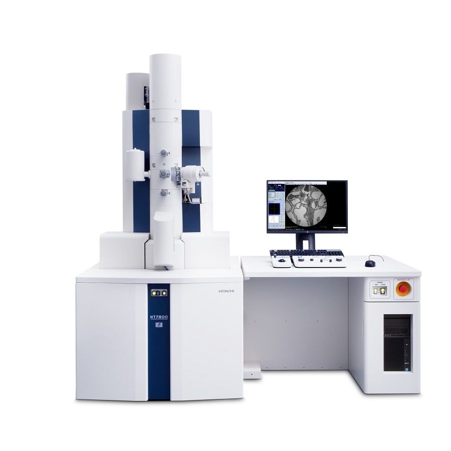
日立科学仪器(北京)有限公司为您提供《生物样品中微观形貌、内部结构检测方案(透射电镜)》,该方案主要用于生物农业中表征检测,参考标准《暂无》,《生物样品中微观形貌、内部结构检测方案(透射电镜)》用到的仪器有日立120kV透射电镜HT7800。
我要纠错
推荐专场
相关方案
 咨询
咨询