

方案详情文
智能文字提取功能测试中
SARTORIUSWhite Paper Sample UsageTime to Results August,2020 Keywords or phrases: Advanced Flow Cytometry; Target ldentification;TargetValidation; Biologics Discovery; Antibody Screening;Antibody Binding Assays; ADCC;Antibody dependent;cellular cytotoxicity;Functional Profiling; Lead Charac-terization; Bioprocessing; Cell Line Development Addressing Challenges inBiologics Discovery WithAdvanced Flow Cytometry Newly developed biopharmaceuticals are being approvedat all time high rates in the US.1 Biopharmaceutical productshave high efficacy and safety, and have the potential to treatdiseases and conditions that have no other treatment options.However, development and production of biopharmaceuti-cals is expensive, time consuming,and requires a great dealof expertise. To make innovation in this sector more viable,companies need to find ways to cut costs and speed up timeto actionable results. Flow cytometry allows researchers to look at proteins onmultiple cell types simultaneously, and works well for cells in suspension. The ability to perform multiplexed experimentson multiple cell types makes flow cytometry an attractive toolfor use in biopharma. Whilst flow cytometry has traditionallybeen considered low throughput,new advances in the tech-nology have simplified workflows and improved the speed ofsample preparation and time to actionable results. Here we outline the use of flow cytometry in four key stagesof biologics development: screening, lead development,bioprocessing, and clinical trials. Flow cytometry in screening Target Identification and Validation Identifying therapeutic targets in basic research is oftenaccomplished using CRISPR or RNAi based screens.Once possible hits have been identified,flow cytometry isuseful in validating the phenotype of each hit after knock-out, knock-in or knock-down. Flow cytometry is particularlyuseful for assaying the phenotype of cells in suspension,such as immune cells, yet throughput has been limited inthe past, making large-scale screens impossible using thismethod. High throughput flow cytometry systems are increasinglyavailable, including instruments with automated loadingof sample plates, which significantly reduce hands-on time(and total time). Furthermore, advances in autosamplingand decreased sample volume requirements have madelargerscreens feasible. Smaller sample sizes reduce boththe quantity of reagents required for the assay and thecost, as well as allowing for use of more easily translatablecell-based models.Flow cytometry is capable of measuringmultiple parameters at once, therefore complex pheno-types can be screened, facilitating identification of novelmechanisms. Aside from the physical aspect of loading andacquiring samples, data acquisition and processing canbe limiting as most systems are designed for smaller-scalelaboratory use rather than high throughput screening.Researchers should look for advanced flow cytometry plat-forms with integrated data analysis that meet the needs oftheir particular assay. After screening, researchers may wish to develop assays ormodel systems to validate hits which can be a time con-suming and daunting task. Flow cytometry can thereforebe used to determine parameters such as the efficiencyof transfecting cells with a fluorescent marker as part ofprocess optimization. Antibody Library Screening Once potential targets have been identified as describedabove, therapeutic agents need to be developed if noneexist for those targets. If the therapy will be antibody-based,appropriate antibodies must be identified that bind to thetarget. This can be achieved either by using display tech-nologies or animal immunization. Display technology-based screening Flow cytometry can help identify which of an existing libraryof antibodies orantibody-like molecules has appropriatespecificity, selectivity, and affinity to the antigen. Withthe ability to measure multiple fluorophores at once,flowcytometry enables multiplexing by combining positive andnegative cell lines and adding "off target"cell lines into asingle experimental well. Gathering data about specificity and selectivity is thus achieved with minimal sample input.Each cell line is identified by a specific fluorescent marker,allowing antibody binding to each cell type to be distin-guished and quantified. Immunization-based Antibody generation and screening Cell lines are generated that express the target, andscreened for high surface expression of the antigen. Flowcytometry allows for simultaneous analysis of both antigenexpression and cell growth, so that the best cell lines can beselected for hybridoma screening. The antigen of interest isused to immunize animals (often mice), leading to produc-tion of the antibody of interest by the animals’B-cells. Thesera from the immunized animals are screened to determinewhich animals produce high levels of antibody against thetarget antigen. B-cells from these specific animals are thenisolated using flow cytometry and fused to a myeloma cellline for hybridoma library generation. Binding Assays As previously mentioned, not every mouse will generatea sufficient quantity of antibody or antibodies that arespecific enough, so flow cytometry can be used to detectwhich mouse sera contains antibodies that bind to theappropriate antigen with high affinity and specificity. Thistype of testing was traditionally done with ELISA, howeverflow cytometry is more rapid. Instead of testing the bindingaffinity of antibodies to target and non-target antigens inturn, different spectrally distinct cell lines can be used ineach sample that express different antigens, effectively col-lapsing the ELISA workflow. Flow cytometry also allows thisscreening to be done in cells, and thus avoids the pitfallsof solid phase methods such as ELISA that may not allowligands to remain in their natural conformations. Althoughtime and sample size can still be limiting, choosing anadvanced flow cytometry platform with high throughput,low sample volume requirements, and the ability to managesheath and waste-while running samples-saves time,reagents, and money, whilst identifying the right mice touse in hybridoma generation.Additionally, ELISA assaysmay miss some antibodies due to their solid phase bindingaspect, therefore flow cytometry can be used on antigenpresenting cells and may more readily identify antibodiesthat will specifically bind antigens in living tissue. Characterization Once antibodies have been identified and are being pro-duced, they must be further characterized and tested forperformance-related characteristics before moving intopreclinical testing. Parameters such as EC50 determine thepotency of each antibody, helping determine which anti-bodies will move forward in development. Isotyping is usedto determine the correct backbone to clone into. Epitopebinning identifies the specific epitopes recognized by theantibodies. Assays for these characteristics are traditionally Comparison of sample usage and time to results in typical biologics workflows using traditional (e.g.ELISA and flow cytomers)and advanced flow cytometry platforms (e.g. IntellicytiQue Screener PLUS). done individually by ELISA, but can instead be performedtogether using an advanced flow cytometry platformcapable of multiplexing beads and cells. Using cells insuspension rather than solid phase assays like ELISA isparticularly useful for epitope binning, which often requiresthe antigen to be in its proper configuration. Not all flowcytometers have the throughput necessary to efficientlyassay the number of dilutions required for epitope binningand affinity assays, however advanced flow cytometryplatforms are available that make this feasible,particularlyif they can multiplex beads and cells, allowing for bothisotyping and quantification in the samewell. Functional Profiling Beyond the ability to bind to an antigen, an effectiveantibody for therapeutic use needs to elicit a response.The desired response may be antibody-dependent cellularcytotoxicity (ADCC),antibody-dependent cellular phago-cytosis (ADCP),antibody-dependent cytokine release(ADCR),antibody internalization, and/or acting as anagonist or antagonist of a signaling pathway.Older meth-ods to quantify these effects relied on population levelmeasurements of cell membrane integrity(cellularhealth)and release of cytokines. Flow cytometry, however, candiscriminate single cells to get information on multiple celltypes within a sample including cell membrane integrity,annexin binding, caspase activation, and specific immuno-phenotype changes. This multiplexing can greatly reducethe time to actionable results. Antibody conservation at thisstage is also critical, so miniaturization of the assay shouldbe considered. Choosing an advanced flow cytometry platform with low volume requirements and the ability toread microwell plates will assist in miniaturization. As thenumber of wells and parameters analyzed increases, thecomplexity of the analysis will also increase, especially asdata is acquired and managed on a per-well basis. It istherefore best to look for systems with integrated, simpli-fied data analysis and management. Flow cytometry in lead development Once a set of lead antibodies has been identified, the leadsmust be further characterized, with the most promisingleads undergoing modification and preclinical testing.Lead characterization reduces the risk of unknowns whenmoving forward. Traditionally this has been expensive, timeconsuming, and required expertise in techniques such aschromatography and surface plasmon resonance (SPR). Not every antibody has the biophysical characteristics tobe produced at scale, so druggability needs to be consid-ered. Protein aggregation is common in drug development,and can reduce the efficacy of an antibody, or even induceunwanted immunogenic responses in patients. Aggrega-tion is influenced by development itself, but also by thebiological characteristics of the monoclonal antibody.Understanding which antibodies are stable and less likely toaggregate, as well as the characteristics of the aggregates(versus the non-aggregated antibodies) before moving for-ward with drug development would be beneficial.At highconcentrations, monoclonal antibodies also have variable self-association behavior, which must also be understood todetermine how the antibody will behave at the high con-centrations required for therapeutic use. Analysis of binding affinities (KD) by SPR technologyrequires a largevolume of antibody. Flow cytometry-basedmeasurements of association and dissociation of antibod-ies for an antigen have been shown to correlate with SPRmeasurements. Once a small group of antibodies have been thoroughlycharacterized, preclinical development can proceed in anappropriate model system.Whatever model system is usedflow cytometry can be employed to determine the efficacy,immunogenicity, and toxicology of the treatment. Therapeutic efficacy requires binding of the antibody to itstarget,which is easily measured by traditional flow cytom-etry, and often requires recruitment of effector functionof the host. Due to its ability to multiplex and characterizecomplex mixtures of cells, flow cytometry is an excellentchoice for measuring this activity. Innate lymphoid cells(ILCs) arenot defined by a single marker, however subsetscan be defined by a combination of markers using flow cy-tometry. ILCs include effector cells, which release cytokinesin response to stimulation. Flow cytometry can thereforebe used to measure the levels of cytokines and ILCs in theblood of a preclinical model organism, in addition to mea-suring the impact that cytokine release has on other cells inthe animal. Introduction of a foreign protein into a human or otheranimal can lead to an unwanted immune response, pos-sibly leading to inactivation of the therapeutic protein byanti-drug-antibodies (and therefore lowered efficacy), oradverse effects for the patient. Although measurement ofimmunogenicity has typically been done by ELISA or SPRassays, flow cytometry can reduce costs using a miniatur-ized assay. Overall, it is a useful technology for understand-ing the impact of a biopharmaceutical in a preclinical modeldue to the ability to simultaneously measure multipleparameters; speed and volume may be limiting, but an ad-vanced flow cytometry platform with high throughput andlow sample volume requirements makes this feasible. Flow cytometry in bioprocessing Once leads have been chosen for further development,optimal clones for producing antibodies should be selectedfor development of cell lines. Analysis of both the level of,antibody production and growth characteristics of cell linesfacilitates development of cell lines that will be productiveand grow robustly, saving money and time. Cell lines usedfor antibody production are often grown in suspension, making flow cytometry a useful technique foranalyzingcharacteristics of these cells. Measuring lgG levels produced by each clone can identifyoptimal antibody producers. Simultaneously measuringmultiple parameters using flow cytometry, (such as lgGlevel using a bead-based assay) as well as cell density andhealth, provides more information about which clonesnot only produce more lgG at the point of measurement,but also which clones grow better, thus improving culture.Having this additional information allows for more informeddecisions about clone selection, which may ultimately im-prove antibody production. Although throughput can stillbea limitation, using an advanced flow cytometry platformcapable of measuring both beads and cells simultaneouslyincreases the speed at which clones can be analyzed. Flow cytometry in clinical trials One of the great strengths of flow cytometry, the ability tomultiplex assays, is beneficial when analyzing the effects ofan antibody or other biopharmaceutical in a clinical trial. As-says of immune cells (and possibly tumor or diseased cells)after in vivo treatment can measure cell-mediated cytotox-icity, cell health, function of immune cells, phenotypes, andcytokine profiling. Flow cytometry enables identification ofcells such as ILCs, which don't have a single unique marker-they can only be identified by a combination of markers.ILCs play an important role in tumor surveillance by produc-ing cytokines 4. Multiplexed assays in flow cytometry give amore complete picture of the effects of the biopharmaceu-tical agent than any individual assay. ( References ) ( 1. FDA drug approvals hit all-time high.(2019).Retrieved f rom https://cen.acs.org/pharmaceuticals/drug-development/FDA-approved-record-number- drugs/97/web/2019/01 ) 2. Cromwell, M., Hilario, E.,&Jacobson,F. (2006). Proteinaggregation and bioprocessing. The AAPS Journal, 8(3),E572-E579. doi:10.1208/aapsj080366 ( 3. Geuijen,C., Clijsters-van der Horst, M . , Cox, F., Rood, P., Throsby, M., &Jongeneelen, M. et al. (2 0 05). Affinity ranking of antibodies using flow cytometry:Application in antibody phage display-based targetdiscovery.Journal of Immunological Methods,302(1-2), 68-77. d oi: 1 0.1016/j.jim.2005.04.016 ) 4. Cerwenka, A., &Lanier, L.(2001). Natural killer cells,viruses and cancer. Nature Reviews Immunology, 1(1),41-49. doi:10.1038/35095564 Sales and ServiceContacts For further contacts, visitWww.sartorius.com Essen BioScience, A Sartorius Companywww.sartorius.com/intellicytE-Mail: info.intellicyt@sartorius.com North AmericaAPACEssen BioScience Inc.Essen BioScience K.K.300 West Morgan Road4th Floor Daiwa ShinagawaAnn Arbor, Michigan, 48108North Bldg.USA1-8-11 Kita-ShinagawaTelephone +1 734 7691600Shinagawa-ku, Tokyo140-0001EuropeJapanEssen BioScience Ltd.Telephone +81 3 6478 5202Units 2 & 3 The QuadrantNewark CloseRoyston HertfordshireSG8 5HLUnited KingdomTelephone +44 (0)1763 227400 Document version 2020|08 Find out more: www.sartorius.com/intellicyt AML(急性髓系白血病)是一种让人谈之色变的绝症,主要的治疗方法是化疗和造血干细胞移植,四十几年来新药寥寥无几,目前靶向药只有针对Bcl2的Venetoclax(维奈克拉),所以对于AML开发新的靶向药显得尤为重要和迫切。引发AML的原因是由于多个基因的突变,导致髓样干细胞分化形成一群成熟白细胞的过程受到阻碍,以及未成熟的异常细胞的增殖。转录因子HoxA9在70%的AML病人体内仍然维持活性,而在髓样干细胞正常的分化过程中该基因必须被关闭。但是到目前为止还未发现针对HoxA9表达调节的抑制剂。美国麻省总医院和哈佛干细胞研究所的研究人员在含有33万个化合物的分子库中筛选这种抑制剂,确定了新的AML的药物靶点,并发表于Cell杂志上[1]。是的!没看错,33万个化合物!不用高通量手段是万万不行的!所以20分钟即可分析一块384孔板的高通量流式iQue®大显身手!iQue® 高通量流式细胞仪药物筛选细胞系(Lys-GFP-ER-HoxA9 cells)的建立研究人员通过基因工程方法建立了能够持续表达HoxA9的AML小鼠细胞髓样细胞模型,将GFP(绿色荧光蛋白)和只有在成熟的白细胞中表达的溶菌素融合表达,当细胞达到成熟状态就会发出绿色荧光。而且为了能让HoxA9持续高效表达,将HoxA9和ER(雌激素受体)进行融合,可以通过ER的激活剂E2来对HoxA9的表达进行调节。E2不存在情况下,HoxA9表达被抑制,该细胞系分化成成熟白细胞,产生GFP荧光,并产生CD11b等成熟白细胞表面Marker。化合物筛选及功能确认研究人员筛选了超过33万个小分子(化合物来自于NIH Molecular Library Program’s Molecular Library Small-Molecule Repository (MLSMR)),希望找到能够让该细胞系产生绿色信号的小分子,绿色信号的出现意味着HoxA9 表达而导致的分化阻断得到了疏通。将每种化合物(4 μM)与细胞(约2500个细胞,共50 μL)混合,加入384孔板中,用ER的拮抗剂fulvestrant作为阳性对照。使用iQue® 对GFP+&CD11b+的药物进行高通量筛选。33万个化合物,一共1000块384孔板,iQue®可以接机械臂,1个小时可以筛选3块384孔板,一天24小时轮转可以测试72块板,5天不到就可以全搞定啦!最终筛选到12种化合物,其中有11种通过抑制一种叫做DHODH的代谢酶起到作用,截至这篇文章发表还没有研究表明DHODH在髓样干细胞分化过程中发挥作用。进一步实验表明抑制DHODH的活性确实能够诱导小鼠和人类AML细胞的分化。DHODH抑制剂Brequinar结构式随后在几种AML小鼠模型中检测了一种已知的DHODH抑制剂Brequinar,发现了一套能够抑制白血病的剂量方案,不但能够延长生存时间,而且不会出现常规化疗导致的副作用。新靶点治疗AML展望目前ASLAN公司的ASLAN003是一种口服的高活性DHODH抑制剂,有潜力成为AML治疗的全球首创(first-in-class)药物。该公司已经证明了ASLAN003对 DHODH 的有效抑制,且相比于第一代抑制剂和其他新的 AML 治疗不存在相关的毒性,在 AML 患者中都有诱导细胞分化和适用性的潜力。ASLAN003被美国食品药品监督管理局(Food and Drug Administration)授予孤儿药物称号。目前根据clinicaltrials.gov提供的信息,已经完成了AML适应症的II期临床试验(NCT03451084)[2]。ASLAN003结构式可以说没有iQue®高通量流式细胞仪,这个研究是完不成的。这款流式中的战斗机为什么牛呢?- 进样就像蜻蜓点水,速度快:5分钟即可完成一块96孔板检测,自然就可以实现高通量,最少仅需几微升样品;- 吸空气间隔样品的骚操作:“混匀-测定”,免洗流程;- 软件功能强大可以让您一饱眼福,什么EC50计算都是小Case,门一变,其他计算瞬间跟着变,边读样边分析。Download下载白皮书《iQue®高通量流式应对生物制剂发现中的挑战》,了解更多智能化高通量筛选的妙招!点击下载 获取全文-参考文献-Inhibition of Dihydroorotate Dehydrogenase Overcomes Differentiation Blockade in Acute Myeloid Leukemia.CellASLAN003, a potent dihydroorotate dehydrogenase inhibitor for differentiation of acute myeloid leukemia | Haematologica
关闭-
1/5
-
2/5
还剩3页未读,是否继续阅读?
继续免费阅读全文产品配置单
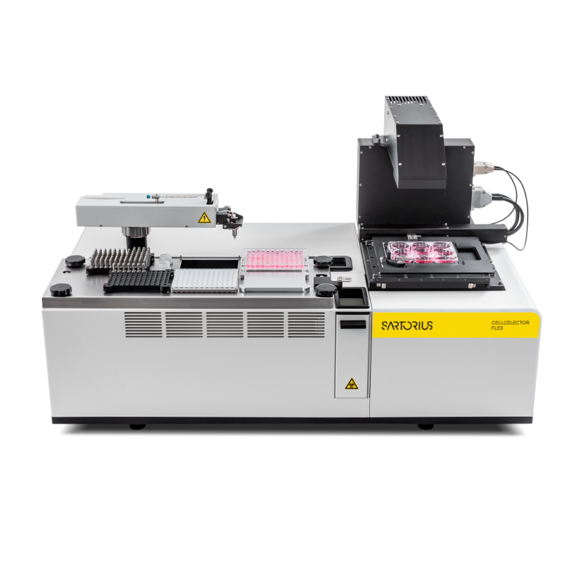
德国赛多利斯集团为您提供《生物制剂中高通量筛选检测方案(流式细胞仪)》,该方案主要用于化药制剂中其他检测,参考标准《暂无》,《生物制剂中高通量筛选检测方案(流式细胞仪)》用到的仪器有赛多利斯 CellCelector 全自动无损细胞分离系统。
我要纠错
推荐专场

相关方案
 咨询
咨询