

方案详情文
智能文字提取功能测试中
Cellular Physiologyand BiochemistryCell Physiol Biochem 2020;54:1231-1248DOI: 1033594/000000311O 2020 The Author(s)Published by Cell Physiol BiochemPress GmbH&Co.KG, Duesseldorfwww.cellphysiolbiochem.comi1231Published online: 17 December 2020This article is licensed under the Creative Commons Attribution-NonCommercial-NoDerivatives 4.0 Interna-tional License (CC BY-NC-ND). Usage and distribution for commercial purposes as well as any distribution ofmodified material requires written permission. Cellular Physiologyand BiochemistryCell Physiol Biochem 2020;54:1231-1248DOI: 10.33594/000000311 O 2020 The Author(s).Published byPublished online: 17 December 2020Cell Physiol Biochem Press GmbH&Co.KG1232Zolotoff et al.: Obstructive Sleep Apnea and Blood-Brain Barrier Cindy Zolotoff10 Rue de la Marandiere, 42270 Saint-Priest-en-Jarez (France)Tel. +33477421452, E-Mail cindy.zolotoff@univ-st-etienne.fr Accepted: 8 December 2020 Original Paper Intermittent Hypoxia and Its Impacton Nrf2/HIF-1a Expression and ABCTransporters: An in Vitro Human Blood-Brain Barrier Model Study Cindy Zolotoffa.b Anne-Cloe VoirinabClementine PuechbFrederic Rocheb,cNathalie Pereka aINSERM, U1059, Sainbiose, Dysfonction Vasculaire et Hémostase, Universite de Lyon, Universite JeanMonnet, Saint-Etienne, France, bEA 4607 Systeme Nerveux Autonome-Epidemiologie PhysiologieIngenierie Sante, Universite de Lyon, Universite Jean Monnet,Saint-Priest-en-Jarez, France, ‘Service dePhysiologie Clinique et de I'Exercice, Centre VISAS, CHU, Saint Etienne, France Key Words Intermittent hypoxia·Oxidative stress·Blood brain-barrier model·ABC transporters·Tightjunction proteins Abstract Background/Aims: Obstructive sleep apnea (OSA) is characterized by repeated episodes ofcomplete or partial obstruction of the upper airways, leading to chronic intermittent hypoxia(IH). OSA patients are considered at high cerebrovascular risk and may also present cognitiveimpairment. One hypothesis explored is that disturbances may be linked to blood-brain barrier(BBB) dysfunction. The BBB is a protective barrier separating the brain from blood flow. TheBBB limits the paracellular pathway through tight and adherens junctions, and the transcellularpassage by efflux pumps (ABC transporters). The aims of this study were to evaluate the impactoflH and sustained hypoxia (SH) on a validated in vitro BBB model and to investigate the factorsexpressed under both conditions. Methods: Exposure of endothelial cells (HBEC-5i) in our invitro model of BBB to hypoxia was performed using IH cycles: 1%02-35 min/18% O2-25 minfor 6 cycles or 6 h of SH at 1%02. After exposure, we studied the cytotoxicity and the level ofROS in our cells. We measured the apparent BBB permeability using sodium fluorescein, FITC-dextran and TEER measurement. Whole cell ELISA were performed to evaluate the expressionof tight junctions, ABC transporters, HIF-1a and Nrf2. The functionality of ABC transporterswas evaluated with accumulation studies. Immunofluorescence assays were also conductedto illustrate the whole cell ELISAs. Results: Our study showed that 6 h of IH or SH induced aBBB disruption marked by a significant decrease in junction protein expressions (claudin-5,VE-cadherin, ZO-1) and an increase in permeability. We also observed an upregulation in ( N. Perek and F. Roche contributed equally to this work. ) ( SNA-EPIS-SAINBIOSE, Faculte de Medecine -Campus Sante Innovations, ) P-gp protein expression and functionality and a downregulation in BCRP. Hypoxia inducedproduction of ROS, Nrf2 and HIF-1a. They were expressed in both sustained and intermittentconditions, but the expression and the activity of P-gp and BCRP were different. Conclusion:Understanding these mechanisms seems essential in order to propose new therapeuticstrategies for patients with OSA. 2020 The Author(s). Published by Cell Physiol Biochem Press GmbH&Co. KG Introduction Obstructive sleep apnea (OSA) is a serious sleep disorder affecting approximately 5%of the adult population over 45 years old and more than 10% after 65. OSA is characterizedby repeated episodes of complete or partial obstruction of the upper airways during sleep,resulting in chronic cyclical intermittent hypoxia (IH) [1]. IH plays a crucial role in the increaseof cardiovascular and cerebrovascular risks [2]. Increasing data support a time relationshipbetween OSA and cognitive impairment [3], but a causal link has yet to be established.A hypothesis has been raised [4] in which IH acts as a biostressor that could disrupt theblood-brain barrier (BBB] via molecular responses. Adaptive responses at the BBB levelmay occur specifically through regulation of BBB transporters, like the ATP binding cassette(ABC) transporter family when altering BBB microvessel permeability. These changes in BBBtransporters may have long-term consequences that alter the brain's microenvironment,leading in the long-term to cognitive impairment [4]. The BBB is a protective barrier separating the brain from blood flow and is composedprimarily of endothelial cells closely related to astrocytes, pericytes and neurons, whichtogether form the neurovascular unit [5]. The BBB has tight [6] and adherens junctions[7] which allow it to control the exchanges between the brain and blood flow and to limitpermeability. Furthermore,the transmembrane proteins that make up the tight junctions (TJ) (zonulaoccludens (ZO)-1, claudin-5, etc.] and adherens junctions (e.g., vascular endothelial (VE)-cadherin), limit the paracellular transfer of molecules [8] and strengthen interactionsbetween endothelial cells. Brain microvessel endothelial cells also express transportersthat will allow blood-brain regulation and adaptation such as ATP ABC transporters, andmore precisely breast cancer resistance protein (BCRP] and/or P-glycoprotein (P-gp] [9].These two latter transporters are the most expressed and involved at the human BBB level.Moreover, P-gp may efflux xenobiotic components or glutamate [4] and BCRP is involvedin the efflux in the blood flow of uric acid [10]. All those microstructural cell elements areessential to ensure the functional integrity of the BBB. Thus, under hypoxic conditions, manyfactors are secreted and induce a modification in the expression of junction proteins or in theactivity of ABC transporters. IH or sustained hypoxia (SH) produces reactive oxygen species(ROS) [11] thatlead to oxidative stress [12,13] and contribute to inflammatory cascades [14].Indeed, the formation and accumulation of ROS in hypoxic cells is a hallmark of hypoxiainvolved in BBB dysfunction [15]. In association with the production of ROS, severalfactors are activated or stabilized. Effectively,hypoxia-inducible factor 1 (HIF-1), and morespecifically HIF-1c, is stabilized [16] and stimulates the transcription of genes involved invarious biological processes such as angiogenesis, cell proliferation, inflammation or cancer[17, 18]. Nuclear factor erythroid 2-related factor 2 (Nrf2] expression is also modulated inresponse to hypoxia [19]. Nrf2 controls the expression of antioxidant defence genes underhypoxic conditions, allowing cells to regulate the oxidative stress mediated ROS species.Nrf2 regulates the expression of these genes by binding to antioxidant response elementsin their promoter regions, which reduce the levels of damaging ROS in the cell [20]. Thosetranscription factors are known to modify the expression and activity of ABC transporters,such as HIF-1a which induces an increase in P-gp expression in hypoxia colon carcinomacells [21]anda decrease in BCRP expression [22] in choriocarcinoma cells also under hypoxia.To determine how IH causes adaptative metabolic response at the BBB level, implying Nrf2, HIF-1a and ABC transporter expressions, we proposed to use a human in vitro BBB modeldeveloped in our laboratory [23] and setting up a model of intermittent hypoxic exposure. Currently, there are few in vitro models to study the effects of IH encountered in OSA.Indeed, there are no standardized protocols. Some studies carried out"short" cycles (3 minof normoxia followed by 15 s of hypoxia) [24,25], others were "longer”(25 min of hypoxiafollowed by 35 min of normoxia [26,27] or 1 h of hypoxia followed by 30 min of normoxia[28]). Each model has advantages and disadvantages.The severity of hypoxia also seems tovary greatly between the different models [29]. We chose 1% oxygen in the environmentof the cell that represents the pathological percentage of oxygen exposure observed inthe in vivo brain. We therefore decided to develop our own model with alternating 35 mincycles of hypoxia at 1%0, followed by cycles of reoxygenation with 18% 0, for 25 minduring 6 cycles on our model of BBB. The 6-h duration is important and representativeof one short night similar to the pathological conditions found in OSA. For the human invitro BBB model, we used HBEC-5i human brain endothelial cells and human astrocyte(HA) conditioned medium [23]. We first looked at the effects of this IH on a human BBBmodel: transendothelial electrical resistance (TEER] and apparent permeability (P) withthe expression ofVE-cadherin, claudin-5 and ZO-1 junction proteins. Then ABC transporter[P-gp, BCRP) expression and functionality were evaluated with accumulation studies andfluorescent selective substrates and inhibitor combinations. We also evaluated the level ofintracellular ROS and the expression of two transcriptional factors expressed under hypoxicconditions, such as Nrf2 and HIF-1a. The purpose of the study was to 1) compare the effects of SH and IH, 2] quantify proteinexpressions of junctions and ABC transporters,3] characterize the effects of IH on the activityof ABC transporters, and 4) identify the expression of Nrf2 and HIF-1a under IH and SH. Materials and Methods Chemicals and reagents Cell culture inserts for 24-well plates (high density pore, 0.4-um pore diameter size, translucentpolyterephthalate ethylene (PET) membrane, culture flasks and companion plates were purchased fromDominique Dutscher (Strasbourg,France).Imaging plates FC, 96 wells, TC-surface were purchased fromMoBiTec (Goettingen, Germany). Rabbit polyclonal to VE-cadherin (VE-cadherin-ab33168] for ELISA and immunofluorescence (IF)were purchased from Abcam (Cambridge, United Kingdom). Rabbit polyclonal anti-ZO-1(ZO-1-ab59720) forIF was purchased from Abcam (Cambridge, United Kingdom). Secondary antibodies for IF: goat anti-rabbitantibodies (A-21127 Alexa Fluor 488), DAPI for microscopy and superFrostTM were obtained from ThermoFisher Scientific (Waltham, USA). Mouse monoclonal IgG anti-ABCG2 (sc-18841), mouse monoclonal anti-rabbit IgG to P-gp [sc-390883), mouse monoclonal anti-claudin-5 (sc-374221] and secondary antibody forcell ELISA mIgG BP-HRP [sc-516102) were purchased from Santa Cruz Biotechnology (Dallas, Texas, USA).Rabbit ZO-1 polyclonal antibody for ELISA [40-2200), Dulbecco’s Modified Eagle Medium/Nutrient MixtureF-12 (11330057), rhodamine-123, secondary antibodies for IF: goat anti-rabbit antibodies (A-11034 AlexaFluorTM 488), DAPI for microscopy,superFrost microscope slide and Coomassie PlusTM protein assay wereobtained from Thermo Fisher Scientific (Waltham, USA). Vectashield Antifade mounting medium (H-1000]iwas purchased from Eurobio Scientific. OxiSelect M Intracellular ROS Assay Kit (green fluorescence,STA-342] and HIF-1a Cell Based ELISA Kit[CBA-281) were purchased from Cell Biolabs (San Diego, USA]. NFE2L2 ELISA Kit (human) (OKCD02754)was purchased from Aviva Systems Biology (San Diego,USA]. Astrocyte medium (AM),astrocyte growth supplement (AGS), fetal bovine serum (FBS) and penicillin-streptomycin solution for astrocytes were purchased from CliniSciences (Nanterre, France). Amphotericin B(30-003-CF) and Trypsin 2.21 mM EDTA were purchased from Corning (Manassas, USA). Penicillin-streptomycin (100X) was purchased from PanReac Applichem (Darmstadt,Germany). All other reagents were purchased from Sigma-Aldrich (St Quentin Fallavier, France). Zolotoff et al.:Obstructive Sleep Apnea and Blood-Brain Barrier In vitro BBB model The BBB model was composed of HBEC-5i endothelial cells (from ATCC-Manassas,VA, USA) and a HAcell line (ScienCell Research Laboratories, Carlsbad,CA,USA) cultured in PET transwells with 0.4-um poresand in plates [23]. Initially, HBEC-5i were cultured in DMEM/F12 HAM, supplemented with 10% FBS, 40 ug/mLendothelial cell growth supplement (ECGS) and 1% antibiotic antimycotic solution. HAs were grown inAM containing 2% FBS, 1% AGS and 1% penicillin-streptomycin solution according to manufacturer’sinstructions. Afterwards, HA were in the same medium as HBEC-5i. In our model,HBEC-5i endothelial cellswere in contact with astrocytic conditioned medium. In fact, in addition to playing a role in the inductionand long-term maintenance of barriers, astrocytes can release chemical factors that modulate endothelialpermeability [30]. The conditioned medium corresponds then to a medium that has remained in contactwith the astrocytes for 48 h. For each condition, the medium was renewed every 2 d. HBEC-5i with HAconditioned medium reached an optimal steady state plateau at day 14 which was maintained for 5 d [23].Cells were cultured at 37°C with 5% CO. Experimental hypoxia and IH protocols For this protocol we used a hypoxic workstation: Baker Ruskinn,Invivo, 300 and the ICO,N,IC AdvancedGas Mixing System (Maine, USA). This hypoxia chamber maintained and controlled temperature, oxygen,humidity and carbon dioxide. The ICO,N,IC provided accurate control over 0,[0.1%-20.9%] and CO, [0.1%-30%). In our model, two incubation conditions were performed: SH or IH. Under SH, cells were incubatedfor a continued period of 6 h at 1% 0,and 5% CO,. Under IH, cells were exposed to repeated hypoxia (35min, 1%O,)/reoxygenation (25 min, 18%0,) for 6 cycles. We decided to use the hypoxia chamber at 1%0,since in the brain under physiological conditions, the oxygen level is close to 5%. Thus, we used the lowestlevel of oxygen possible in which we had no cytotoxic effects on the cells but where a response was observed. For each condition,DMEM/F12 was conditioned the day before at 1% 0, in this hypoxic chamber. Inorder to ensure that the oxygen balance was achieved, we used an oximeter (Multi 3510 IDS, FDO 925, WTW,Germany) to control the percentage of dissolved oxygen within the medium. The conditioned medium of HAwas also conditioned according to the two conditions of the model. Control normoxic cells were maintainedin normal atmospheric gas pressure. Cytotoxicity assays MTT assay was employed to measure cellular metabolic activity during IH and SH. Cells were seededat a density of 10,000 cells/well in permeable 96-well plates and cultured at 37℃ with conditioned mediumof HA. Once at confluence, cells were exposed at IH or SH for 6 h. Control cells under normal atmosphericgas pressure also received the MTT solution. Formazan production was assessed at 570-nm (MultiskanTMRC, Thermo Fisher Scientific, France). Metabolic activity was measured by comparing results from normalatmospheric gas pressure conditions, considered as 100% metabolically active. Then we also evaluated cell death using a LDH activity method to confirm the least cytotoxicconcentrations given by MTT results. Cells were seeded at a density of 10,000 cells/well in permeable 96-well plates and cultured at 37°C with conditioned medium of HA. Once at confluence, cells were exposed atIH or SH for 6 h. Control cells under normal atmospheric gas pressure also used in the LDH test. In this kit,LDH reduces NAD to NADH, which is specifically detected by a colorimetric assay (450-nm). Imaging plates we use are covered with a sheet of high-performance fluorocarbon film. This film allowsvery high gas transfer rates. Thus, physiological experiments on cells with exact control of the oxygen partialpressure in the cell microenvironment are possible. Determination ofintracellular ROS levels To determine ROS activity in our model, we employed The OxiSelectM Intracellular ROS Assay Kit.Cells were seeded with conditioned medium of HA in permeable 96-well plates and then incubated withdichlorodihydrofluorescein diacetate (DCFH-DA] at 37°C for 1 h. Then cells were exposed to IH or SH for 6h. After that, fluorescence was read with a fluorimetric plate reader at 480-nm/530-nm. ROS results weredetermined by comparison with a dichlorodihydrofluorescein (DCF) standard curve. Zolotoff et al.: Obstructive Sleep Apnea and Blood-Brain Barrier Quantification ofHIF-1a and Nrf2 and during hypoxia and IH For both factors, cells were cultured into permeable 96-well plates with conditioned medium of HAfor 6 h of IH or SH. Then HIF-1a and Nrf2 expressions were measured with a cell-based ELISA assay (CellBiolabs and Aviva Systems Biology) according to the manufacturer's instructions. Barrier properties P was assessed using hydrophilic fluorescents molecules: sodium fluorescein (Na-Fl MW=376 Da]and fluorescein isothyocyanate dextran (FITC-dextran, MW=4kDa). Ringer HEPES with 10 ug/mL of Na-Fl was loaded onto the apical side of the insert and incubated at 37°C for 1 h. Fluorescence was measuredwith a fluorescence spectrophotometer (Fluoroskan Ascent M, Thermo Fisher Scientific, France] at 485-nmexcitation and 530-nm emission wavelengths. P is expressed in cm.s- and was calculated using theformula used in our previous work (Puech, et al. [23]). TEER was recorded using an EVOMresistance meter with STX-2 electrodes to characterize theformation of a tight endothelial cell monolayer. One electrode was placed on the luminal side and the otherelectrode on the abluminal side. The measurement of a blank filter (without cells) was performed and thesignal was subtracted from that recorded for the filter with cells. Then the resulting value was reportedto the membrane area to obtain the TEER measurement in Q.cm². HBEC-5i with HA conditioned mediumreached an optimal steady state plateau at day 14 which was maintained for 5 d. Measurements were thenconducted at this time [23]. Whole cell ELISA Cells were seeded into permeable 96-well permeable plates at a density of 10,000 cells/well andcultured at 37°C with conditioned medium of HA. At confluence, cells were exposed to IH or SH. Then cells were fixed with 4% paraformaldehyde and permeabilized with methanol with H,0,.Claudin-5, ZO-1, VE-cadherin, P-gp and BCRP expressions were evaluated in our model with primarypolyclonal antibodies diluted respectively at 2 ug/mL, 5 ug/mL, 1/200 and 2 ug/mL for P-gp and BCRP andincubated at room temperature for 2 h or overnight at 4°C. Secondary IgG antibody diluted 1/2500 or1/500was incubated at room temperature for2 h. Tetramethylbenzidine (TMB) substrate was added into eachwell. At the end, the reaction was stopped by addition of 1 N hydrochloric acid and was measured at 450-nm. Our results were normalized with the total protein content as assessed by Coomassie blue total proteinassay,according to the manufacturer’s instructions. Immunofluorescence assays Firstly, cells of inserts were fixed and then permeabilized with glacial methanol (placed at-20°C) for 6min. The nonspecific sites were then blocked for 30 min at 37°C. The primary antibody was diluted 1/200in the blocking solution for 1 h at 37°C. The secondary antibody was diluted 1/500 in the blocking solutionfor 45 min at 37°C. Finally, the nuclei were detected using DAPI diluted 1/1000 in the blocking solution for30 min at room temperature. The inserts were then placed on glass slides and covered with a Vectashieldmounting medium, and glass coverslips were placed on the inserts. The junction staining was performedusing an epifluorescence microscope (ZEISS, Axioskop 40) equipped with ZEN software. ABC transporter functional assay by accumulation The functionality of two ABC transporters, BCRP and P-gp, was evaluated with accumulation studies.The transport of specific substrates, rhodamine-123 (10 _M), with or without inhibitors of BCRP and P-gpwas quantified. Cells were cultured with conditioned medium of HA and submitted to normal atmosphericgas pressure, 6 h of IH or SH. Then cells were pretreated with inhibitors: KO-143 (10 uM) for BCRP andverapamil (100 pM) for P-gp, 15 min at 37°C. Rhodamine-123 was then diluted in each inhibitor solutionand incubated for 1 h at 37°C. At the end, cells were washed with cold PBS and lysed with sodium dodecylsulfate with sodium borate.Fluorescence was measured with a fluorescence spectrophotometer at 485-nmexcitation and 530-nm emission wavelengths. Results were expressed as optical density (OD) per mg of totalprotein. Zolotoff et al.:Obstructive Sleep Apnea and Blood-Brain Barrier Statistical analysis Data were analyzed using Graphpad Prism 6 software (San Diego, CA, USA). These results are tested byone-way ANOVA (nonparametric) using Tukey's post hoc tests and P<0.05 was considered as significantlydifferent from controls. Results are presented as sample mean±SEM. Results Cytotoxicity assays Before conducting experiments on our model of BBB, we verified that SH and IH did notinduce cell death. Cellular toxicity was evaluated with a MTT test and then the LDH activitywas measured to evaluate cell death. We chose to perform cycles of IH and SH at 1% 0,since under normal oxygenation conditions the brain is close to 4.6%0, [31]. Indeed, inbrain cancers (glioblastoma), considered as highly hypoxic tissue, the oxygenation rate ofthe tissue is close to 0.7-0.8%O,[29]. Actually, there is no standardized protocol for an IHmodel reflecting the conditions seen in OSA. There are fast cycles [24, 25] and longer cycles[26, 28, 32]. Based on several published papers, we decided to use the following cycles: 35min hypoxia at 1%0, followed by 25 min of reoxygenation at 18% 0, repeated for 6 cycles.Since no significant cytotoxic effects were observed (Fig. 1), these parameters were chosenfor all subsequent experiments. Impact ofIH on HBEC-5i model TEERandPappmeasurements. We could observe the effects of IH and SHon our BBB modelcomposed of endothelial cells cultivated with astrocyte conditioned medium. According toPuech et al. [23], HBEC-5i with HA conditioned medium reached an optimal steady stateplateau at day 14 which was maintained for 5 d. The experiments were done when theoptimal TEER was obtained. The impact of IH on TEER is shown in Fig. 2A. For IH cycles,TEER decreased significantly by 16.4%, values varied from 40.8±0.7 to 34.1±0.5 Q.cm²(p<0.001). Compared to 6 h of SH, we also observed a significant decrease of 19.2% withvalues from 40.8±0.7 to 32.9±0.5 Q.cm²(p<0.001). P (Fig. 2B] was measured after 6 h of IH or of SH. For 6h of IH, a significant increasein coefficient permeability of 93.5% was observed with values from 8x10±0.5x10⁶ to16.2x10-6±0.08x105 cm.s"1(p<0.001) for Naf-Fl. If we compare this result with 6 h of SH, Fig. 1. Cytotoxicity effects for 6 h of IH or 6 h of SH in our model of blood-brain barrier measured by a MTTassay (A) and LDH activity method (B). Values are presented as mean value ± SEM [n=6, N=2 for MTT assayand n=10, N=2 for LDH activity). SH: sustained hypoxia, IH: intermittent hypoxia, control: normal atmo-spheric gas pressure. Fig. 2. Transendothelial electrical resistance (TEER) (A) and apparent permeability (Papp] for Na-Fl (B] orFITC-dextran (C) after 6 h of IH or SH in our model of blood-brain barrier. Results are represented as meanvalue±SEM(n=9,N=3], ***ps0.001 compared to control condition. SH: sustained hypoxia, IH: intermittenthypoxia, control: normal atmospheric gas pressure. we also observed a significant increase of 85% with values between 8x10-±0.5x10⁶ to15.51x10-6±0.10x10⁻⁵cm.s-1 (p<0.001) for Na-Fl. Our results with FITC-dextran confirmedour previous data. We observed a significant increase in coefficient permeability of 89%with values between 3.8x10⁻⁶±1.2x10⁻⁶ to 7.2x10⁻⁶±1.2x10 cm.s1 ((p<0.001) at 6 hof SH. At 6 h of IH, we also observed a significant increase of 84% with values between3.8x10-6±1.2x10⁻⁶to 7x10⁶±1.13x1075 cm.s-1(p<0.001). Expression oftight (claudin-5, ZO-1] and adherens (VE-cadherin) junctions. We evaluatedhow the impact on the tightness of BBB was correlated with an alteration of tight andadherens junctions. VE-cadherin,Z0-1 and claudin-5 are the most represented cell junctionsin our model. After 6 h of IH or SH in our in vitro BBB model, cell junction expressions wereevaluated with whole cell ELISA assay (Fig.3]. For IH, we observed a significant decrease inthe expression of all candidate proteins (VE-cadherin, ZO-1 and claudin-5). Expression ofVE-cadherin (Fig. 3A] was significantly decreased by 19.7% (p<0.017). For tight junctions,expression of ZO-1 (Fig. 3B) was significantly decreased by 23% (p<0.045) and claudin-5expression (Fig.3C) showed a decrease of 27% (p<0.047). For comparison with 6 h of SH, we observed a higher decrease in expression of each celljunction than IH. Expression of VE-cadherin decreased by 57%, 58% for ZO-1 and 50% forclaudin-5 (p<0.001). In order to illustrate and confirm the results obtained with cell ELISA, we performedimmunofluorescence assays on VE-cadherin and ZO-1 junctions. ImmunofluorescenceVE-cadherin and ZO-1 assays (Fig. 4A and 4B) confirmed whole cells assays; BBB showedimportant disruptions at cell-cell junction zones at 6 hofIHand6 hof SH (white arrows,Fig.4)compared to control. Fig. 3. Expressions of VE-cadherin (A], Z0-1 (B] and claudin-5 (C) after exposure of cells to 6 h of IH or SHin our model of blood-brain barrier. Results are represented as mean value±SEM [n=9,N=3), **p≤0.01,***ps0.001 compared to control conditions. SH: sustained hypoxia, IH: intermittent hypoxia, control: normalatmospheric gas pressure. Expressions ofABC transporters: BCRP and P-gp In several studies, a dysregulation of transporters was observed in response to IH.For this reason,the expression of two main transporters (BCRP and P-gp] were evaluatedby whole cell ELISA (Fig. 5). Expression of BCRP (Fig. 5A) significantly decreased by 50%after 6 h of IH (p<0.001). Moreover, after 6 h of SH, a significant decrease of 22% was alsoobserved (p<0.002). In contrast, expression of P-gp (Fig. 5B) showed a significant increaseof 68% after 6 h of IH. For 6 h of SH exposure, an even more significant increase of 104% wasobserved (p<0.001) for this protein. Functionality ofABC transporters: accumulation studies We verified the functionality of ABC transporters in our endothelial cells. For this,rhodamine-123 which is a substrate for both P-gp and BCRP was studied. We looked at theaccumulation of rhodamine-123 in endothelial cells with and without inhibitors of BCRPand P-gp (K0143 and verapamil, respectively). Inhibitor concentration and cytotoxicity weremeasured in previous studies [23]. Inhibitors allowed a better accumulation of Rhodamine123, proving that these two ABC transporters are functional (Fig. 6. After IH or SH, weobserved a global decrease in Rhodamine 123 accumulation, which is more important inSH conditions than IH. This proved that ABC transporters were more active after hypoxia.The use of verapamil allowed an important Rhodamine-123 accumulation in line with amore important expression and functionality of P-gp after hypoxia. Moreover, with KO 143,Rhodamine-123 is more accumulated only after SH conditions, no differences were shownafter IH. This discrepancy is in line with a lower BCRP expression after IH reflected thefunctionality of this protein. Zolotoff et al.:Obstructive Sleep Apnea and Blood-Brain Barrier Fig.4. Immunostaining of VE-cadherin (A] or ZO-1 (B] after exposure of cells to 6 h ofIH or SH in our modelof blood-brain barrier. SH: sustained hypoxia, IH: intermittent hypoxia, control: normal atmospheric gaspressure. Scale bar=100 um. Fig. 5. Expressions of BCRP [A] and P-gp (B] after 6 h of IH or SH in our model of blood-brain barrier Resultsare represented as mean value ± SEM[n=9,N=3),*p≤0.05,**ps0.01,***ps0.001 compared to control con-ditions. SH: sustained hypoxia, IH: intermittent hypoxia, control: normal atmospheric gas pressure, BCRP:breast cancer related protein,P-gp: P-glycoprotein. O 2020 The Author(s). Published by Published online: 17 December 2020Cell Physiol Biochem Press GmbH&Co.KG Fig. 6. Functionality of rhodamine-123 transportby accumulation test with or without specific in-hibitors of BCRP (KO 143) and P-gp (verapamil),after exposure of cells to 6 h of SH or IH. Resultsare represented as mean value ± SEM [n=10, N=2),*ps0.05 between rhodamine and rhodamine + KO143, ***p≤0.001 between rhodamine before inhi-bition versus rhodamine with inhibitors. SH: sus-tained hypoxia, IH: intermittent hypoxia, control:normal atmospheric gas pressure, BCRP: breastcancer related protein, P-gp: P-glycoprotein. The measurement of the activity on BCRP was performed despite a significant decreasein its expression in both conditions in order to see if the proteins, although in smallerquantities, would be active or not. In the presence of the competitive BCRP inhibitor, K0143,we observed an increase of 22% in rhodamine-123accumulation (p=0.023). In comparison,in the presence of verapamil, the competitive P-gp inhibitor, we observed an increase of 38%in rhodamine-123 accumulation (p<0.001). First, we showed that after exposure to 6 h of SH, we had a decrease of 23% inrhodamine-123 accumulation (p=0.012], showing that ABC activity reflected by a moreimportant efflux was more important after SH. Competitive inhibition with K0143 allowedan increase of 44% in rhodamine-123 accumulation.Competitive inhibition with verapamilallowed a more important increase of 68% (p<0.001) in accumulation. For 6 h of IH, the rhodamine accumulation was the same as observed in controlcondition. In the presence of K0143, there was a slight but nonsignificant decrease in theaccumulation of rhodamine-123,probably related to the underexpression observed in BCRPexpression after 6 h of IH. However, with verapamil, we observed a significant increase of63% in rhodamine-123 accumulation (p<0.001), in line with an increase of P-gp expression. Expressions ofROS, HIF-1a and Nrf2 in response to IH and SH ROS levels. As a first step, we verified if IH and SH induced ROS production in our modelof BBB with the DCFH-DA assay. The DCFH-DA diffuses through the membrane cell and isthen intracellularly converted to hydrophilic DCF. Once in the cells, DCF reacts with RoSallowing its fluorescence to be measured at 570-nm. As shown in Fig. 7, significant increasein fluorescence intensity was detectable after 6 h of SHor IH. For SH, ROS generation showedan important 4-fold increase from 181.28±10.9 to 722.6±46.1 relative fluorescence units(RFU)(p<0.001). Under IH conditions, ROS production showed a 3-fold increase from181.3±10.9 to 520±34.3 RFU (p=0.0012]. HIF-1a and Nrf2. IH and SH also regulate several transcription factors such as HIF-1a andNrf2. First, we analyzed a key mediator of the cellular response to hypoxia, HIF-1α [Fig. 8A].IH exposure increased HIF-1o expression of endothelial cells by 92%. Whereas SH produceda 161% increase in expression of HIF-1. Secondly, we analyzed Nrf2 transcription factor,which has an important role in the cellular response to ROS (Fig. 8B]. In IH conditions, Nrf2expression increased by 152%. For SH, an even greater increase was observed with anincrease of 200% (all p<0.001) in the expression of Nrf2. zolotoff et al.:Obstructive Sleep Apnea and Blood-Brain Barrier Fig. 7.ROS production was measured by DCF fluo-rescence after 6 h of normal atmospheric gas pre-ssure,IH or SH in our model of blood-brain barrier.1000 M H,0, was used as a positive control. DCFfluorescence levels were measure after these con-ditions. Values are presented as mean value ± SEM(n=9,N=3),* ps0.05, ** p≤0.01, *** p≤0.001 com-pared to control condition. SH: sustained hypoxia,IH: intermittent hypoxia, control: normal atmo-spheric gas pressure, ROS: reactive oxygen species,DCF: dichlorodihydrofluorescein. Fig. 8.Expressions of HIF-1o (A], and Nrf2 (B) after exposure of cells 6 h of IH or SH in our modelof blood-brain barrier. Results are represented as mean value ± SEM [n=9, N=2],*** p≤0.001 compared to controlcondition. SH: sustained hypoxia, IH: intermittent hypoxia, control:normal atmospheric gas pressure, HIF-1c: hypoxia-inducible factor 1 alpha, Nrf2: nuclear factor erythroid 2-related factor 2. Discussion OSA is a major health problem characterized by repeated episodes of hypoxia-reoxygenation. IH is associated with high cardiovascular or cerebrovascular risk andcognitive disorders. A hypothesis has emerged the last years that cognitive impairment maybe linked to the opening of the BBB and leading to neuronal alteration [4]. In order to betterunderstandthe cellular mechanism under IH conditions, an in vitro model appeared suitable.We have used the human BBB model developed in our laboratory to study effects of IH. The BBB, under physiological conditions, protects the brain from blood circulation. Tightandadherentjunctions which composed them limitin particularthe paracellular passage [33].Maintaining the homeostasis of the BBB is also possible thanks to the presence of ABCtransporters like P-gp or BCRP. In order to better characterize IH effects we have comparedthese effects with SH effects, which were better established in the literature. Under SH,tight junctions are known to be altered and less expressed at the BBB [34, 35],leading toan increase in paracellular flux. In our study, we could characterized this disruption underSH as expected and published by others [36, 37]. However, few studies have examinedthe effects of IH on tight and adherens junctions of BBB. After 6 h of IH, we observed anincrease in membrane permeability allowing the passage of small molecules such as Na-Fl or Dextran (Fig. 2). This increased permeability is now known to be associated witha decreased expression of claudin-5, VE-cadherin and ZO-1 (Fig. 3 and 4). After repeatedhypoxia-reoxygenation processes,claudin-5 [38] and ZO-1[39] were altered and showedevidence of an increased permeability of the endothelium of the cerebral microvessels. We then evaluated the effects of IH on ABC transporters, especially BCRP and P-gp. Adecrease in BCRP expression and an increase in P-gp expression were observed for bothhypoxic conditions (Fig. 5). Similar data are found in the literature for both SH [22, 40]and IH conditions [41, 42]. Moreover, on ABC transporters there are sites that bind and/orhydrolyze cytoplasmic ATP. Hydrolyzed ATP provides the energy necessary to transport asubstrate in one direction against a concentration gradient. To study this effect, we performedfunctionality tests with a common substrate for BCRP and P-gp using rhodamine-123. Twodistinct ways of response were observed and are presented in Fig. 5. Indeed, we observed anincrease in activity of P-gp and BCRP in SH correlating with the study of [43]. In contrast, weobserved an isolated increase in P-gp activity for IH. This latter result is probably in line withthe underexpression of BCRP after 6 h of IH as shown in other study [44]. This decrease wasnot linked to ATP level because we had observed an increase in activity of P-gp at the sametime, and we did not observe the same effects between SH and IH. Moreover, there is strongcooperation between P-gp and BCRP. It is recognized that in the absence of activity of one ofthem, the other membrane protein is sufficient to prevent the entry of compounds into thebrain [45]. In response to hypoxia, many mechanisms are activated like stress oxidative and hypoxiapathway. In our work, we evaluated two transcription factors Nrf2 and HIF-1a. Initially,oxidative stress is observed, leading to the production of ROS [46-48]. Regulation of ROSis essential for maintaining cellular homeostasis. In response to the production of ROS, thetranscription factor Nrf2 increases and gives the cell the ability to produce antioxidant factorswhich reduce the damaging impact of intracellular ROS [20]. We demonstrated that IH likeSH could also induce transcription factors sensitive to the oxygen content of endothelial cells,hypoxia inducible factors and in particular HIF-1. Furthermore, Nrf2 and HIF-1a signallingare both regulated by the presence of ROS [49]. These two conditions allowed a significantincrease in ROS (Fig. 7). Our results are in agreement with the literature: an increase in ROSin acute hypoxic conditions in the endothelial cells of BBB [50] or in other tissues such asnasal polyp-derived fibroblasts [51]. The same observation is found in conditions of IH inthe BBB [52-54]. Furthermore, this increase in ROS is directly correlated with an increasein HIF-1o and/or Nrf2 activity (Fig.8).Indeed, the expression of HIF-1o known to increaseunder hypoxic conditions is correlated with ROS production [32].Thus, in line with theliterature we observed a higher expression of this factor in SH compared to IH conditions[551. The production of ROS under SH or IH conditions is also known in the literature toincrease the expression of Nrf2 [56], which is essential for regulating responses to oxidativestress. Moreover, the longer the duration of hypoxia, the greater is the expression ofNrf2 [57]. In our study, the expression of junction proteins is much lower under SH conditions,which can be explained by the higher ROS production in our model. Indeed, the productionof ROS is known to alter BBB functioning. Schreibelt et al.[58] showed that ROS activatea cascade leading to a rearrangement of the cytoskeleton and the disappearance of tightjunctions including claudin-5 in the BBB. Moreover, disorganization/breakdown of thesejunctions is associated with excessive production of ROS found in many neurodegenerativedisorders like Alzheimer's or Parkinson’s diseases [59]. In addition, HIF-1a also a candidateto BBB alteration is more expressed in SH than in IH, which may explain the expression ofmore altered junction proteins in SH [36]. In addition, a downstream gene of HIF-1c thatwould be very interesting to look at is the vascular endothelial growth factor (VEGF). Indeed,this factor increases in hypoxic conditions through the HIF-1a pathway and leds notably toan increase in permeability of the BBB, associated with an alteration of the junctions [37].Conversely, Nrf2 is significantly increased in both conditions and allows protection of the Zolotoff et al.: Obstructive Sleep Apnea and Blood-Brain Barrier Fig. 9. Representation of the tight, adherens junctions within the endothelial cells in our model of blood-brain barrier. Tightly joined and functional endothelial cells under normal atmospheric gas pressure.Opening of the paracellular pathway under SH and IH, and alteration in P-gp-BCRP expression/activity.BCRP: breast cancer related protein, P-gp:P-glycoprotein. BBB tight junctions and explain protein expression observed under IH. IH as well as SH aredeleterious for the organization and the functionality of brain microvascular endothelialcells (Fig.9]. Regarding ABC transporters, under both hypoxic conditions there is an increase in theexpression and functionality of P-gp. Nrf2 and HIF-1a are known to upregulate this proteinin different biostress conditions such as tumorogenesis or post epileptic stress [21, 60, 61].By contrast, Nrf2 positively regulates BCRP under hypoxia [62]. In our model, expressionsof BCRP are reduced in both hypoxic conditions, but the decrease is more severe under IH.The activity of BCRP is preserved under SH but not in IH, suggesting that other adaptativemechanisms associated with SH are involved. Indeed, a downregulation of BCRP expressionis also observed under inflammatory conditions, particularly when placentas are exposed toviral or bacterial infection [63].Acutelow-grade systemic inflammation is present in patientswith OSA and may contribute to BCRP suppression at the protein level in the BBB [42].In addition, a recent study published by our laboratory on the effects of serum from patientswith sleep apnea showed a downregulation of the expression and functionality of BCRP inour BBB model [44] (Fig.10). According to various studies, HIF-1a and Nrf2 are closely involved in the mutualpromotion and stabilization of their expression during hypoxia [64]. A recent studydemonstrated a link between Nrf2 and HIF-1. The activation of the Nrf2 factor would beresponsible for both the expression of HIF1a and its stabilization. In addition, two genesproduced by Nrf2 would also be able to increase its stability [49]. In another study, it isthe expression of the HIF-1a gene that is involved in the promotion of Nrf2 [57]. Therefore,the coordinated increase in the expression of Nrf2 and HIF1a triggered notably by theproduction of ROS in IH and SH showed a crosstalk between these two factors in our study. The methodological limitations of our study are essentially linked to the relativeslowness of adaptation of the system regulating hypoxia for the conditions of IH. These 2020 The Author(s). Published by Published online: 17 December 2020Cell Physiol Biochem Press GmbH&Co.KG Zolotoff et al.:Obstructive Sleep Apnea and Blood-Brain Barrier Fig. 10. One hypothesis of regulation of BCRP and P-gp transporters by HIF-1a and Nrf2 under IH andSH. ROS: reactive oxygen species, HIF-1o: hypoxia-inducible factor 1 alpha, Nrf2: nuclear factor erythroid2-related factor 2, BCRP: breast cancer related protein, P-gp: P-glycoprotein, IH: intermittent hypoxia, SH:sustained hypoxia. transitions are slower than in OSA clinical situations. In addition, the cyclicity of thephenomena of desaturation and reoxygenation are much longer with our device than in theclinical situation of obstructive apnea. Indeed, our cycles duration is long, but this representa first interesting approach of the delayed effect between sustained hypoxia and intermittenthypoxia. In humans as in animal models it is very difficult to know precisely the partialpressure of oxygen present during apneic phenomena in the cerebral microvascular bloodcompartment. Estimates are made from brain tissue PO2 measurements which are not veryprecise. However, our results on BBB cellular models are consistent with brain imaging dataobtained in apneic patients. These very reproducible results confirm in our opinion in IH aswell in SH an opening of the BBB (at least one functional alteration) involving the proteintight junctions as well as the ABC transporters. Conclusion Our model of IH tested on an in vitro model of the human BBB is innovative forunderstanding the underlying mechanisms affecting the brain in the pathology of sleepapnea. IH generated by our repeated cycles induced the expression of various factors duringhypoxic stress,including expression modulations ofNrf2 which has been little explored underIH conditions. The consequences of the stress are an opening of the paracellular pathwayand a dysregulated activity of the ABC transporters. Finally, these results also highlight thatthe mechanisms involved in response to IH differ from those under SH and are important inorder to propose new strategies to limit the potential cerebral consequences of respiratoryrelated sleep disorders. Zolotoff et al.: Obstructive Sleep Apnea and Blood-Brain Barrier Acknowledgements Author Contributions Cindy Zolotoff: Conceptualization, Methodology, Investigations, Formal analysis,Writing-Original Draft; Anne-CloeVoirin: Methodology, Formal analysis; Clementine Puech:Methodology, Formal analysis; Frederic Roche and Nathalie Perek: Conceptualization,Validation, Supervision, Writing-Review & Editing. Statement ofEthics The authors have no ethical conflicts to disclose. Disclosure Statement The authors have no conflicts of interest to declare. References 1 Guilleminault C, Tilkian A, Dement WC: The sleep apnea syndromes. Annu Rev Med 1976;27:465-484. 2 Semenza GL:Hypoxia-Inducible Factor 1 and Cardiovascular Disease. Annu Rev Physiol 2014;76:39-56. 3 Iyalomhe O, Swierczek S, Enwerem N, Chen Y, Adedeji MO, Allard J, Ntekim O, Johnson S, Hughes K, KurianP, Obisesan TO: The Role of Hypoxia-Inducible Factor 1 in Mild Cognitive Impairment. Cell Mol Neurobiol2017;37:969-77. 4 Lim DC, Pack AI: Obstructive sleep apnea and cognitive impairment: Addressing the blood-brain barrier.Sleep Med Rev 2014;18:35-48. 5 Dyrna F, Hanske S, Krueger M, Bechmann I: The Blood-Brain Barrier J Neuroimmune Pharmacol2013;8:763-773. 6 Wolburg H,Noell S, Mack A, Wolburg-Buchholz K, Fallier-Becker P: Brain endothelial cells and the glio0-vascular complex. Cell Tissue Res 2009;335:75-96. 7 Wolburg H, Lippoldt A: Tight junctions of the blood-brain barrier: development, composition andregulation. Vascul Pharmacol 2002;38:323-337. ( 8 Anderson JM, Van Itallie CM: Tight junctions and the molecular basis for regulation of paracellularpermeability. Am J Physiol 1995;269:G467-G475. ) 9 Loscher W, Potschka H:Blood-Brain Barrier Active Efflux Transporters: ATP-Binding Cassette Gene Family.NeuroRx 2005;2:86-98. 10 Komori H, Yamada K, Tamai I: Hyperuricemia enhances intracellular urate accumulation via down-regulation of cell-surface BCRP/ABCG2 expression in vascular endothelial cells. Biochim Biophys ActaBiomembr 2018;1860:973-980. 11 Kondoh M, Ohga N, Akiyama K, Hida Y, Maishi N, Towfik AM, Inoue N, Shindoh M, Hida K: Hypoxia-Induced Reactive Oxygen Species Cause Chromosomal Abnormalities in Endothelial Cells in the TumorMicroenvironment. PLOS One 2013;8:e80349. ( 12 Debevec T, Millet GP, PialouxV: Hypoxia-Induced Oxidative Stress Mo d ulation with Physical Activity. FrontPhysiol 2017 Feb 13;8:84. ) 13 Tafani M, Sansone L, Limana F, Arcangeli T, De Santis E, Polese M, Fini M, Russo MA: The Interplay ofReactive Oxygen Species, Hypoxia, Inflammation, and Sirtuins in Cancer Initiation and Progression. OxidMed Cell Longev 2016;2016:3907147. 14 Mittal M, Siddiqui MR, Tran K, Reddy SP, Malik AB: Reactive Oxygen Species in Inflammation and TissueInjury.Antioxid Redox Signal 2014;20:1126-1167. ( 巧 Pun PBL, LuJ, Moochhala S: Involvement of ROS in BBB dy s function. Free Radic Res 2009;43:348-364. 珀 ) ( Guzy RD, Hoyos B, Robin E, Chen H, Liu L, Mansfield KD, Simon MC, Hammerling U, Schumacker PT:Mitochondrial c o mplex III i s required for hypoxia-induced ROS production and cellular oxygen sensing. CellMetab 2005;1:401-408. ) 17 Carmeliet P, Dor Y, Herbert JM, Fukumura D, Brusselmans K, Dewerchin M, Neeman M, Bono F, AbramovitchR, Maxwell P, Koch CJ, Ratcliffe P, Moons L, Jain RK, Collen D, Keshert E, Keshet E: Role of HIF-1alpha inhypoxia-mediated apoptosis,cell proliferation and tumour angiogenesis. Nature 1998;394:485-490. 18 Liu W, Shen SM, Zhao XY, Chen GQ: Targeted genes and interacting proteins of hypoxia inducible factor-1.Int J Biochem Mol Biol 2012;3:165-178. 19 Itoh K, Chiba T,Takahashi S, Ishii T, Igarashi K, Katoh Y, Oyake T, Hayashi N, Satoh K, Hatayama I, YamamotoM, Nabeshima Y: An Nrf2/small Maf heterodimer mediates the induction of phase II detoxifying enzymegenes through antioxidant response elements. Biochem Biophys Res Commun 1997;236:313-322. 20 Kaspar JW, Niture SK, Jaiswal AK: Nrf2:INrf2 (Keap1) signaling in oxidative stress. Free Radic Biol Med2009;47:1304-1309. 21 Ding Z, Yang L, Xie X, Xie F, Pan F, Li J, He J, Liang H: Expression and significance of hypoxia-induciblefactor-1 alpha and MDR1/P-glycoprotein in human colon carcinoma tissue and cells. JCancer Res Clin0ncol 2010;136:1697-1707. 22 Francois LN, Gorczyca L, Du J, Bircsak KM, Yen E, Wen X, Tu MJ, Yu A, Illsley NP, Zamudio S, Aleksunes LM:Down-regulation of the placental BCRP/ABCG2 transporter in response to hypoxia signaling. Placenta2017;51:57-63. 23 Puech C, Hodin S, Forest V, He Z, Mismetti P, Delavenne X, Perek N: Assessment of HBEC-5i endothelialcell line cultivated in astrocyte conditioned medium as a human blood-brain barrier model for ABC drugtransport studies. Int J Pharm 2018;551:281-289. 24 Kumar GK, Kim DK, Lee MS, Ramachandran R, Prabhakar NR: Activation of tyrosine hydroxylase byintermittent hypoxia: involvement of serine phosphorylation. JAppl Physiol (1985) 2003;95:536-544. 25 Yuan G, Adhikary G, McCormick AA, Holcroft JJ, Kumar GK, Prabhakar NR: Role of oxidative stress inintermittent hypoxia-induced immediate early gene activation in rat PC12 cells. J Physiol 2004;557:773-783. 26 Dyugovskaya L, Polyakov A, Lavie P, Lavie L: Delayed neutrophil apoptosis in patients with sleep apnea. AmJ Respir Crit Care Med 2008;177:544-554. 27 Gozal E, Sachleben LR, Rane MJ, Vega C, Gozal D: Mild sustained and intermittent hypoxia induce apoptosisin PC-12 cells via different mechanisms. Am J Physiol Cell Physiol 2005;288:C535-C542. 28 Toffoli S, Feron O, Raes M, Michiels C: Intermittent hypoxia changes HIF-1alpha phosphorylation patternin endothelial cells: unravelling of a new PKA-dependent regulation of HIF-1alpha.Biochim Biophys Acta2007;1773:1558-1571. 29 Hunyor I, Cook KM: Models of intermittent hypoxia and obstructive sleep apnea: molecular pathways andtheir contribution to cancer. Am J Physiol Regul Integr Comp Physiol 2018;315:R669-R687. 30 Abbott NJ: Astrocyte-endothelial interactions and blood-brain barrier permeability. J Anat 2002;200:629-638. 31 Carreau A, El Hafny-Rahbi B, Matejuk A, Grillon C, Kieda C: Why is the partial oxygen pressure of humantissues a crucial parameter? Small molecules and hypoxia. J Cell Mol Med 2011;15:1239-1253. 32 Martinive P, Defresne F,Quaghebeur E, Daneau G, Crokart N, Gregoire V, Gallez B, Dessy C, Feron O: Impactof cyclic hypoxia on HIF-1a regulation in endothelial cells - new insights for anti-tumor treatments. FEBS J2009;276:509-518. 3Daneman R, Prat A: The Blood-Brain Barrier. Cold Spring Harb Perspect Biol 2015;7:a020412.升 Fischer S, Wobben M, Marti HH, Renz D, Schaper W: Hypoxia-Induced Hyperpermeability in BrainMicrovessel Endothelial Cells Involves VEGF-Mediated Changes in the Expression of Zonula Occludens-1.Microvasc Res 2002;63:70-80. 35 Wu LW, Yin F, Peng J, Wang W-D, Gan N: The tight junction proteins ZO-1, occludin and actin participate inthe permeability increasing of blood-brain barrier induced by hypoxia-ischemia. Zhongguo Dang Dai Er KeZa Zhi 2008;10:513-516. 36 Engelhardt S, Al-Ahmad AJ, Gassmann M, Ogunshola 00: Hypoxia selectively disrupts brain microvascularendothelial tight junction complexes through a hypoxia-inducible factor-1 (HIF-1) dependent mechanism.J Cell Physiol 2014;229:1096-1105. 37 Yeh WL, Lu DY, Lin CJ, Liou HC, Fu WM: Inhibition of Hypoxia-Induced Increase of Blood-Brain BarrierPermeability by YC-1 through the Antagonism of HIF-1a Accumulation and VEGF Expression. MolPharmacol 2007;72:440-449. 38 Zhao H, Zhang Q. Xue Y, Chen X, Haun RS: Effects of hyperbaric oxygen on the expression of claudins aftercerebral ischemia-reperfusion in rats. Exp Brain Res 2011;212:109-117. 39 Jiao H, Wang Z, Liu Y, Wang P, Xue Y: Specific Role of Tight Junction Proteins Claudin-5,Occludin, and ZO-1 ofthe Blood-Brain Barrier in a Focal Cerebral Ischemic Insult. J Mol Neurosci 2011;44:130-139. 40 Chatard M, Puech C, Roche F, Perek N: Hypoxic Stress Induced by Hydralazine Leads to a Loss of Blood-Brain Barrier Integrity and an Increase in Efflux Transporter Activity. PLoS One 2016;11:e0158010. 41 Dopp JM, Moran JJ, Abel NJ, Wiegert NA, Cowgill JB, Olson EB, Sims JJ: Influence of intermittent hypoxia onmyocardial and hepatic P-glycoprotein expression in a rodent model. Pharmacotherapy 2009;29:365-372. 42 Poller B, Drewe J, Krahenbuhl S, Huwyler J, Gutmann H: Regulation of BCRP (ABCG2] and P-glycoprotein(ABCB1) by cytokines in a model of the human blood-brain barrier. Cell Mol Neurobiol 2010;30:63-70. 43 Krishnamurthy P, Schuetz JD: The ABC Transporter Abcg2/Bcrp: Role in Hypoxia Mediated Survival.Biometals 2005;18:349-358. 44 Voirin AC, Celle S, Perek N, Roche F: Sera of elderly obstructive sleep apnea patients alter blood-brainbarrier integrity in vitro: a pilot study. Scientific Reports 2020;10:11309. 45 Agarwal S, Hartz AMS, Elmquist WF, Bauer B: Breast Cancer Resistance Protein and P-glycoprotein in BrainCancer: Two Gatekeepers Team Up. Curr Pharm Des 2011;17:2793-2802. Lavie L: Obstructive sleep apnoea syndrome--an oxidative stress disorder. Sleep Med Rev 2003;7:35-51. Li L, Ren F, Qi C, Xu L, Fang Y, Liang M, Feng J, Chen B, Ning W, Cao J: Intermittent hypoxia promotesmelanoma lung metastasis via oxidative stress and inflammation responses in a mouse model ofobstructive sleep apnea. Respiratory Research 2018;19:28. 48 Wang Y, Zhang SXL, Gozal D: Reactive oxygen species and the brain in sleep apnea. Respir Physiol Neurobiol2010;174:307-316. 49 Lacher SE, Levings DC, Freeman S, Slattery M: Identification of a functional antioxidant response element atthe HIF1A locus. Redox Biol 2018;19:401-411. 50 Al Ahmad A, Gassmann M,Ogunshola 0O: Involvement of oxidative stress in hypoxia-induced blood-brainbarrier breakdown.Microvascular Research 2012;84:222-225. 51 Moon YM, Kang HJ, Cho JS, Park IH, Lee HM: Nox4 mediates hypoxia-stimulated myofibroblastdifferentiation in nasal polyp-derived fibroblasts. Int Arch Allergy Immunol 2012;159:399-409. 52 Lochhead JJ, McCaffrey G, Quigley CE, Finch J, DeMarco KM, Nametz N, Davis TP: Oxidative stress increasesblood-brain barrier permeability and induces alterations in occludin during hypoxia-reoxygenation. J CerebBlood Flow Metab 2010;30:1625-1636. 53 Won S, Sayeed I, Peterson BL, Wali B, Kahn JS, Stein DG: Vitamin D Prevents Hypoxia/Reoxygenation-Induced Blood-Brain Barrier Disruption via Vitamin D Receptor-Mediated NF-kB Signaling Pathways. PLOSOne 2015;10:e0122821. 54 Zehendner CM, Librizzi L, Hedrich J, Bauer NM, Angamo EA, de Curtis M, Luhmann HJ: Moderate HypoxiaFollowed by Reoxygenation Results in Blood-Brain Barrier Breakdown via Oxidative Stress-DependentTight-Junction Protein Disruption. PLOS One 2013;8:e82823. 55 Ryan Silke, Taylor CormacT, McNicholas Walter T: Selective Activation of Inflammatory Pathways byIntermittent Hypoxia in Obstructive Sleep Apnea Syndrome. Circulation 2005;112:2660-2667. 56 Kolamunne RT, Dias IH, Vernallis AB, Grant MM, Griffiths HR: Nrf2 activation supports cell survival duringhypoxia and hypoxia/reoxygenation in cardiomyoblasts; the roles of reactive oxygen and nitrogen species.Redox Biol 2013;1:418-426. 57 Ji W, Wang L, He S, Yan L, Li T, Wang J, Kong ANT, Yu S, Zhang Y: Effects of acute hypoxia exposurewith different durations on activation of Nrf2-ARE pathway in mouse skeletal muscle. PLOS One2018;13:e0208474. 58 Schreibelt G, Kooij G, Reijerkerk A, van Doorn R, Gringhuis SI, van der Pol S, Weksler BB, Romero IA,Couraud PO, Piontek J, Blasig IE, Dijkstra CD, Ronken E, de Vries HE: Reactive oxygen species alter brainendothelial tight junction dynamics via RhoA, PI3 kinase, and PKB signaling. FASEB J 2007;21:3666-3676. 59 Maiuolo J, Gliozzi M, Musolino V, Scicchitano M, Carresi C, Scarano F, Bosco F, Nucera S,Ruga S, Zito MC,Mollace R, Palma E, Fini M, Muscoli C,Mollace V: The"Frail" Brain Blood Barrier in NeurodegenerativeDiseases: Role of Early Disruption of Endothelial Cell-to-Cell Connections. Int J Mol Sci 2018;19:2693. 60 Jeddi F, Soozangar N, Sadeghi MR, Somi MH, Shirmohamadi M, Eftekhar-Sadat AT, Samadi N: Nrf2overexpression is associated with P-glycoprotein upregulation in gastric cancer. Biomed Pharmacother2018;97:286-292. Zolotoff et al.: Obstructive Sleep Apnea and Blood-Brain Barrier 61 Merelli A, Ramos AJ, Lazarowski A, Auzmendi J: Convulsive Stress Mimics Brain Hypoxia and Promotes theP-Glycoprotein (P-gp] and Erythropoietin Receptor Overexpression. Recombinant Human ErythropoietinEffect on P-gp Activity. Front Neurosci 2019;13:750. 62 Ibbotson K, Yell J,Ronaldson PT: Nrf2 signaling increases expression of ATP-binding cassette subfamily CmRNA transcripts at the blood-brain barrier following hypoxia-reoxygenation stress. Fluids Barriers CNS2017;14:6. 63 Lye P, Bloise E, Nadeem L, Peng C, Gibb W, Ortiga-Carvalho TM, Lye SJ, Matthews SG: Breast CancerResistance Protein (BCRP/ABCG2) Inhibits Extra Villous Trophoblast Migration: The Impact of Bacterialand Viral Infection. Cells 2019;8:1150. 64 Toth RK, Warfel NA: Strange Bedfellows: Nuclear Factor, Erythroid 2-Like 2 (Nrf2] and Hypoxia-InducibleFactor 1 (HIF-1) in Tumor Hypoxia. Antioxidants (Basel) 2017;6:27. 文章题目:Intermittent Hypoxia and Its Impact on Nrf2/HIF-1α Expression and ABCTransporters: An in Vitro Human BloodBrain Barrier Model Study间歇性缺氧对Nrf2/HIF-1α表达及ABC 转运体的影响:体外人血脑屏障模型研究文章出处:Cellular Physiology and Biochemistry, 2020, 54(6):1231-1248.法国里昂大学血管功能障碍和止血科工作站使用情况:Invivo2 300使用气体浓度:低氧(1% O2,18% O2)摘要:阻塞性睡眠呼吸暂停(OSA)的特点是反复发作上呼吸道完全或部分阻塞,导致慢性间歇性缺氧(IH)。阻塞性睡眠呼吸暂停综合征(OSA)患者被认为有较高的脑血管风险,也可能存在认知障碍损害。其中一个假设是,这种紊乱可能与血脑屏障有关(BBB)功能障碍。血脑屏障是一种保护屏障,将大脑与血液流动隔开。血脑屏障通过紧密和粘附的连接限制细胞旁通路,并通过外排泵(ABC 转运体)限制细胞外通路。本研究的目的是评估IH 和持续缺氧(SH)对验证的体外血脑屏障模型的影响,并研究两种条件下表达的因子;研究表明,6 h 的IH 或SH 可诱导血脑屏障破坏,连接蛋白表达(claudin-5,VE-cadherin, ZO-1)表达降低,血脑屏障表观通透性增加,外排转运体相关蛋白P-gp 蛋白表达上调及BCRP 蛋白表达下调;此外缺氧诱导ROS、Nrf2 和HIF-1α的产生,P-gp 和BCRP 在持续和间歇条件下均有表达,但其表达和活性不同。本研究为阻塞性睡眠呼吸暂停(OSA)患者提出新的治疗策略似乎至关重要。Fig. 4. Immunostaining of VEcadherin (A) or ZO-1 (B) after exposure of cells to 6 h of IH or SH in our model of blood-brain barrier. SH: sustained hypoxia, IH:intermittent hypoxia, control: normal atmospheric gas pressure. Scale bar=100 μm.Fig. 9. Representation of the tight, adherens junctions within the endothelial cells in our model of bloodbrain barrier. Tightly joined and functional endothelial cells under normal atmospheric gas pressure. Opening of the paracellular pathway under SH and IH, and alteration in P-gp - BCRP expression/activity.BCRP: breast cancer related protein, P-gp: P-glycoprotein. 体外血脑屏障模型中,内皮细胞(HBC-5I)暴露于低氧环境中,IH:1% O2-35 min/18% O2-25min,6 个周期;SH:1%O2 诱导6h; 6 h 的IH 或SH 可诱导血脑屏障破坏,降低连接蛋白表达VE-cadherin, ZO-1 表达(图4); 在血脑屏障模型中,正常大气压下内皮细胞紧密连接功能正常;SH 和IH 作用下,细胞旁通路打开,紧密连接蛋白表达降低,P-gp 、BCRP 表达活性改变,SH 下P-gp 及BCRP 表达上调,IH下P-gp 表达上调,BCRP 表达下调(图9),表明IH 和SH 对脑微血管内皮细胞的组织和功能是有害的。
关闭-
1/18
-
2/18
还剩16页未读,是否继续阅读?
继续免费阅读全文产品配置单
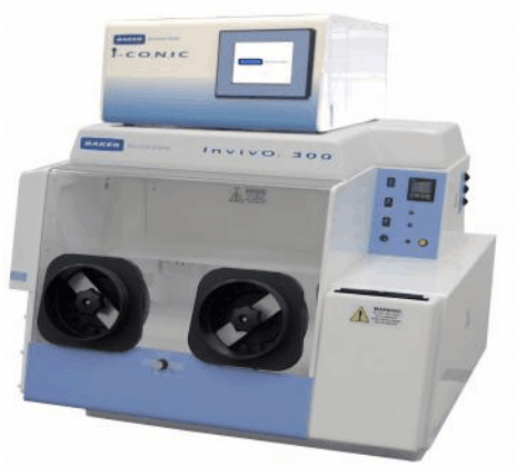
北京隆福佳生物科技有限公司为您提供《内皮细胞中P-gp 、BCRP 蛋白表达检测方案(其它生化设备)》,该方案主要用于其他中生化检验检测,参考标准《暂无》,《内皮细胞中P-gp 、BCRP 蛋白表达检测方案(其它生化设备)》用到的仪器有Invivo2 300低氧工作站(低氧培养箱)。
我要纠错
相关方案
 咨询
咨询