方案详情文
智能文字提取功能测试中
长柔毛薯蓣叶和茎树皮提取物及其银纳米颗粒的细胞毒性研究Cytotoxic Potential of Diospyros villosa Leaves and Stem Bark Extracts and Their Silver Nanoparticlesplants 2 of 13Plants 2023, 12,769 https://doi .org/10.3390/ plants12040769 Academic Editors: Juei-Tang Cheng,I -Min Liu and Szu-Chuan Shen Received: 28 December 2022 Revised: 3 February 2023Accepted: 6 February 2023 Published: 8 Februar y 2023 Copyright: o 2023 by t he authors.Licensee MDPI, Basel, Switzerland.This article is an open access article distributed under the terms and conditions of the Creat i ve Commons A t t r i bution (CC BY ) l icense (https://creativecommons.org/licenses/by/4.0/) Oluwatosin Temilade Adu 1, Yougasphree Naidoo 1, Johnson Lin 2, Depika Dwarka 3, John Mellem 3,Hosakatte Niranjana Murthy 4D:and Yaser Hassan Dewir 5,*D Department of Biological Sciences, School of Li f e Sciences, College of Agriculture, Engineering and Science,University of KwaZulu-Natal, Westvil l e Campus, Private Bag X54001,Durban 4000, South Africa 2 Department of Microbiology , School of L i fe Sciences, College of Agriculture, Engineering and Science,University of KwaZulu-Natal, Private Bag X54001, Durban 4000, South Africa 3 Department of Biotechnology and Food Technology, Durban University of Technology, P.O. Box 1334,Durban 4000, South Africa 4 Department of Hor t icultural Science, Chungbuk Nat i onal Universi t y, Cheongju 28644, Republic of Korea 5 Plant Production Department, Col l ege of Food & Agricul t ure Sciences, King Saud University, Riyadh 11451, Saudi Arabia Correspondence: ydewir@ksu.edu.sa Abstract: Diospyros villosa is traditionally used for an anti-bacterial property. Its cytotoxic effects have not been studied before. Therefore, this study aimed to examine the nutritional properties as wel l the cytotoxic effects of D. villosa. The leaves and stem barks were subjected to three different extraction methods (methanol, chloroform and hexane) and thei r nanoparticles were synthesized a t two different temperatures (room temperature and at 80°C). Thereafter, extracts were assessed using the associated AOCC protocols, for t hei r nutrit i onal content (moisture, f ibre, proteins, l ipid,ash and hydrolysable carbohydrates). Diospyros villosa extracts and t heir corresponding nanoparticles were then incubated overnight with cancerous and noncancerous cell lines to evaluate thei r cytotoxic potential. The nutritional analysis revealed that both young and mature l eaves were rich sources of protein having values of 14.95% and 11.37% respectively. The moisture content was observed to be higher i n all t he l eaf types (8.54±0.75%,9.67±0.98% and 7.40±0.80%) compared to the stem (2.13±0.07%) respectively. The MTT cytotoxicity assay showed that the cell viabil i ty of MCF-7 cell lines was significantly lower when exposed to hexane and chloroform leaves extracts of D. v i llosa (ICso of 26.64 and 26.07 ug mL-1) respectively, compared to camptothecin (36.54 ug mL-1). Simi-larly, the MCF-7 cell viability was observed to be significantly lower when exposed to hexane and chloroform stem extracts of D. villosa (ICso of 24.57 and 3.92 ug mL-1), compared to camptothecin (36.54 ug mL -1). The cel l viability of A549 cell l ines was also found lower when exposed to the hexane and chloroform extracts (IC50 of 7.76 and 4.59 ug mL-1) compared to camptothecin (IC50of 19.26 ug mL -1). Furthermore, the viability of A549 cell lines was found lower when exposed to hexane and chloroform stem extracts of D. villosa (IC50 of 10.67 and 5.35 ug mL-1) compared to camptothecin (19.26 ug mL -1). T he biosynthesized nanoparticles further displayed an anticancer activity with an ICso value of 4.08 ug mL-1 when compared to the control (36.54 ug mL-1). However,the HEK293 cel l viabi l ity was observed to be significantly higher on exposure to hexane stem extracts of D. villosa (ICso of 158.5 ug mL-1) compared t o camptothecin (ICso of 14.77 ug mL-1). Therefore,Diospyros villosa leaves, stem bark and nanoparticles synthesized showed high potential for being considered as a candidate for an anti-cancer regimen. Keywords: cell viabil i ty; cytotoxicity; camptothecin; Diospyros villosa; nanoparticles 1. Introduction Cancer is a complex disease of uncontrolled growth of tumour cells due to signalling failure of oncogenic expressions resulting i n many different types of cancers based on the origin of tumours in the particular organs [1]. An estimated 19.3 million new cancer cases and almost 10 mil l ion cancer deaths were reported i n 2020 [2]. In fact, almost 11.7% of all the new cancer cases were reported to be female breast cancer and identified to be t he most commonly diagnosed cancer in women [3]. Up to now, efforts are made to develop efficient approaches not only to diagnose cancer but also to t reat t he disease. A variety of therapeutic approaches including chemotherapy [4], molecularl y t argeted t herapy [5], gene therapy [6],radiotherapy [7], i mmunotherapy [8] phototherapy [9], and embolotherapy [10] have been extensively applied to treat cancers in clinic. All these t herapeutic measures present severe effects on the patients. However, there i s still a need to secure a more reliable, cheaper and readily available therapeutic measure, with l imited side effects. There are many medicinal plants with therapeutic properties that have been used traditionally i n many countries and are also being researched by various groups in the form of extracts against different types of cancer for possible t reatments [11-13]. Addi-tionally, dietary supplementation of phytonutrients i s an emerging trend that provides a multifaceted defensive mode against various maladies such as cancer by limiting tumour development by binding to the cancer cell membrane or their receptors, thereby initiating cytotoxic i ty and apoptosis i nhibiting tumour growth [14]. These phytonutrients possess certain key advantages over alternative chemotherapy agents such as their affinity and level of t issue penetration, strong target specificity and low toxicity [15] Medicinal plant therapeutic agents also contribute i ndirectly by activating the endogenous defence systems一 by modulating cellular signalling processes [16] and thereby enhancing the overall health status. There is an abundance of medicinal shrubs, vegetables and trees in South Africa that are yet to be prodded meticulously for their health-promoting properties. Similarly,nanopart i cles synthesized f rom green plants were reported to possess unique biological properties and hence become useful in therapeutics and drug delivery [17]. The biosynthe-sized nanoparticles eradicate cancer cells by f low and penetration to different regions of tumours through blood vessels into the target cells [18]. Diospyros villosa (L.) De Winter (D. villosa) is an African plant which naturally occurs in southern parts of the continent. D. villosa root was reported to be used by a group of herbalists found in the botanically diverse Western Cape of South Africa to t reat gastroin-tes t ina l complaints, worms, and flatulence [19]. Also, the root of the D. villosa plant was used i n the rura l community of northern Maputaland to t reat pain and dysmenorrhea [20].Diospyros ferrea (Wild.) leaves nanoparticles were reported for their anti-cancer activities against MCF-7 cancer cell l i nes [21]. Hence, this research study was then geared towards making a signi f icant contribution to the present search being carried out to ascertain the nutritive contents of D. villosa leaves and stem bark and to investigate the anti-cancer properties of D. villosa leaves and stem bark as well as its nanoparticles on breast can-cer cell lines (MCF-7), human embryonic kidney i mmortalized cell l ines (HEK 293), and adenocarcinomic human alveolar basal epithelial cancer cells (A549). 2. Results The yield of different extracts of D. villosa leaves and stem bark is given i n Table 1. It was observed that methanol extraction i n the leaves produced a maximum yield of 10.8%,whereas chloroform and hexane extraction in the leaves yielded 8.4% and 7.1% respectively.Similarly, the methanol extraction i n t he stem bark produced a yield of 7.2% meanwhile,chloroform and hexane extraction yielded 7.9% and 10.3% respectively. The yield obtained from leaves nanoparticles at room temperature and 80 °C was observed to be 7.4% and 5.5% respectively. Also, the percentage yield f rom stem nanoparticles at room temperature and 80 °C was found to be 4.0% and 3.95% respectively. The proximate analysis of different extracts of D. villosa leaves and stem bark is given in Table 2. I t was observed t hat the protein content of mature leaves of D. villosa was found significantly (p<0.05) higher compared to the stem F(3,11)=51.45, p=0.0009. Similarly,the moisture content of the leaves (emergent, young and adult) was found significantly (p <0.05) higher compared to the stem. Also, t he moisture content in the leaves was found significantly higher compared to the stem bark. Meanwhile, the moisture content was not as much as the reported value (14.83%) i n the leaves of Diospyros mespiliformis-a member of the same family (Ebenaceae) by Ebbo et al . [22]. The protein content in the mature leaves (14.95%) was slightly higher than t he reported values (11.49%) [22]. Furthermore , t he crude fibre content in the leaves and stem bark was higher compared to the reported value in Diospyros mespiliformis leaves. Table 1. Yield of extracts of D. villosa leaves, stem bark and nanoparticles. Methanol(%) Chloroform (%) Hexane (%) Nanoparticle at RT Nanoparticle at 80°C (g/g of Dry Plant Material) (g/g of Dry Plant Material) Leaves 10.8 8.4 7.1 0.07 0.055 Stem bark 7.2 7.9 10.3 0.04 0.039 Table 2. Nutritional content (%) of i nvestigated D. villosa leaves and stem bark. Literature Emergent Leaves (%) Young Leaves (%) Mature Leaves (%) Stem (%) Leaves [22] Stem [22] Protein 8.50±1.61 11.37±0.68 14.95±0.83* 5.22±0.72 11.49±0.10 5.51±0.10 Lipids 11.97±_1.36 11.39±1.77 14.37±0.16 13.34±0.28 3.00±0.01 1.83±0.16 Crude fibre 29.73±2.71* 36.4±3.49* 29.60±2.77* 40.17±3.63* 2.66±0.16 6.83±0.33 Ash 8.33±0.44 6.33±0.60 6.67±0.67 5.33±0.60 11.16±0.44 22.66±0.33 Moisture 8.54±0.75 9.67±0.98 7.40±0.80 2.13±0.07 14.83±0.44 11.33±0.60 Carbohydrate 32.93±0.62 24.84±0.91 27.01±0.44 33.81±0.35 55.03±0.01 50.96±0.25 Values are mean ± SD of carefully conducted triplicate experiments. * p <0.05. The MT T assay was used to determine the cytotoxicity of t he D. villosa leaves and stem using different extraction media as well as the biosynthesized AgNPs at different temperatures (RT and 80 °C) on cancerous and non-cancerous cell l ines. For the extract to be anticancer, it should display toxicity on MCF-7, or A549 cancer cells and mild reactivity to HEK293 with further supporting evidence of ICso. The lower the ICso values indicated ,the higher the cytotoxic activity in cancerous cells. Among the different leaf extracts, hexane extract showed a noteworthy cytotoxic effect on the MCF-7 cell line (ICs0 26.64 ug mL-1) and chloroform extract showed a significant cytotoxic effect (ICs0 26.07 ug mL -1) (Table 3 and Figure 1). However, the methanolic leaf extract showed the best cytotoxic effect (ICso 7.09 ug mL-1) in MCF-7 cells. The hexane,chloroform and methanol l eaf extract demonstrated greater anti-cancer activity than the standard camptothecin (ICso 36.54 ug mL-1). The cytotoxicity of MCF-7 cells by both hexane (IC s o 24.57 ug mL-1) and chloroform stem extracts (ICso 3.919 ug mL -1) (Table 4 and Figure 2) was much greater compared to camptothecin (ICso value of 36.54 ug mL-1). However, the greatest anticancer activity produced in MCF-7 cells was demonstrated by the methanolic stem extract (ICs0 0.17 ug mL -1). The Diospyros villosa stem nanoparticles biosynthesized at RT showed a significant toxic effect on MCF-7 (4.08 ug mL-1) compared to Camptothecin (36.54 ug mL-1) (Table 5 and Figure 3). In addition, the leaves nanoparticle synthesized at RT showed a significant toxic effect on MCF-7 cell l ines (ICso 2.03 ug mL-1) The ICso of t he l eaves and stem nanoparticles synthesized at 80 °C was found to be 2.53 and 5.11 ug mL -1 respectively. Table 3. IC s o values of methanol, chloroform and hexane extract of D. villosa leaf against MCF-7 cell. Methanol Leaf Extr. Chloroform Leaf Extr. Hexane Leaf Extr. Camptothecin IC50 0.16 26.07 26.64 36.54 E88883 ML CL HL c Camptothecin Figure 1. Cell viability of MCF-7 cancer cel l l ine treated with different concentrations of D. villosa leaves .* (Camptothecin vs. CL); (Camptothecin vs. ML); p<0.05.**(Camptothecin vs. ML), p<0.01. Table 4. ICso values of methanol , chloroform and hexane extract of D. villosa stem against MCF-7 cell. Methanol Leaf Extr. Chloroform Leaf Extr. Hexane Leaf Extr. Camptothecin ICs0 0.16 26.07 26.64 36.54 Concentration (ugmL-) Figure 2. Cell viability of MCF-7 cancer cell lines treated with different concentrations of D. villosa stem extract . ** (Camptothecin vs. MSB), p <0.01. Table 5. ICso values of D. villosa leaves and stem bark nanoparticles at both RT and 80 °C against MCF-7 cell. Leaves (RT) Stem (RT) Leaves (80°C) Stem (80°C) Camptothecin IC50 2.03 4.08 2.53 5.11 36.54 Concentration (ug mL-1) Figure 3. Cell viability of MCF-7 cancer cell l ines t reated with different concentrations of nanopar-ticles synthesized from D. villosa leaves and stem extract at room temperature and 80 °C.*(ML vs.Camptothecin), p< 0.05; *** (ML vs. Camptothecin), p<0.001. The viability of the HEK 293 cell line was observed t o be higher on exposure to methanolic leaves extract of D. villosa (IC50 41.85 ug mL-1), chloroform leaves extract (ICso of 198.5 ug mL-1) and hexane l eaf extract (ICs0 of 158.5 ug mL-1) compared to camptothecin (IC50 of 14.77 ug mL-1) (Table 6 and Figure 4). Table 6. ICso values of methanol , chloroform and hexane extract of D. villosa leaf against HEK293 cell. Methanol Leaf Extr. Chloroform Leaf Extr. Hexane Leaf Extr. Camptothecin IC50 158.5 198.5 41.85 14.77 Concentration (ug mL-1) Figure 4. Cell viabil i ty of HEK293 cancer cell lines treated with different concentrations of D. villosa leaves extract. * (ML vs. Camptothecin);* (CL vs. camptothecin), p<0.05. The viability of HEK293 cells was further observed to be greater when exposed to hexane stem extract of D. villosa (ICso of 45.13 ug mL-1) (Figure 5). However, the viability of the HEK293 cells was largely affected when treated with the chloroform (ICso of 3.93 ug mL-1)and methanolic (ICso of 0.10 ug mL -1) extracts of D. villosa (Figure 5). Camptothecin produced an IC50 of 14.77 ug mL-1 (Table 7). Figure 5. Cell viability of HEK293 cancer cell l ines treated with dif f erent concentrations of D. villosa stem extract. Table 7. ICso values of methanol, chloroform and hexane extract of D. villosa stem against HEK293 cell. Meth. Stem Extr. Chloroform Stem Extr. Hexane Stem Extr. Camptothecin ICs0 45.1 3.93 0.10 14.77 Diospyros villosa leaves nanoparticles biosynthesized at RT showed a significant toxic effect on the HEK293 cel l l ine (ICso of 4.77 and 7.09 ug mL-1) compared to camptothecin (14.77 ug mL-1) (Table 8 and Figure 6). The viability of HEK293 cells was higher when exposed to leaves and stem nanoparticles (ICs0 of 333.8 and 51.36 ug mL-1) of D. villosa (synthesized at 80°C). The cell viability of A549 cells on exposure to chloroform (ICs0 of 4.592 ug mL-1) and hexane leaves extract (ICso of 7.76 ug mL -1) showed a greater anti-cancer effect compared to camptothecin (IC50 of 19.26 ug mL-1) (Table 9 and Figure 7). Table 8. ICso values of D. villosa leaves and stem bark nanoparticles at both RT and 80 °C against HEK293 cell. Leaves (RT) Stem (RT) Leaves (80°C) Stem (80°C) Camptothecin IC50 4.77 7.09 333.80 51.36 14.77 Concentration (ug mL-1) Figure 6. Cel l viabi l ity of HEK 293 cancer cell l i nes t reated with different concentrations of nanoparti-cles synthesized from D. villosa leaves and stem extract a t room temperature and 80 °C. * (Leaves RT vs. Camptothecin); p<0.05. *** (Leaves RT vs. Camptothecin), p < 0.001; *** (Leaves 80 °℃ vs.Camptothecin), p <0.001. Table 9. ICso values of methanol , chloroform and hexane extract of D. villosa l eaf against A549 cell. Concentration (ug mL-1) Figure 7. Cell viabi l ity of A549 cancer cel l l ines treated with different concentrations of D. villosa leaves extract . *** (ML vs. Camptothecin), p<0.001. In addition, the cell viability of A549 cells was observed to be lower on exposure to hexane stem extract (with ICs0 value of 5.35 ug mL-1), chloroform s t em extract (IC50 of 10.67 ug mL -1) and methanolic extract (13.48 ug mL-1) of D. villosa compared t o control (IC50 of 19.26 ug mL-1) (Table 10 and Figure 8). Table 10. ICso values of methanol, chloroform and hexane extract of D. villosa stem against A549 ceFll. Concentration (ugmL-1) Figure 8. Cell viability of A549 cancer cell l ines treated with different concentrations of D. villosa stem extract. *** (MSB vs. Camptothecin), p <0.001. The viability of A549 cells was observed to be l ower on exposure to D. villosa stem nanoparticles at 80 °C and RT (ICso values of 5.03 and 4.93 ug mL -11 respectively) compared to control (19.26 ug mL-1) (Table 11 and Figure 9). Table 11. ICso values of D. villosa leaves and stem bark nanoparticles at both RT and 80 °C against A549 cell. Concentration (ug mL-1) Figure 9. Cel l viability of A549 cancer cel l l ines t reated with different concentrations o f nanoparticles synthesized from D. villosa leaves and stem extrac t at room temperature and 80 °C. ***(Leaves RT vs.Camptothecin),p< 0.001. *** (Leaves 80 °C vs. Camptothecin), p<0.001. 3. Discussion Diospyros villosa leaves have long been recognized as a traditional medic i nal plant.However, t he putative anti-cancer effects of D. villosa leaves and t heir mechanisms of action have not been scientifically evaluated previously. As illustrated in Figures 1 and 7, viability assays revealed that MCF-7 and A549 cells were more vulnerable to the plant extracts of D. villosa leaves than HEK293 cells. Similarly, a same t rend was observed with D. villosa stem bark (Figures 2 and 8). MCF-7 and A549 cel l s were more vulnerable to stem extracts of D. villosa. Thumbrain et al . [28] pinpointed that if an extract should be an anticancer agent,it should display toxicity on the A549 and MCF-7 cell l ines while being somewhat l ess toxic to HEK293 cells. In t his study, both l eaves and stem bark exhibited strong cytotoxicity against MCF-7 and A549 cells but with disproportionate t rend towards HEK293 cells. This shows that D. villosa extracts may not be toxic t o normal cells, which makes i t an i deal anticancer agent. The exact mechanism of action through which t he plant extracts exhibited i ts t oxicity on cells was not established in this study. Earlier studies further demonstrated that most proteins in typical African diet come from high quality plant protein [29]. Elevated l evels of protein and essential amino acids can i nhibit the cancer progression and growth. This i s in agreement with Gao e t al . [30] who reported that plant proteins activated IGF-1 insulin signalling in order to regulate cancer growth and autophagy but to a very little extent. In addi t ion, the presence of considerable amount of protein i n t he plant may be considered an avenue for building a complex compound with them embedded f unctional phytochemicals,some of which are thought to stop carcinogenesis through their antioxidant properties by interfering with oxidative stress signalling pathway and suppressing DNA damage.This is also in agreement with Kim et al . [31] where i t was pointed out that a member of Diospyros genus (Diospyros kaki) exhibited cell death via activation of platelet-derived growth factor receptors (PDGFRs) which serve as the active binding site for membrane auto-phosphorylation. Our experimental findings support the notion t hat the i ncorporation of D. villosa protein into supplements may play a role in cancer inhibition and retrogression. The results further showed that D. villosa leaves, stem and nanoparticles synthesized at room temperature were able to inhibit cell growth in vitro with high efficiency. Even more,the extracts showed more potency i n MCF-7 cancer l ines, displaying high cytotoxicity. This is in l ine with Park et al. [32] where Diospyros kaki (T humb.) suppressed t he proliferation of human cancer cell lines by decreasing cyclin Di expression. Although, the mechanism of D. villosa cancer i nhibition may not have been achieved through cyclin D1 expression,the excellent display of ICso may be considered. I n t his present study, the D. villosa l eaves, stem and biosynthesized nanoparticles with the highest anticancer activity presented IC50showing potent i nhibitory effect on the growth of MCF-7 and A549 cel l l i nes. The results showed that the D. oillosa plant presented the lower range of IC50 compared to a referenced anticancer medication, showing the higher potency. On the other hand, the methanolic extract of D. villosa plant showed the l owest potenc y i n inhibiting cell growth i n MCF-7 and A549 cells. Taking in account that the beneficial properties of Diospyros plants are related to a variety of bioactive components that enhance antioxidant capacity and consequently anticancer activity [33,34]. Diospyros villosa may be strong candidates for future cancer studies, having high antioxidant and anticancer activity. The activities of both hexane leaves and stem extracts were quite promising as effective anticancer agents. The hexane stem extract did not just only inhibit the growth of MCF-7 cells but also possess a l ower ICso compared to standard. The lowest value of ICso as produced by methanolic l eaves extracts would have been considered the best, but higher percentage viability of MCF -7cells further explained t hat the methanolic leaves extract may r ather be considered a strong antioxidant/antibacterial than anticancer agent. Also, the hexane stem extract displayed both lower ICso value compared to the standard and a low percentage viability of A549cells. In fact, the less toxicity of hexane leaves extract to HEK293 was observed as t he IC50values was quite higher. Both hexane stem extract and the synthesized stem nanoparticles at room temperature showed marked anticancer activities. 4. Materials and Methods 4.1. Plant Collec t ion Fresh samples of mature leaves and stem bark of D. villosa were collected f rom KwaZulu-Natal, Durban, South Africa (29°84'33.6"S, 31°4’12" E). The plant was i dentified and a voucher specimen was deposited in the Ward Herbarium (01/18257) at the School of Life Sciences, University of KwaZulu-Natal, Westville campus. The collected plant parts (leaves and stem bark) were washed, air-dried and pulverized into fine powder. The powdered samples were kept in a cool dry place for extraction purposes. 4.2. Plant Extraction Powdered samples of t he plant weighing 8 g were heated to a temperature of 40 °C for 15 min with 100 mL of 95% methanol in a round bottom flask attached to a Soxhlet apparatus.The crude extract was retained and the process was repeated thrice. Successive extractions using chloroform and hexane, respectively, were carried out after 30 min intervals. The condensate was further evaporated to dryness under reduced pressure at 40°C in a rotary evaporator. The crude extract was stored at 4°C and used within 48 h for further tests. 4.3. Proximate Analysis The proximate analysis of leaves and stem samples i ncluding the moisture, crude fibre, proteins, l i pids, ash content and hydrolysable carbohydrates were assessed. Moisture content was determined by drying the leaves and stem samples at 80 °C in an oven until a constant weight was obtained [35]. The crude fibre was determined by the loss in weight on ignition of dried residue following the digestion of fat-free samples with 1.25% each of sulphuric acid and sodium hydroxide solutions [35]. The total protein content (N×6.25)was estimated by t he macro-Kjeldahl nitrogen assay method using a digestion apparatus combined with the photo-colourimetric method described by Baethgen and Alley [36]. The total lipids content was determined according to AOAC [37], by n-hexane extraction using an automatic Soxhlet analyzer (Soxtherm 2000 Automatic, C. Gerhardt, Northants, UK). 4.4. Synthesis of Silver Nanoparticles (AgNPs) Silver nitrate (1 M AgNOs; Sigma Aldrich, South Africa) was prepared by dissolving 0.17 g in 100 mL of distilled water. Following this, 1 mM of AgNo3 was prepared by diluting 10 mL i n distilled water (90 mL). The reduction of Ag* was achieved by adding 5 mL of each D. villosa aqueous extrac t (leaves or stem bark) to 20 mL of 1 mM AgNO3.The mixtures were incubated for 24 h in the l ight at room temperature (RT; 24°C). The procedure was repeated with the i ncubation at 80°C by heating the extracts i n a water bath for 60 min. The colour change from light yellow to dark brown was indicative of the presence of AgNPs [38,39]. Syntheses were performed i n triplicate. 4.5. Quantification of AgNPs Each AgNP solution was subjected to centrifugation using an Eppendorf microcen-tri f uge (5804/5804R,Sigma-Aldrich, Inc ., St. Louis, MO, USA). The treatment solutions (leaves and stem bark at room temperature and 80°C) were separately transferred i nto pre-weighed microcentrifuge t ubes and purified for 2 h at 1650×gand 4C. The supernatant from each solution was decanted and the insoluble residue was reconstituted in 20 mL sterile distilled water and centrifuged repeatedly three more times for effective removal of unreacted materials. Samples were t hen oven-dried at 40 °C for 24 h after which the tubes were re-weighed to obtain the yield of the synthesized AgNPs. 4.6. Measurement of Cell Viability Human embryonic kidney (HEK293), breast cancer (MCF-7) and human lung cancer (A549) cells were donated by the Department of Biotechnology, Durban University of Technology. Cells were grown at 37 °C in a humidified incubator under 5% CO2 i n Dulbecco's modified Eagle's medium (DMEM) containing 10% Foetal Bovine Serum (FBs)and antibiotics (Penicillin; 10,000 U mL-1 and Streptomycin sulphate; 10,000 U mL-1[Penicil l in/Streptomycin]). Antibiotics change the phenotype and morphology of cells;therefore, the use of the antibiotics should be in very low concentrations, thus for t his study, 1% Penicillin/Streptomycin was used . The cells were grown until 80% confluence was reached with media replaced as necessary. After confluence was reached, cel l s were trypsinized and sub-cultured. The 3-(4,5 dimethylthiazol-2-yl)-2,5-diphenyltetrazolium bromide (MTT) assay was used to determine the cytotoxicity of the extracts. The MTT assay was conducted according to Dwarka et al . [40] with minor modifications. Briefly,cells (50 uL of 1×10² cells mL -1), as well as 50 uL of DMEM, were seeded into a 96-well f l at bottom plate and incubated (37 °C for 24 h) in a humidified incubator under 5% CO2.Cells were then treated with 50 uL of extracts at varying concentrations (7.8-1000 ug mL -1)prepared in 5% DMSO and incubated for 24 h. Camptothecin was used as a positive control.MTT reagent (20 uL, 5 mg mL -1) was added to t he cells and incubated at 37 °C for 4 h. One hundred microliters of DMSO was then added to each wel l to solubilise t he formazan salt formed, and absorbance was read at 570 nm on a microplate spectrophotometer (Mul t iscan Go, Thermo Scientific, Waltham, MA, USA) for both treated and untreated cells. The percentage viability was calculated using t he following f ormula: 4.7. Statistical Analysis Data are displayed as means ± SEM. Statistical analysis was performed using Graph Pad Prism 5 (Graph Pad Software Inc., San Diego, CA, USA). The results were compared using one-way ANOVA followed by Bonferroni post hoc t ests. Also, two-way ANOVA f ollowed by Bonferroni post hoc tests was used where necessary. Effects were considered statistically significant at p-value <0.05. The l ower the half maximal i nhibitory concentration (ICs0) of t he cancerous cell l i nes, the more the potency of the extract as an anticancer agent. 5. Conclusions In this work, we have explored the nutritive contents and anticancer effect of the D. villosa leaves and stem bark as well as the nanoparticles against three cancer cell lines.The study revealed that D. villosa had high fibre and protein contents. Diospyros villosa may therefore be considered a plant with grea t potential as food supplement. I t is further possi-ble to conclude that D. villosa extracts potentially inhibited the viability of human breast carcinoma and lung carcinoma cells. Hence, its incorporation i nto nutritive supplements may provide a prophylactic regimen to both breast and lung cancer. Author Contributions: Conceptualization, O.T.A. and Y.N.; methodology, O.T.A., Y.N. and J.L.;formal analysis, O.T.A., Y.N., J.L ., D.D. and J.M.; i nvestigation, O.T .A., Y.N. and J .L .; formal analysis,O.T.A., Y.N., J.L., D.D. and J.M.; data curation, O.T .A., Y.N. and J .L.; formal analysis, O.T.A., Y.N.,J.L., D.D. and J.M.; validation, H.N.M. and Y.H.D.; writing-original draft preparation, O.T.A., Y.N.,J.L., D.D. and J .M.; wri t ing-review and editing, O.T.A., Y.N., H.N.M. and Y.H.D.; visualization,H.N.M. and Y.H.D.; supervision, Y.N. All authors have read and agreed to the publ i shed version of the manuscript. Funding: The authors acknowledge Researchers Supporting Project number (RSP2023R375), King Saud University, Riyadh, Saudi Arabia and TWAS-National Research Foundation (NRF), South Africa. Data Availability Statement: All data are presented in t he article. Acknowledgments: The authors acknowledge Researchers Supporting Project n umber (RSP2023R375),King Saud University, Riyadh, Saudi Arabia. The authors are t hankful to the TWAS-National Re-search Foundation (NRF ) for their f inancial support and University of KwaZulu-Natal for providing research facilities for this work. Conflicts of Interest: The authors declare no conflict of interest. References 1. Anusewicz, D.;Orzechowska, M.; Bednarek, A.K. Lung squamous cel l carcinoma and l ung adenocarcinoma differential gene expression regulation through pathways of Notch, Hedgehog, Wnt, and ErbB signal li ng. Sci. Rep. 2020,10,1-15.[CrossRef ] 2. Sung , H.; Ferlay, J .; Siegel, R.L.; Laversanne, M.; Soerjomataram, I .; Jemal, A.; Bray, F . Global cancer statist i cs 2020: GLOBOCAN estimates of incidence and mortal i ty worldwide for 36 cancers in 185 countries. CA Cancer J . Clin. 2021,71,209-249.[CrossRef ][PubMed ] 3. Bray, F; Ferlay, J.; Soerjomataram, I.; Siegel , R.L.; Torre, L.A.; Jemal , A. Global cancer statistics 2018: GLOBOCAN estimates of incidence and mortality worldwide for 36 cancers in 185 countries. CA Cancer J. Clin. 2018, 68, 394-424. [CrossRef] [PubMed ] 4Dickens, E.; Ahmed, S. Principles of cancer t r eatment by chemotherapy. Surgery 2018,36,134-138. 5 Piawah, S.; Venook, A.P. Targeted therapy for colorectal cancer metastases: A review of current methods of molecularly t argeted therapy and the use of tumor biomarkers in the treatment of metastatic colorectal cancer. Cancer 2019, 125,4139-4147. [CrossRef ][PubMed ] 6. Carrillo, M.A.; Zhen, A.; Kitchen, S.G. The use of the humanized mouse model in gene t herapy and immunotherapy for HIV and cancer. Front . Immunol. 2018, 9, 746.[CrossRef ] 7. Brundha, M.P; Pathmashri, V.P; Sundari, S. Quantitative changes of red blood cells in cancer patients under palliative radiotherapy-a retrospective study. Res. J. Pharm. Technol. 2019,12, 687-692. [CrossRef ] 8Dobosz, P; Dzieciatkowski, T. The intriguing history of cancer immunotherapy. Front. I mmunol. 2019,10,2965. [CrossRef ]9 Chang , M.; Wang, M.; Wang, M.; Shu, M.; Ding, B.; Li, C.; Pang, M.; Cui, S.; Hou, Z.; Lin, J. A multifunctional Cas-cade bioreactor based on hollow-structured Cu2 MoS4 for synergetic cancer chemo-dynamic t herapy/starvation ther-apy/phototherapy/immunotherapy with remarkably enhanced efficacy. Adv. Mater. 2019,31, e1905271. [CrossRef] 10. Kishore, S.A.; Bajwa, R.; Madoff, D.C. Embolotherapeutic strategies for hepatocellular carcinoma: 2020 update. Cancers 2020,12,791. [CrossRef ] 11. Kamble, S.S.; Gacche, R.N. Evaluation of anti-breast cancer, anti-angiogenic and antioxidant propert i es of selected medicinal plants. Eur. J. I nteg. Med. 2019,25,13-19. [CrossRef ] 12. Goodarzi , S.; Tabatabaei, M.J .; Jafari, R.M.; Shemirani , F; Tavakoli, S.; Mofassseri, M.; Tofighi , Z. Cuminum cyminum fruits as source of luteolin-7-O-glucoside, potent cytotoxic flavonoid against breast cancer cell l ines. Nat. Prod. Res. 2018, 34,1602-1606.[CrossRef ] [PubMed ] 13. Aumeeruddy, M.Z.; Mahomoodally, M.F. Global documentation of traditional l y used medicinal plants in cancer management:A systemat ic review. S. Afr. J. Bot 2021, 138,424-494. [CrossRef ] 14. Chen, H.; Liu, R.H. Potential mechanisms of action of dietary phytochemicals for cancer prevention by targeting cellular s ignaling transduction pathways. J. Agric . Food Chem. 2018,66,3260-3276.[CrossRef] [PubMed ] 15. Soldati, L.; Renzo, L.D.; Jirillo, E.; Ascierto, P.A.; Marincola, F.M.; Lorenzo, A.D. The i nfluence of diet on anti-cancer immune responsiveness. J. Transl . Med. 2018, 16,75. [CrossRef ] 16. Marabini , L.; Melzi, G.; Lolli, F; Dell'Agli , M.; Piazza, S.; Sangivanni , E.; Marinovich, M. Effects of Vitis vinifera L. leaves extract on UV radiation damage in human kerat i nocytes (HaCaT). J . Photochem. Photobiol . B 2020, 204, 111810. [CrossRef ] 17. Chenthamara, D.; Subramaiaam, S.; Ramakrishnan, S.G.; Krishnaswamy, S.; Essa, M.M.; Lin, F.H.; Qoronfleh, W. Therapeutic efficacy of nanoparticles and routes of administration. Biomat. Res. 2019,23,20. [CrossRef ] Philander, L.A. An ethnobotany of Western Cap 28e Rasta bush medicine. J. Enthnopharmacol. 2011, 138,578-594. [CrossRef] de Wet, H.; Ngubane, S.C. Traditional herbal remedies used by women in a rural communi t y i n northern Maputaland (South Africa) for the treatment of gynaecology and obstetric complaints. S. Afr. J . Bot. 2014,94,129-139. [CrossRef ] 22. Ebbo, A.A.; Sani, D.; Suleman, M.M.; Ahmed, A.; Hassan, A.Z. Phytochemical composi t ion, proximate analysis and antimicrobial screening of methanolic extract of Diospyros mespiliformis Hochst ex a. Dc (Ebenaceae). Pharmacogn. J. 2019,11,362-368.[CrossRef ] 23. Abdullah, S.; J an, S.E.; Kwak, M.K.; Chong, K.P. Ganoderma boninense mycel i a for phytochemicals and secondary metabolites wi t h antibac t erial activ i ty. J . Microbiol . 2020,58,1054-1064. [CrossRef] 24. Tuncturk, M.; Eryigit , T.; Sekeroglu, N.; Ozgokee, F. Chemical composition of some edible wild plants grown i n Eastern Anatolia.AJEONP 2015,2,31-34. 25. Unuofin, J.O.; Otunola, G.A.; Afolayan, A.J. Nutritional evaluation of Kedrostis africana (L.) Cogn: An edible wild plant of South Africa . Asian Pac. J. Trop. Med. 2017,7,443-449. [CrossRef ] 26. Nyamwamu, N.C.; Okari, O.J.; Gisesa, W.N.O. A survey of medic i nal plants used by the gusii community in the treatment of digestive disorders and other inflammatory conditions. J. Med. Plants 2020,8,21-33. 27. Kim, H.; Lee, K.; Rebholz, C.M.; Kim, J . Plant-based diets and incident metabolic syndrome: Results from a South Korean prospective cohort study. PLoS Med. 2020,17, e1003371.[CrossRef ] 28. Thumbrain, D.; Dwarka, D.; Gerrano, A.S.; Mellem, J.J. Antioxidant and apoptotic potent i al of protein i solates derived f rom Vigna unguiculata (L .) Walp. Int. J . Food Sci. 2020,55,2813-2823. 30. Gao, X.; LaValley, M.P.; Tucker, K.L. Prospective studies of dairy product and calcium intakes and prostate cancer risk:A meta-analysis. J. Nat l . Cancer Inst . 2005, 97, 1768-1777.[CrossRef] 31. Kim, H.S.; Suh, J .S.; J ang, Y.K.; Ahn, S.H.; Raja, G.; Kim, J.C.; Jung, Y.;Jung, S.H.; Kim, T.J . Anti-cancer potential of persimmon (Diospyros kaki) l eaves via the PDGFR-Rac-JNK pathway. Sci. Rep. 2020, 10, 18119.[CrossRef ] 32. Park, S.B.; Park, G.H.; Song, H.M.; Son, H.J.; Um, Y.; Kim, H.Y.; Jeong, J.B. Anticancer activ i ty of calyx of Diospyros kaki Thunb.through downregulation of cyclin D1 via inducing proteasomal degradation and t ranscriptional inhibition in human colorectal cancer cells. BMC Complement . Altern. Med. 2017,17,1-10.[CrossRef ] [P ubMed ] 33. Galo, Y.S.; Dauda, S.; Emmanuel, A. Phytochemical screening and antimicrobial activity of leaf and stem-bark aqueous extracts of Diospyros mespiliformis. Int. J . Biochem. Res. Rev. 2018, 22, 1-8. 34. El-Hawary, S.S.; Tadros, S.H.; AbdelMohsen, M.M.; Mohamed, M.S.; Sheikh, E.E.; Nazif, N.M.; ElNasr, M.S. Phyto-and Bio-Chemical evaluation of Diospyros kaki L. cultivated i n Egypt and i ts biological activi t ies. Braz. J . Biol. 2020, 80, 295-304.[CrossRef ] 35. AOAC. Official Methods of Analysis of Association of Analytical Chemists, 17th ed.; Association of Official Agricul t ural Chemists International: Arlington, VA, USA, 2000. 36. Baethgen, W.E.; Alley, M.W. A manual colorimetric procedure for measuring ammonium nitrogen i n soil and plant kje l dahl digests. Commun. Soil Sci . Plant Anal. 1989, 20, 961-996. [CrossRef ] 37. AOAC. Official Methods of Analysis of the Association of Official Analytical Chemists, 15th ed.; Sec. 985.29.; T he Associat i on: Ar l ington,VA, USA, 1990; Volume II. 39. Sharma, S.; Kumar,S.; Bulchandani, B.D.; Taneja, S.; Banyal , S. Green synthesis of silver nanopart i cles and thei r antimicrobial activity against Gram positive and Gram-negative bacteria. Int . J. Biotechnol. Bioeng. Res. 2013, 4, 711-714. 40. Dwarka, D.; Thaver, V.; Naidu, M.; Koorbanally, N.A.; Baijanath, H. In vitro chemo-preventative activity of Strelitzia nicolai aril extrac t containing bilirubin. Afr . J. Tradit. Complement. Altern. Med. 2017,14,147-156.[CrossRef ] Disclaimer/Publisher’s Note: The statements, opinions and data contained i n all publications are solely those of t he individual author(s) and contributor(s) and not of MDPI and/or the editor(s). MDPI and/or the edi t or(s) disclaim responsibility for any i njury to people or property resulting from any ideas, methods, instructions or products referred to in the content.
关闭-
1/13
-
2/13
还剩11页未读,是否继续阅读?
继续免费阅读全文产品配置单
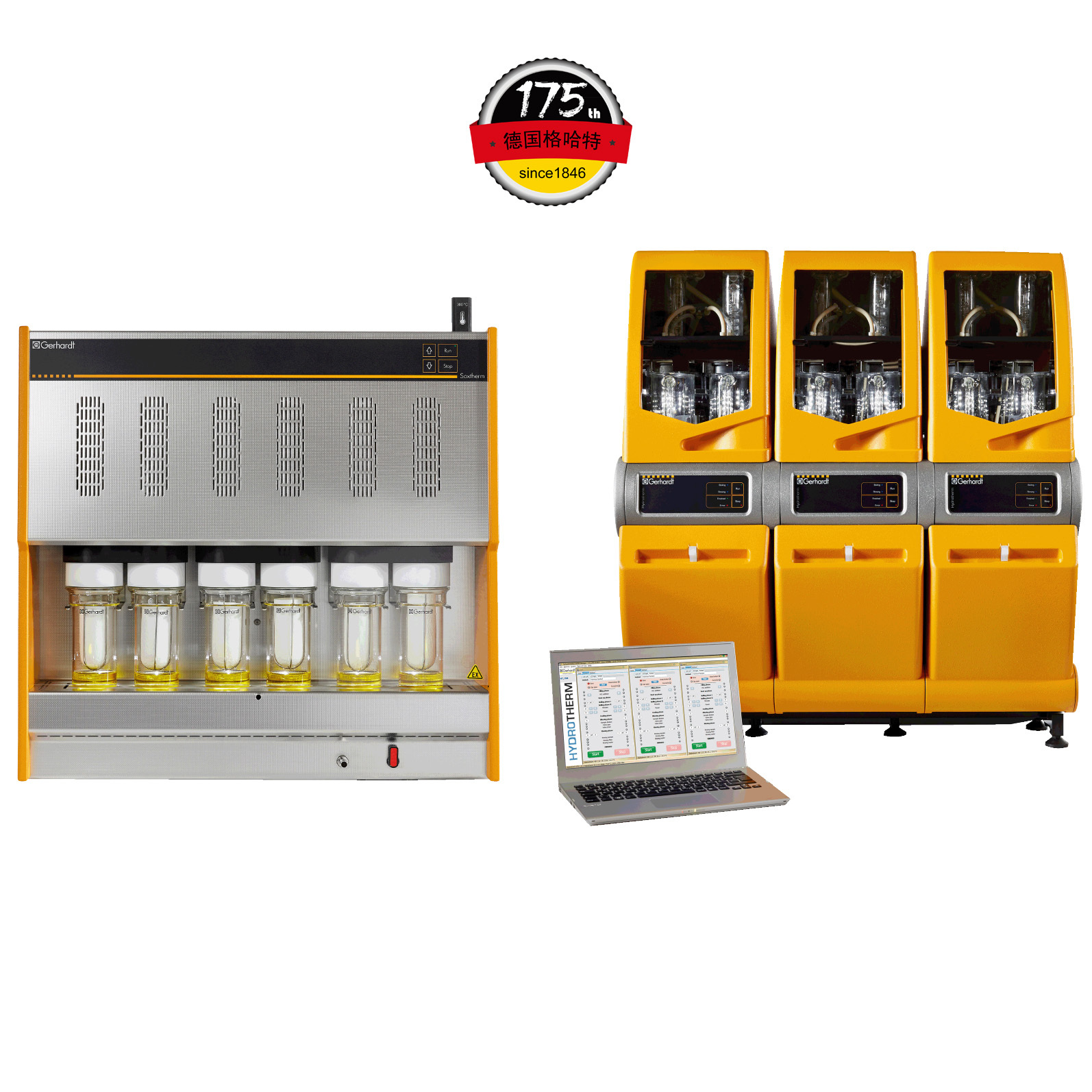
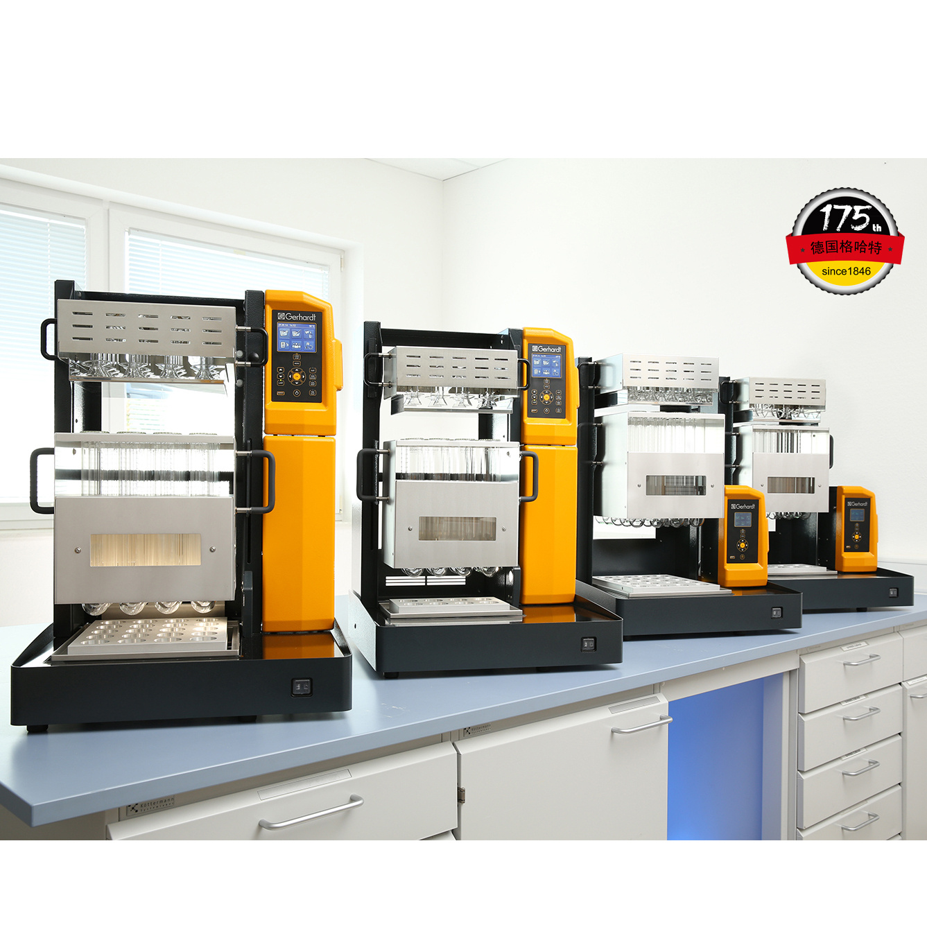
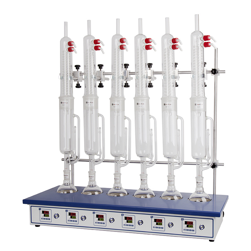
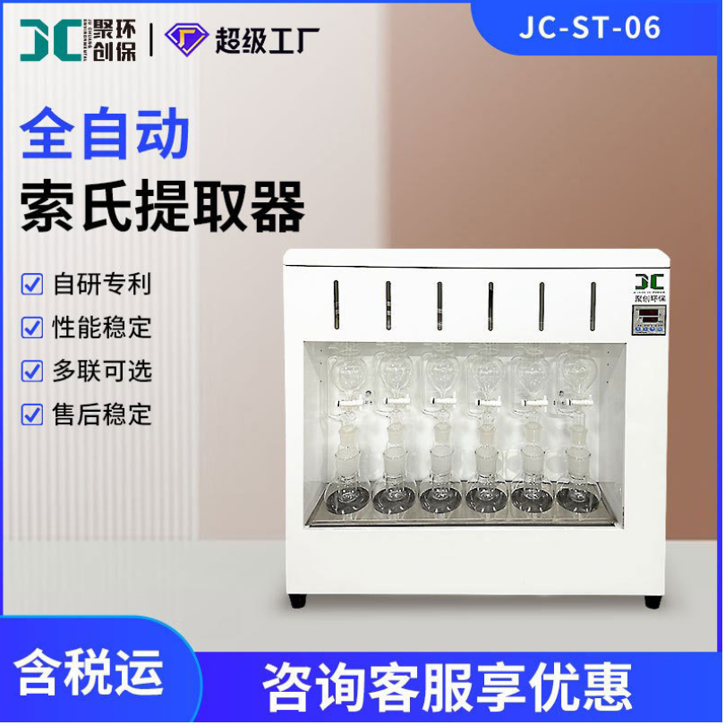
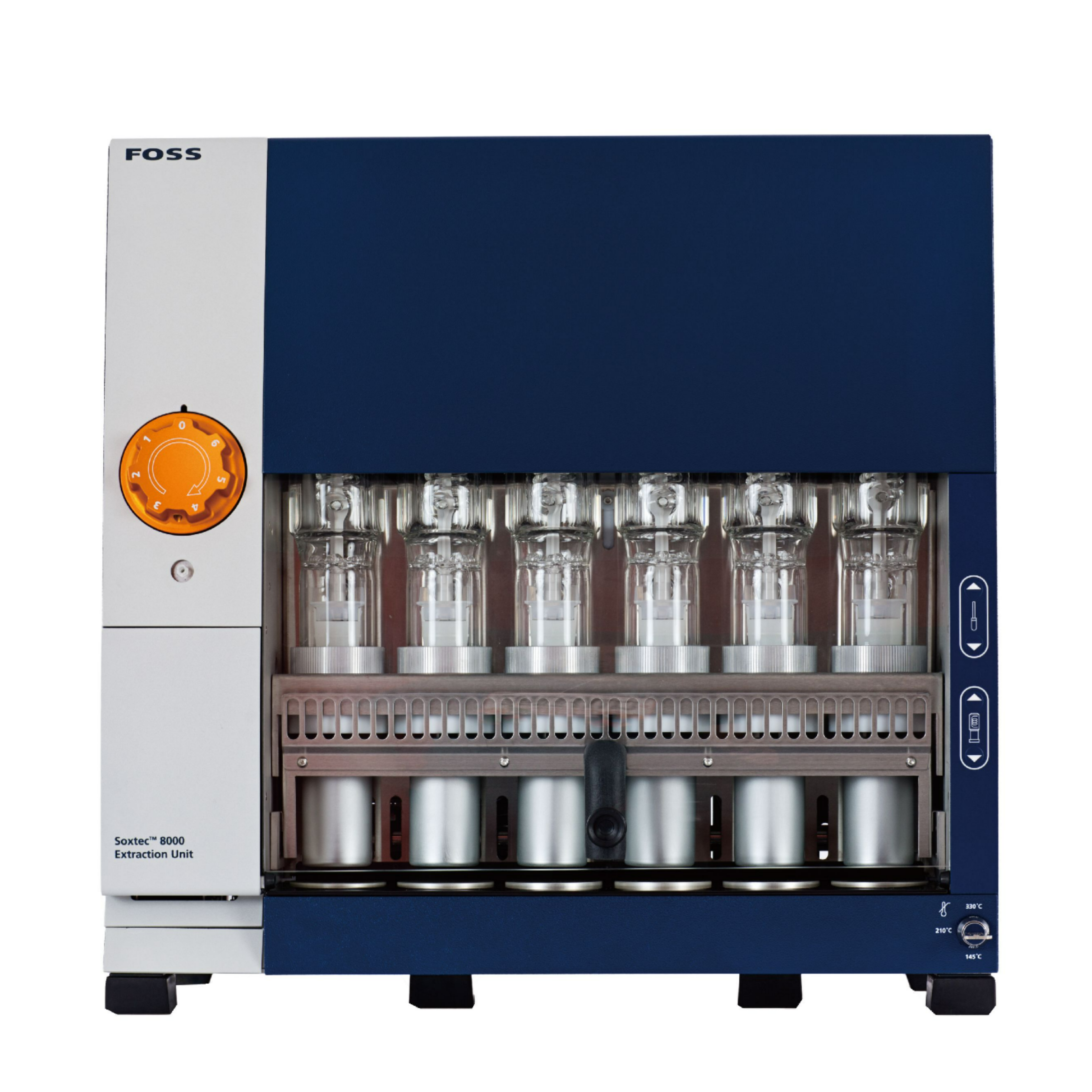
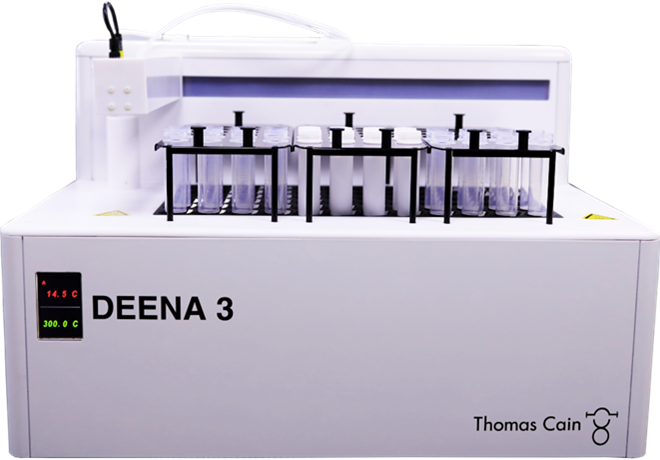
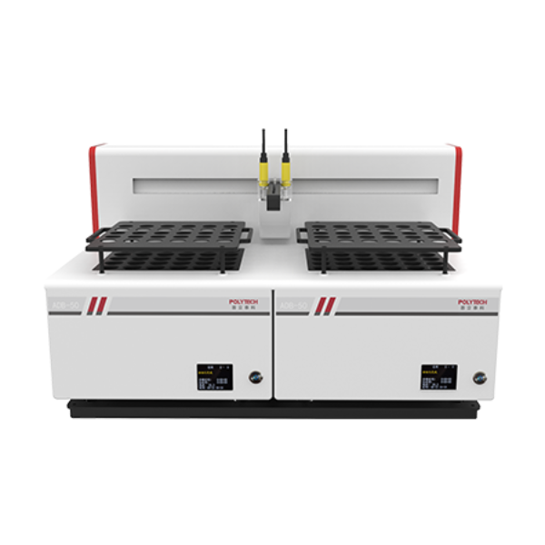
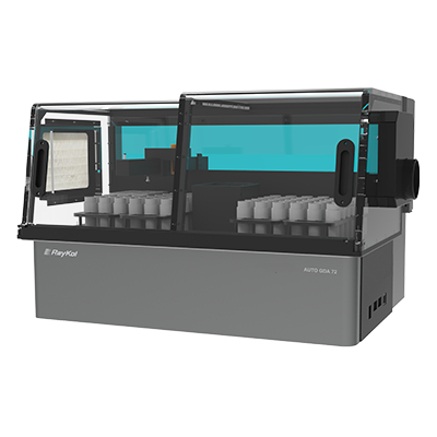
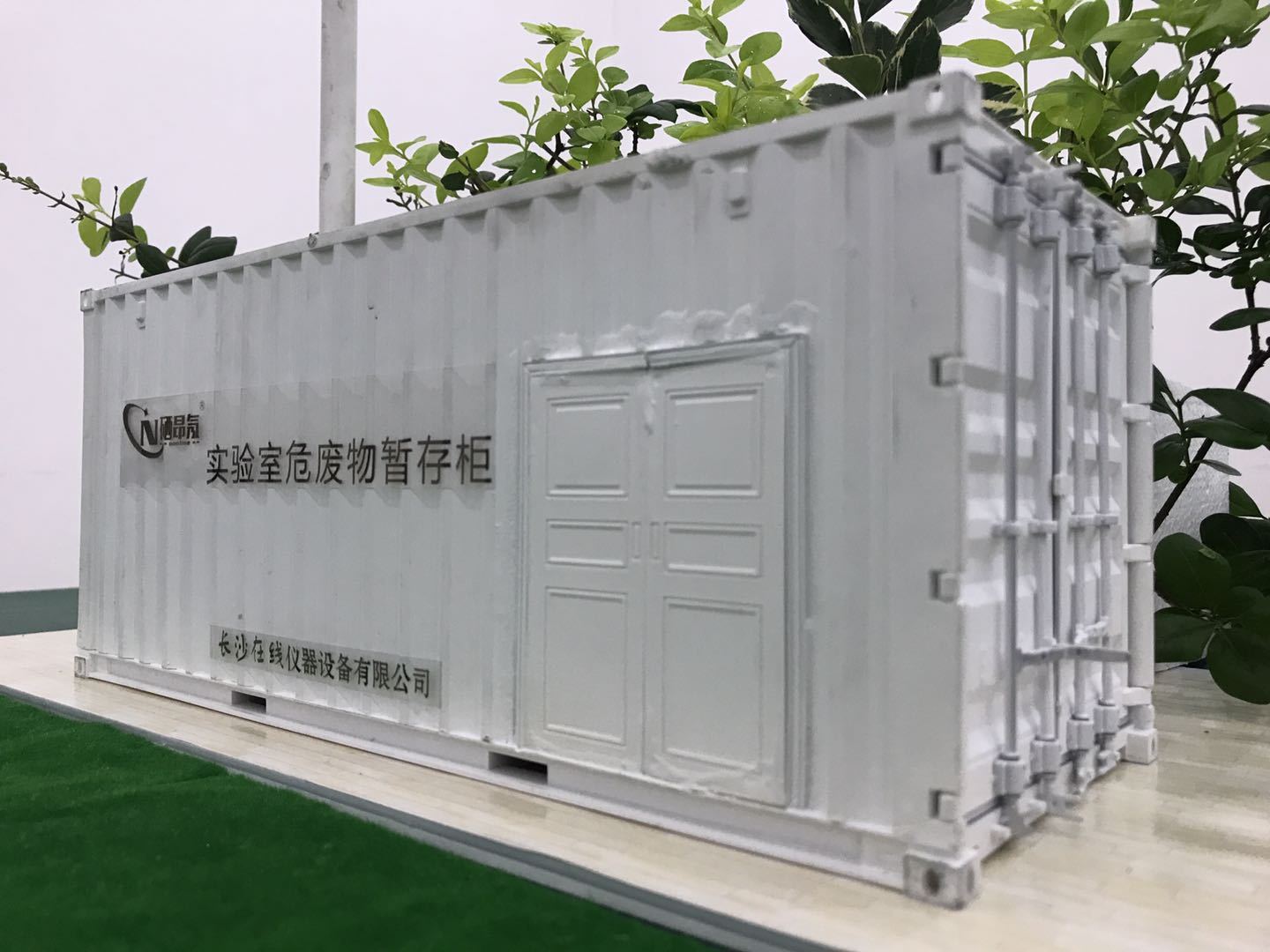
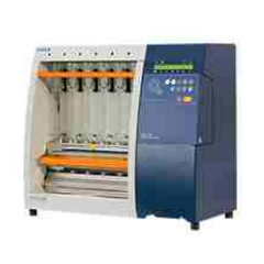
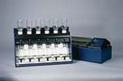
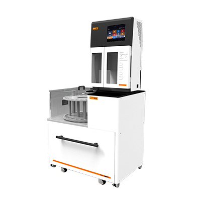
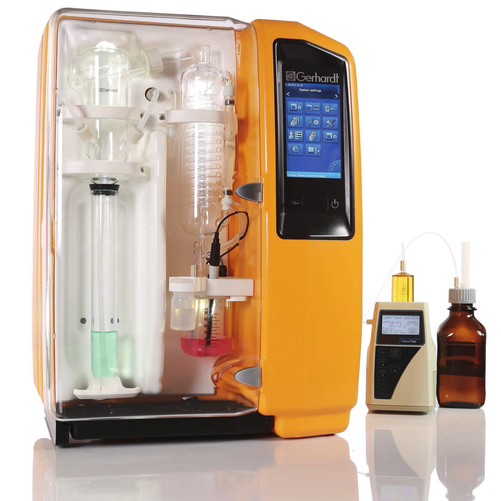
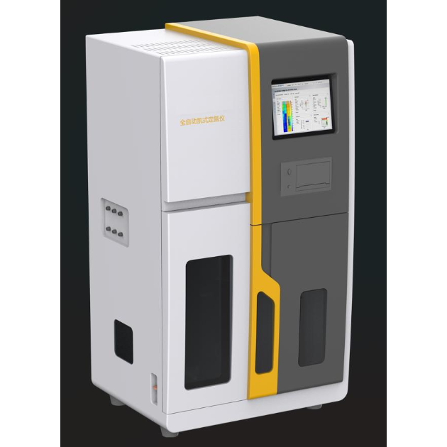
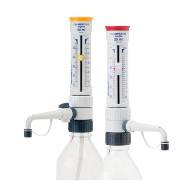
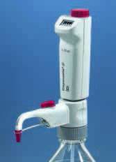
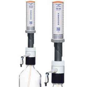
中国格哈特为您提供《长柔毛薯蓣叶和茎树皮总蛋白质、总脂肪、粗纤维含量》,该方案主要用于中药材和饮片中含量测定检测,参考标准《暂无》,《长柔毛薯蓣叶和茎树皮总蛋白质、总脂肪、粗纤维含量》用到的仪器有格哈特全自动超级总脂肪测定系统HT6+SOX416、格哈特凯氏消化系统KT8S、格哈特维克松废气实验室废物处理系统涤气VS、格哈特全自动型纤维分析仪FT12、格哈特带自动进样器凯氏定氮仪VAP500C、德国加液器MM、凯氏定氮催化片。
我要纠错

相关方案
 咨询
咨询