方案摘要
方案下载| 应用领域 | 医疗/卫生 |
| 检测样本 | 其他 |
| 检测项目 | |
| 参考标准 | 0 |
Raman image has been obtained with confocal SERS microspectroscopy from a single, live K562 cancer cells treated with an anticancer drug mitoxantrone, at a concentration of 10-7 M. SERS spectra have been recorded from the inside of the cell in which silver colloidal particles have been introduced to generate the SERS effect. The potential of confocal SERS microspectroscopic imaging at the single cell level is very important for the selective analysis of drugs inside these cells.
Raman image has been obtained with confocal SERS microspectroscopy from a single, live K562 cancer cells treated with an anticancer drug mitoxantrone, at a concentration of 10-7 M. SERS spectra have been recorded from the inside of the cell in which silver colloidal particles have been introduced to generate the SERS effect.
The potential of confocal SERS microspectroscopic imaging at the single cell level is very important for the selective analysis of drugs inside these cells.
文献贡献者


荧光光谱+蛋白质检测+荧光特性
荧光光谱+智能形致变色荧光材料+荧光特性
荧光光谱+荧光探针+精准医疗
相关产品
HORIBA XploRA Nano原子力-拉曼联用系统
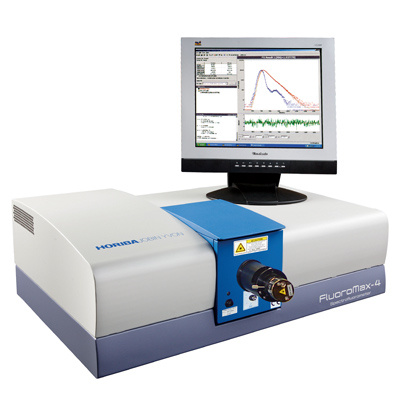
HORIBA高灵敏一体式FluoroMax-4荧光光谱仪
HORIBA Fluorolog®-3科研级荧光光谱仪
HORIBA XploRA INV多功能拉曼及成像光谱仪
HORIBA EMGA-Expert 氧氮氢分析仪
HORIBA EMGA-Pro氧氮氢分析仪
HORIBA 离心式粒度分析仪Partica CENTRIFUGE CN-300
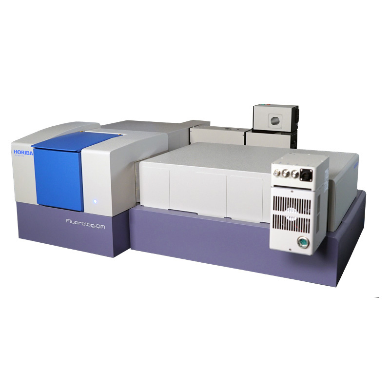
HORIBA Fluorolog-QM模块化科研级稳瞬态荧光光谱仪
ViewSizer 3000 纳米颗粒追踪分析仪
HORIBA LabRAM Odyssey 高速高分辨显微共焦拉曼光谱仪
HORIBA LabRAM Soleil™高分辨超灵敏智能拉曼成像仪
HORIBA NANO Raman
HORIBA XGT-9000 X射线显微分析仪
HORIBA Duetta荧光及吸收光谱仪
HORIBA MacroRAM 台式一体化拉曼光谱仪

关注

拨打电话

留言咨询