方案摘要
方案下载| 应用领域 | 生物产业 |
| 检测样本 | 其他 |
| 检测项目 | |
| 参考标准 | 暂无 |
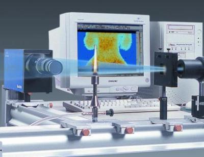
采用LaVision公司的PIV系统,配置使用Imager Pro X 型CCD相机,测量脂质覆盖疏水纳米粒子通过磷脂双层自发迁移的速度矢量场

Hydrophobic nanoparticles introduced into living systemsmay lead to increased toxicity, can activate immune cells, or
can be used as nanocarriers for drug or gene delivery. It is generally accepted that small hydrophobic nanoparticles are
blocked by lipid bilayers and accumulate in the bilayer core, whereas big nanoparticles can only penetrate cells
through slow energy-dependent processes, such as endocytosis, lastingminutes. In contrast to expectations, we demonstrate
that lipid-covered hydrophobic nanoparticles may translocate through lipid membranes by direct penetration
within milliseconds. We identified the threshold size for translocation: nanoparticles with diameters smaller
than 5 nm stay trapped in the bilayer, whereas those with diameters larger than 5 nm insert into the bilayer, opening
pores in the bilayer. The direct proof of this size-dependent translocation was provided by an in situ observation of a
single event of a nanoparticle quitting the bilayer. This was achieved with a specially designed microfluidic device
combining optical fluorescence microscopy with simultaneous electrophysiological measurements. A quantitative
analysis of the kinetic pathway of a single nanoparticle translocation event demonstrated that the translocation is
irreversible and that the nanoparticle can translocate only once. This newly discovered one-way translocation mechanism
provides numerous opportunities for biotechnological applications, ranging from targeted biomaterial elimination
and/or delivery to precise and controlled trapping of nanoparticles.
文献贡献者


在一个双稳湍流涡旋火焰中,对间歇性动态的时间-频率定位
Particle-laden Taylor-Couette流:高阶转变和径向局部波浪涡旋的证据
7根杆束的流体-结构相互作用:用实验数据对比数值模拟
相关产品
汽车光学检测系统AIS
PT403型波长可调谐皮秒激光器
ANL系列高能量高重复频率DPSS纳秒激光器
APL4206 系列高能量皮秒激光放大器
UltraFlux FF/FT 5000 高能量可调谐飞秒激光器系统
UltraFlux FT300型波长可调谐飞秒激光器系统
Ekspla 超高功率激光器系统SYLOS 2A
LaVision PTUx 可编程时间控制单元
LaVision 用于粒子成像测速PIV的相机
LaVision StrainMaser 全场应变测试系统组件
LaVision 用于数字图像相关DIC的相机
LaVision StrainMaster 数字图像相关分析软件包
LaVision 标定板
LaVision StrainMaster 形变应变成像测量系统
LaVision StrainMaster DVC 体视全场应变测量系统

关注

拨打电话

留言咨询