

方案详情文
智能文字提取功能测试中
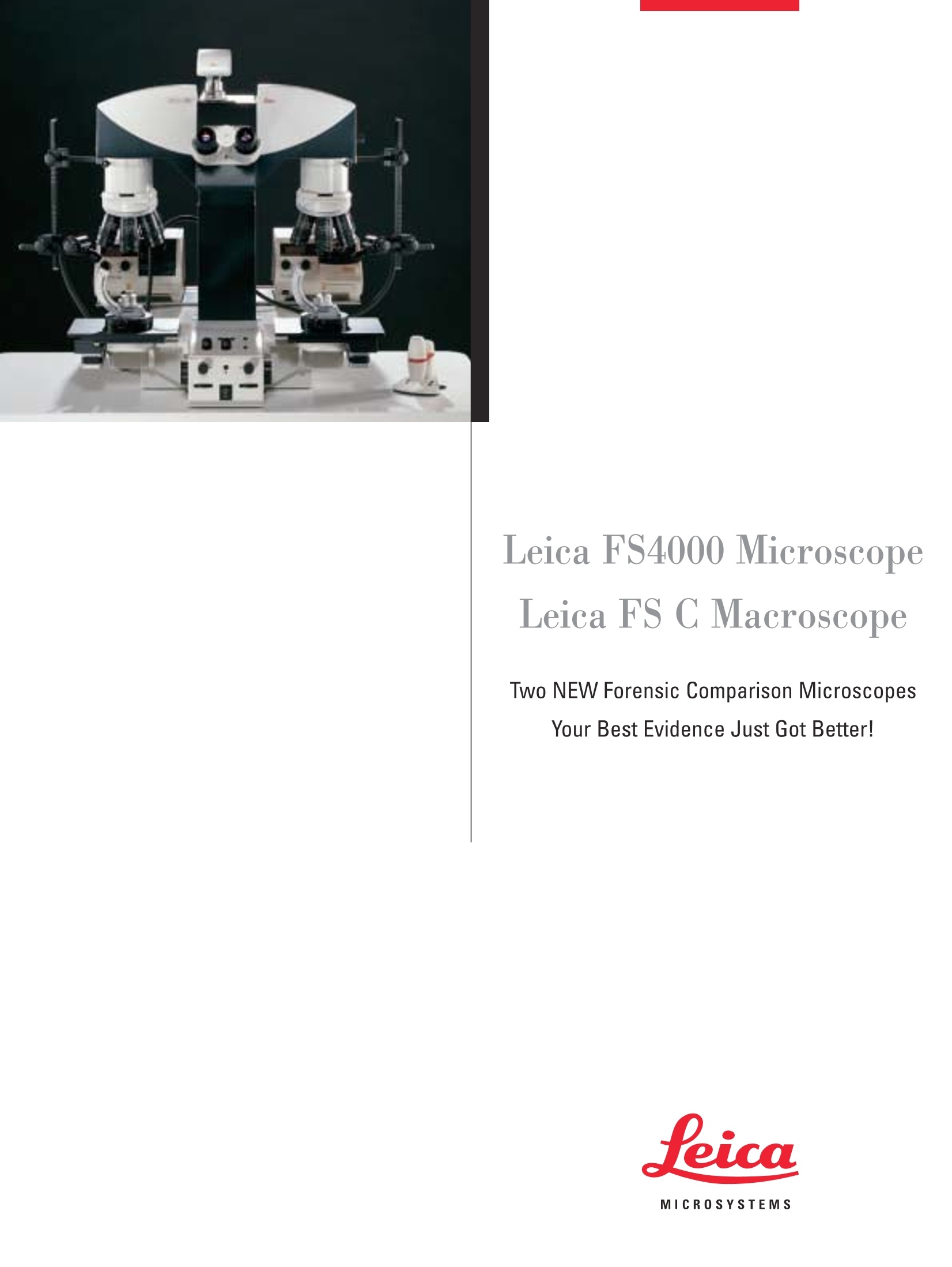
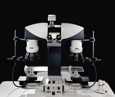
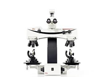
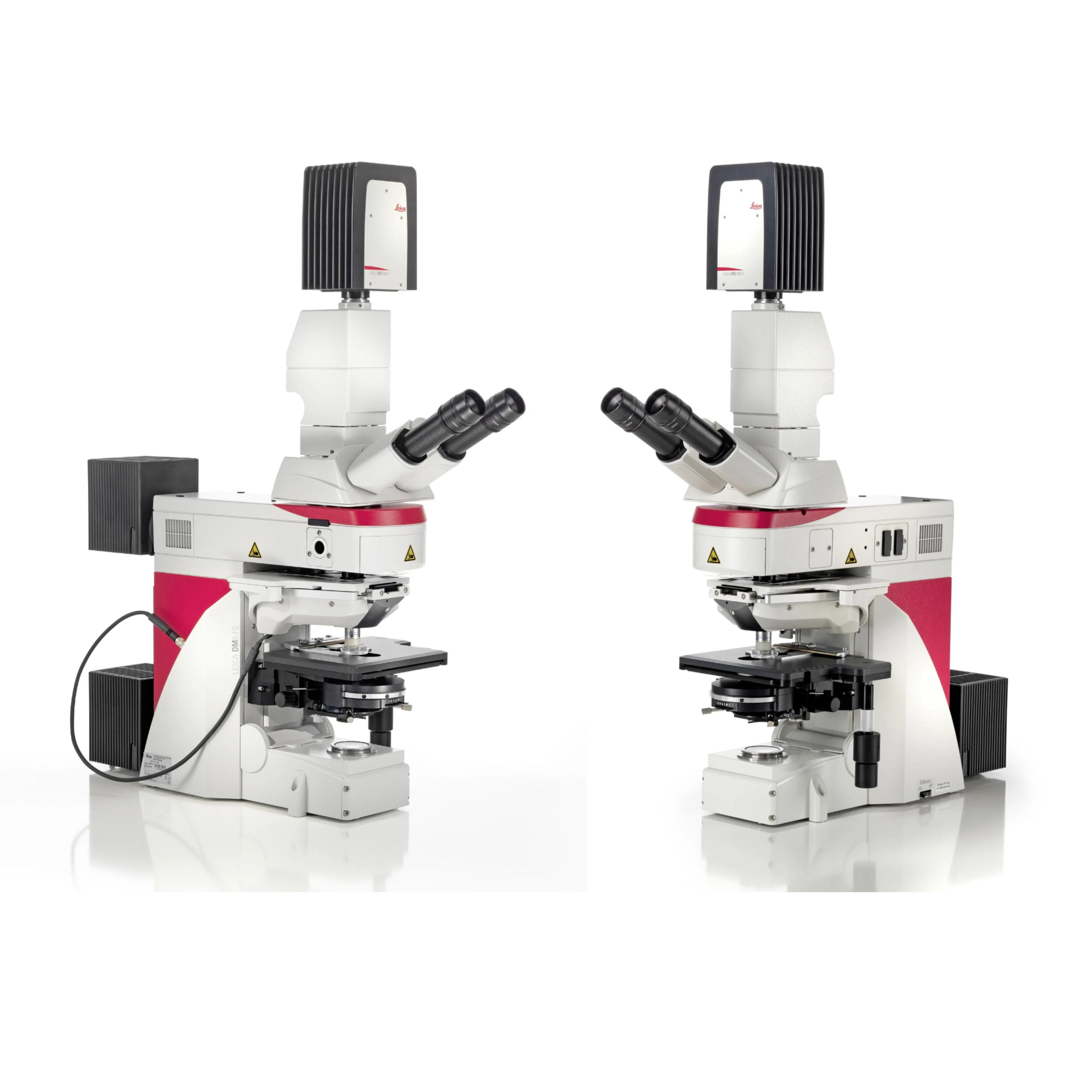
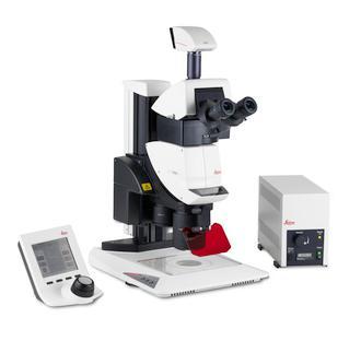
Leica FS4000 MicroscopeLeica FS C Macroscope Two NEW Forensic Comparison MicroscopesYour Best Evidence Just Got Better! MICROSYSTEMS Leica Microsystems Has Already HelpedConvictMany Criminals Since the pioneering days of forensic evidence comparison, onename has always stood for quality - Ernst Leitz GmbH. Over thecourse of nine decades, Leitz comparison microscopes weredeveloped in cooperation with leading forensic scientists fromaround the world. The company name has since changed, butthe quality remains the same. Leica Microsystems continues theLeitz tradition of designing and producing innovative comparisonmicroscopes, and now introduces the Leica FS4000 Forensic So-lution Microscope and Leica FS C Forensic Solution ComparisonMacroscope. 486 23 The forensic scientist's expertise plus Leica's innovationmake a perfect pair The forensic scientist's expert knowledge paired with a Leica mi-croscope is an unbeatable combination. Clear evidence imagesdown to the smallest detail, and unmistakable, reproducible re-sults -Leica’s comparison optics provide all this and more. Thescientist can identify ambiguous evidence and draw expert con-clusions with ease using Leica's newest microscope solutions.Leica comparison microscopes provide state-of-the-art technol-ogy and superior ergonomics to make comparison microscopyeasier and allow the examiner to concentrate on the work athand. Leica Microsystems AG -Winner of the World's First Innovation Award: German Business Innovation Award 2002 The Principal Innovation -A Fully Integrated Comparison Bridge Whether the evidence is fiber analysis, toolmark comparison orballistics, Leica’s new fully integrated, automated comparisonbridge provides better and faster evidence assessment throughoptimized comparison techniques. Both the Leica FS C and theFS4000 feature the new comparison bridge design. The followingobservation modes can be selected at the touch of a button: FullScreen left, FullScreen right Split-image with adjustable dividing line Split-image with adjustable strip for partial superimposed image Color contrasting of non-conforming structures Superimposed image comparison The new comparison bridge's use of complementary color filtersrenders unmatched sample portions in color. The overlaying detailsonly appear in their original color in those places where there isno structural deviation. Side-by-side or superimposed image comparison Leica's bridge design offers new freedom in side-by-side compar-ison. The width and position of the dividing line can be adjustedas the user desires or set as a wide strip, in which both objectscan be overlaid according to the superimposed image mode de-scribed above. Central control unit The central control unit, which operates the comparison bridge, isconveniently and ergonomically located at the base of the stand.The FS C's control unit is located at the bottom of the instrument,and the FS4000 features a separate control box that can be freelypositioned anywhere on the work surface. Light path -Twice as much light The light path is two times more efficient than predecessor systems.The new beam-splitter prism ensures complete color balance andidentical image quality for the viewing tube and documentationport, which greatly contributes to this new level of efficiency. Easy zoom compensation function Comparing deformed specimens or temperature-sensitive materialsmay require that the magnification of one side of the light path beadapted. The new bridge allows a zoom adjustment of +/-4% onthe right side. Zooming is easy and reliable. The calibrated matchedconfiguration is easily reproducible and indicated by an LED. Impression (Microsil) of stamp traces on automobile license platecompared using parallel distribution of light (butterfly view).Image courtesy of the German Federal Bureau of Criminal Investigation Leica Design by Christophe Apotheloz Trace evidence may consist of hair, fiber, paint chips or othermicroscopic items. With magnifications up to 1000x and a widevariety of optical contrast options, such as fluorescence and po-larization, the forensic scientist is properly equipped for everykind of case with the Leica FS4000 microscope. For matching evi-dence, the Leica FS4000 provides identical magnification, repro-ducible illumination, and color balance of the left and rightmicroscope image. The Leica FS4000 features two new Leica DM4000 B microscopes,with fully automatic light management and integrated Leica Va-riolux color module, which allows continuous variable color ad-justment on both microscope stands. Carefully selected opticalpairs and reproducible illumination assure accurate comparison.The desired contrast method is available at the touch of a button.Leica offers a wide variety of documentation options, such as photosystems and cameras with dedicated software, high-output imagerecording, as well as processing and archiving possibilities. The core benefits of the Leica FS4000: Incredible optical performance ●Automized Koehler illumination for each magnification.●Constant Color Intensity Control (CCIC) maintains colortemperature regardless of illumination intensity.●New 1.25x scanning overview system lens. Unparalleled reproducibility · One button activates each new contrast method. ·Automatic switching of required components. · Reproducible field diaphragm and aperture diaphragmsettings at the touch of a button. Manual override and fine-tuning of illumination settingsaccording to personal preferences. Ergonomics is the science of comfort The Leica FS4000 achieves an ideal balance between opto-me-chanical design and ergonomy. The shape, position and layout ofmicroscope controls are optimized to promote a natural, relaxedposture at the microscope. The Leica FS C Macroscope-Simultaneous Comparison In addition to excellent ergonomics and a stable design, the LeicaFS C offers true technical innovations such as motorized, syn-chronous control of both stages. This allows the simultaneouscomparison of evidence in x and y while maintaining a completeoverview of both partial images. Even evidence samples on inclinedsurfaces can be viewed easily with the simultaneous z adjust-ment, as refocusing using individual focus drives is no longerrequired. The Leica FS C ensures fatigue-free microscopy, even over longperiods of time. All operating elements, such as the selection but-tons for observation modes, the focus buttons, the x-y dial enco-ders for the stages, etc., are within easy reach of the user. Split-image comparison of breech face. The core benefits of the Leica FS C: Incredible optical performance Whether it's the structure of a DVD or the impression mark of acrowbar, Leica’s optics are up to any comparison task. With mag-nification deviation of less than 0.1%, the objective pairs providethe highest degree of certainty for optical comparisons. ·New long working distance (60 mm) Plan Apochromatically-corrected objectives provide clear, crisp, high contrastimages with the highest color correction characteristics inthe industry. · The new 1x, 2x, and 4x telecentric objectives provide exactmagnifications no matter what the z position of theobjectives are relative to the sample. · Higher magnification capabilities are achieved through acompatible interface, which allows the use of a varietyof Leica DM microscope objectives. Built-in iris diaphragms in each objective provide aconvenient way to increase contrast and depth of fieldthat is optimized for each sample. ●5 individual click stops for the aperture diaphragm alloweasy reproduction of image settings. Unparalleled reproducibility The Leica FS C system provides accurate, reproducible measure-ments and settings. The new illumination arms that support the fiberoptic illumination system are graduated for rapid set-up and reviewof already examined cases, so that peer review and case follow-upat a later date can be set up more efficiently and accurately. Theexact position of the illumination can be reproduced from the leftside of the instrument to the right side for rapid set-up. Split-image comparison of a striker impression mark. Split-image comparison of breech face markings. · Fixed magnification objective and built-in 1.5x magnificationchanger allow exact replication of magnifications. Intelligent automation with a coded nosepiece allows theimage acquisition database to automatically collectmicroscope data. ●Optional software interface for the Palm OS@ PDA permitsmeasurements to be stored or read directly to the device. Total human engineering Leica's ergonomically designed forensic workstation is a harmo-nious blend of form and function. The settings of the macroscope,work table and chair are easily adjusted to adapt to any body sizeor position. The result: relaxed work, even for many hours at a time. · Features integrated tilting eyetubes from 5° to 35°. ●Motorized z column provides convenient height adjustmentfor tall and short samples. Motorized z table can be adjusted for each examiner’sheight at the push of a button or foot pedal. LLow positioned, centrally located controls for all bridgefunctions, are activated by push button or convenientknobs. Control of left, right or synchronous movement of the stageis provided via Leica's "SmartMoveTM" remote 3D controldevice. Motorized control of x/y/z for single and synchronous movement. The new apochromatic macro-objectives-You can believe your eyes The apochromatically-corrected objectives provide you with bril-liant, bright images in extremely high resolution. Exact opticalmeasurements can be performed at 8 fixed magnification levelsand at object field sizes from 2.6 mm to 55 mm. For higher magni-fication levels, Leica's extensive range of microscope objectivesis available. These objectives can be used without modification tothe Leica FS C. View evidence in perfect light To ensure homogeneous, reflection-free illumination, Leica usesremote-controllable cold light sources with fiber optics and fil-ters. There is also a wide range of accessories to choose from.Additionally, special lighting fixtures are available such as UVlamps for document examination. The intensity of the cold lightsources can be conveniently adjusted using a rotary encoder atthe base of thestand. Oblique incident light - Now with reproducible settings Gone is the time-consuming task of adjusting the oblique light il-lumination devices. Leica’s new oblique incident light holders (in-cluded) allow reproducible settings in 4 axes. Never before has itbeen so easy to perfectly match the illumination of the left andright comparison beam paths.The holders are attached to the ob-jective turrets. When moving the samples, the light cone alwaysremains exactly oriented to the object field. Coaxial illumination for reflection-free images For clear and reflection-free display of fine markings on highly re-flective metallic surfaces or plastics, the Leica FS C comparisonmicroscope can be equipped with newly designed coaxial light il-lumination. You will see details which were previously difficult toimage, such as trace evidence on toolmarks, bullets, adhesive tape,and audio or videotapes. Transmitted light for more transparency Transmitted light is perfectly suited for samples such as paint lay-er cross-sections, foils or textile specimens. Leica’s cold lightsource, using fiber optic bundles, ensures uniform illumination ofsamples in field diameters between 5 mm and 50 mm. Four axis oblique light holder. Convenient Documentationand Archiving Modular, versatile system From a 4.5 mm bullet to a cartridge case, from a stamp to a bill ofsale, from a picked lock to a crowbar, the Leica FS C is ready forall real-time comparisons. Documentation and long-term archi-..ving of comparison samples and the creation of detailed reportswith integrated images are as important as the actual compari-son. Both of Leica's new comparison instruments offer a variety ofdocumentation solutions and are compatible with a wide range ofperipheral devices. Integration- A single vendor, a complete imaging solution Leica digital camera. From high-fidelity image capture to low light fluorescence, Leica’sdigital cameras,software, and microscopes provide the basis tocreate seamless solutions for your image comparison needs.Based on a common software platform, each Leica component,be it hardware or software, works in close concert to provideoptimal system performance. Microphotography The Leica MPS30 and Leica MPS60 microphotography systemsprovide quick and sure documentation. Modular accessoriesrange from a standard small photo cartridge (35 mm) to Polaroidand large-format attachments. Split-image comparison of ejector marks. The extensive range of Leica DC cameras seamlessly integrateswith Leica's image management software. Recorded images areimmediately available and ready to process electronically, e-mailor archive. Software and data transfer The perfect complement to your analog or digital camera for im-age archiving,measuring, analyzing or direct reporting is the net-work-compatible image management software. Every Leica FS C comparison macroscope and FS4000 compari-son microscope comes with Leica software, including an imageviewer and controls for the automated microscope functions.With the click of a mouse, data is transferred to the image man-ager and is available for processing. The modular structure of theimage manager allows the creation of custom image databases(i.e., for ballistics investigations or toolmark comparison). The Leica FS C facilitates completely new software applications,such as image assembly in which multi-field images are assem-bled into a single larger mosaic image. Another software applica-tion, multi-focus module, takes a 3D object at multiple focuspoints and renders only the in-focus elements into a single image. Leica Image Management software IM showing image compare module. 22 caliber shell casing. Comparison Bridge Motorized comparison bridge with integrated ergonomic tube: ·For combination image or split-image comparisonswith adjustable dividing line ·Variable width of the dividing line ·Combination of side-by-side image and superimposedimage possible · Color differentiation of abnormal markings duringsuperimposed image observation · Magnification adjustment (zoom) of the right beam pathpossible (+/-4%) ·Distance between optical paths: 400 mm For macro system only: · Locking screw for rotation of the oblique incident light holder· Slot for insertion of filter slides Tube factor: 1x, 1.5x with magnification changer Field number: 22 Image orientation:Upright, non reversed Leica FS4000 Stand Power supply: Stabilized, multi range (90V-250V), integrated in stand Display: Information display (3.7x7.7 cm) Transmitted light axis illumination:12V/100W halogen lamp Automation: ·Contrast and Light Manager (adjustment of light intensity, fieldand aperture diaphragm), selection of contrast method · Constant Color Intensity Control (CCIC) Condensers Automation: ·Motorized condenser top ·Motorized condenser turret (7 pos.) optional Leica FS4000 (cont.) Contrast techniques: ·BF (brightfield) ·PH (phase contrast) · DF(darkfield) · POL (polarization) · FLL(fluorescence) Fluorescence axis illumination: 100W Hg lamp; 50W Hg lamp Automation: ·FIM (fluorescence intensity management) techniquefor reducing the light intensity in 5 steps ·Boosterlens for increasing the light intensity (optional)。 Circular and rectangular field diaphragms for eyepieceor camera viewing with automatic adjustment Motorized filter turret: 5-position Objective nosepiece: 6-position M25, coded Stages: · Ceramic-coated · Without rack on y drive ·Adjustable torque ·Telescopic stage drive ·With and without rotation ·Left-hand version on request Optional motorized work table: · Height adjustment range: 619 mm plus 300 mm movement ·Lifting capacity: 2000 N · Lifting speed: approx. 12 mm/sec. ·Load capacity:200 kg Control system: integrated toggle switch and by foot pedal · Table area: 1200 mm x 560 mm ·Universal power supply · Leica FS C Stand: · Stable, warp-resistant cast stand with motorized heightadjustment for the comparison bridge carrier (movement255 mm), motorized stages and motorized focus Motorized synchronous motion of the stage and the focusingunit over the entire distance · Built-in power supply for all motorized functions Focusing: 2 motorized focus drives · Focus speed adjusted to the current magnification ·Movement: 25 mm Stages:·Motorized x/y stages (stage surfaces 220 mm x160 mm) with openings of 80 mm x 80 mm, removable glass inlays,ergonomic operating buttons for transverse and synchronizedmovement (50 mm x 50 mm) with optional“SmartMoveTM"remote control module ·Bore holes for positioning the adjustable holder, rotatingstages, large sample stage or bullet holder Stackable rotating stages: ·(0118mm) with openings (0 50 mm), removable glass inlays,clamping device for the stage rotation · Stage carrier with receptacles for revolving polarizer Inclining rotating stages: (0 75 mm) with locking device Inclined up to 45°on every side ·Ridged surfaces Large sample stages: (210 mm x300 mm) with metal plates and glass inserts for thin objects(i.e., documents, can be placed on x/y stages) Objectives: Macro-objectives 0.4x, 1x, 2x, 4x (with iris diaphragms)andmicro-objectives Eyepieces: HC PLAN S 10x/22 Magnification and object fields: 2/3"camera FWD 10x eyepiece 0.63x C-mount Objective FOV Tot.Mag. FOV Tot. Mag. 0.4x 60 55.0 4.00 43.60 10.10 0.6x 60 36.6 6.00 29.00 15.20 1x 60 22.0 10.00 17.44 25.40 1.5x 60 14.6 15.00 11.60 37.90 2x 60 11.0 20.00 8.70 50.60 3x 60 7.3 30.00 5.80 75.90 4x 60 5.5 40.00 4.40 101.30 6x 60 3.60 60.00 2.90 152.00 Dimensions without camera: · Height 785 mm (maximum; Z column extended) ·Width 1035 mm (oblique incident light with maximum extensionof articulated arms) · Depth 530 mm (including front operating buttons) Weight:45 kg (with basic equipment)Optional motorized work table:·Height adjustment range: 619 mm plus 300 mm movement1:· Lifting capacity: 2000 N· Lifting speed: approx. 12 mm/sec.·Load capacity: 200 kg·Control system: integrated toggle switch and foot pedal (optional)· Table area: 1200 mm x 560 mm ·Universal power supply (90V-250V) Illumination: ·Oblique light -4.5 mm randomized bifurcated light guide - 4 axis illumination arms with scales for reproducible settings-UV light source (180W) -Cold light bar for homogeneous broad light illumination with bifurcated 9 mm light guide ·Light source . Color neutral cold light source (250W) with remote control·Transmitted light -9 mm bifurcated randomized light guide - Motorized condenser lens -Illumination field g 5-50 mm ·Coaxial illumination - 9 mm bifurcated randomized light guide with A/4 plate -Polarization system for reflexfree images - Simultaneous coaxial and oblique illumination Leica Microsystems -the brand for outstanding products Leica Microsystems' mission is to be the world’s first-choice provider of innovativesolutions to our customers’ needs for vision, measurement, lithography and analysisof microstructures. Leica, the leading brand for microscopes and scientific instruments, developed fromfive brand names, all with a long tradition: Wild, Leitz, Reichert, Jung and CambridgeInstruments. Yet Leica symbolizes innovation as well as tradition. Leica Microsystems- an international companywith a strong network of customer services and representatives of Leica Microsystemsin more than 100 countries. The companies of the Leica MicrosystemsGroup operate internationally in five businesssegments, where we rank with the marketleaders. Microscopy Systems Our expertise in microscopy is the basis for allour solutions for visualization, measurementand analysis of microstructures in life sciencesand industry. ·Imaging Systems With confocal laser technology and imageanalysis systems, we provide three-dimensionalviewing facilities and offer new solutions forcytogenetics, pathology and materials sciences. · Specimen Preparation We provide comprehensive systems and ser-vices for clinical histo- and cytopathology ap-plications, biomedical research and industrialquality assurance. Our product range includesinstruments, systems and consumables for tis-sue infiltration and embedding, microtomesand cryostats as well as automated stainersand coverslippers. ● Medical Equipment Innovative technologies in our surgical micro-scopes offer new therapeutic approaches inmicrosurgery. With automated instruments forophthalmology, we enable new diagnosticmethods to be applied. Semiconductor Equipment Our automated,leading-edge measurement andinspection systems and our E-beam lithographysystems make us the first choice supplier forsemiconductor manufacturers all over the world.
关闭-
1/14
-
2/14
还剩12页未读,是否继续阅读?
继续免费阅读全文产品配置单
徕卡显微系统(上海)贸易有限公司为您提供《公安比对显微镜解决方案》,该方案主要用于其他中null检测,参考标准《暂无》,《公安比对显微镜解决方案》用到的仪器有公安比对显微镜 Leica FSC。
我要纠错
推荐专场
相关方案
 咨询
咨询