

方案详情文
智能文字提取功能测试中
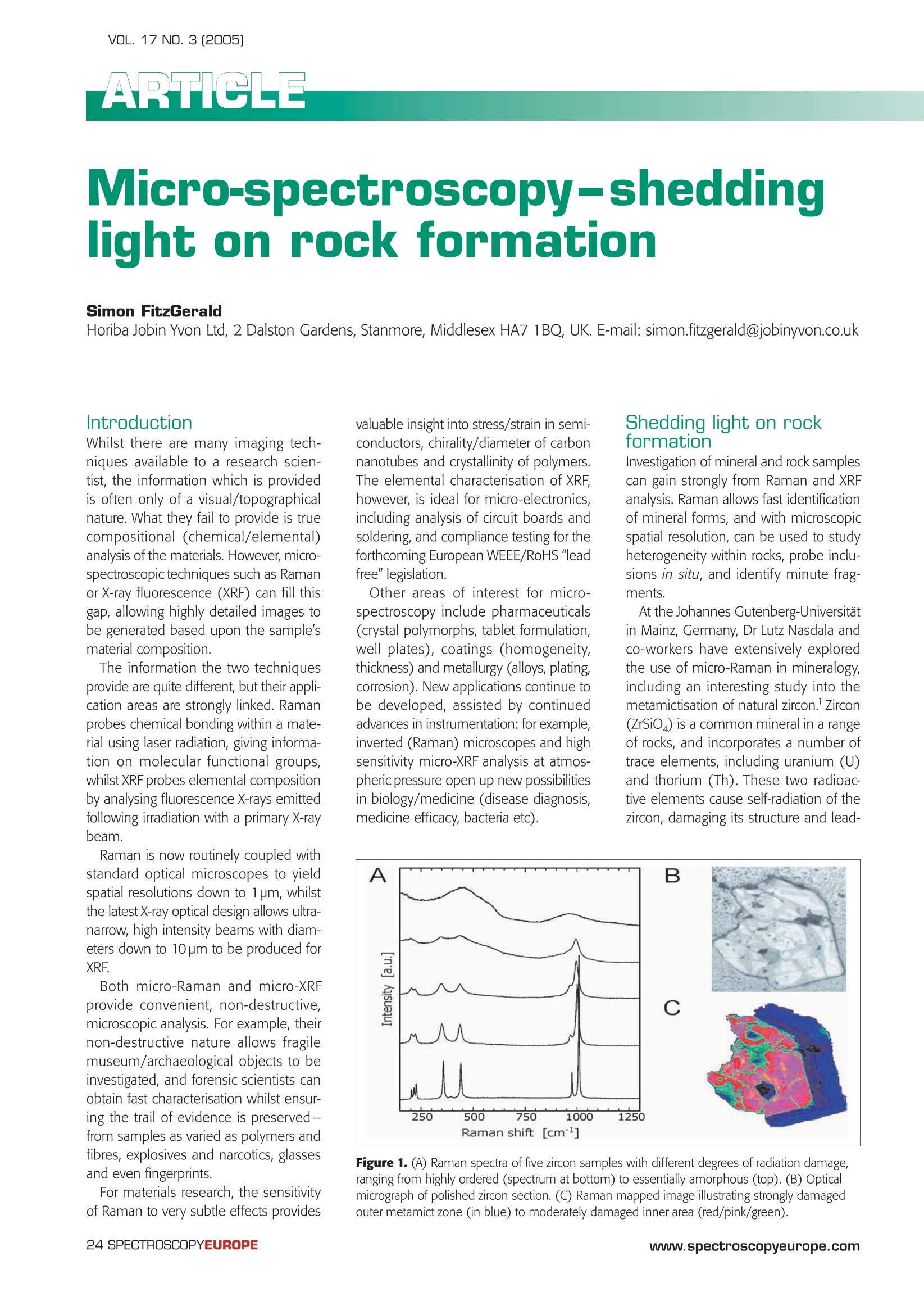
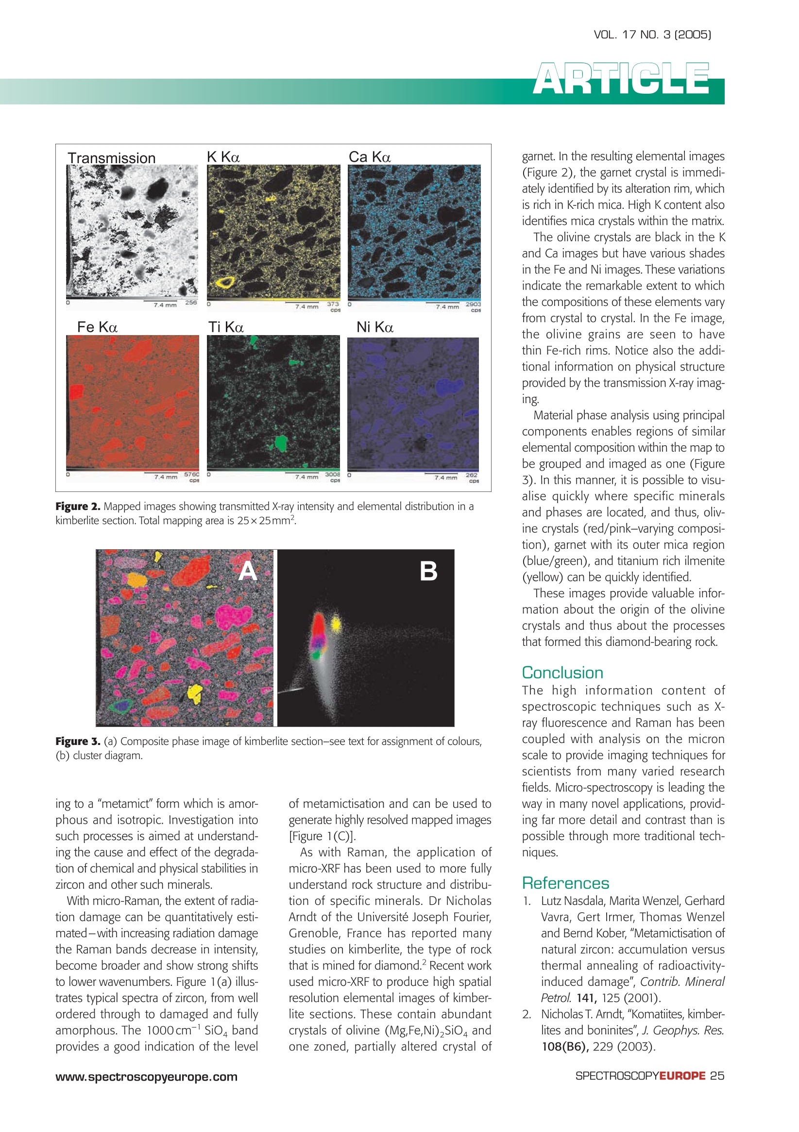
VOL. 17 NO. 3 [2005] Micro-spectroscopy-sheddinglight on rock formation Simon FitzGerald Horiba Jobin Yvon Ltd, 2 Dalston Gardens, Stanmore, Middlesex HA7 1BQ, UK. E-mail: simon.fitzgerald@jobinyvon.co.uk Introduction Whilst there are many imaging tech-niques available to a research scien-tist, the information which is providedis often only of a visual/topographicalnature. What they fail to provide is truecompositional (chemical/elemental)analysis of the materials. However, micro-spectroscopic techniques such as Ramanor X-ray fluorescence (XRF) can fill thisgap, allowing highly detailed images tobe generated based upon the sample'smaterial composition. The information the two techniquesprovide are quite different, but their appli-cation areas are strongly linked. Ramanprobes chemical bonding within a mate-rial using laser radiation, giving informa-tion on molecular functional groups,whilst XRF probes elemental compositionby analysing fluorescence X-rays emittedfollowing irradiation with a primary X-raybeam. Raman is now routinely coupled withstandard optical microscopes to yieldspatial resolutions down to 1pm, whilstthe latest X-ray optical design allows ultra-narrow, high intensity beams with diam-eters down to 10pm to be produced forXRF. Both micro-Raman and micro-XRFprovide convenient, non-destructive,microscopic analysis. For example, theirnon-destructive nature allows fragilemuseum/archaeological objects to beinvestigated, and forensic scientists canobtain fast characterisation whilst ensur-ing the trail of evidence is preserved-from samples as varied as polymers andfibres, explosives and narcotics, glassesand even fingerprints. For materials research, the sensitivityof Raman to very subtle effects provides valuable insight into stress/strain in semi-conductors, chirality/diameter of carbonnanotubes and crystallinity of polymers.The elemental characterisation of XRF,however, is ideal for micro-electronics,including analysis of circuit boards andsoldering, and compliance testing for theforthcoming European WEEE/RoHS "leadfree"legislation. Other areas of interest for micro-spectroscopy include pharmaceuticals(crystal polymorphs, tablet formulation,well plates), coatings (homogeneity,thickness) and metallurgy (alloys, plating,corrosion). New applications continue tobe developed, assisted by continuedadvances in instrumentation: for examplee,inverted (Raman) microscopes and highsensitivity micro-XRF analysis at atmos-pheric pressure open up new possibilitiesin biology/medicine (disease diagnosis,medicine efficacy, bacteria etc). Shedding light on rockformation Investigation of mineral and rock samplescan gain strongly from Raman and XRFanalysis. Raman allows fast identificationof mineral forms, and with microscopicspatial resolution, can be used to studyheterogeneity within rocks, probe inclu-sions in situ, and identify minute frag-ments. At the Johannes Gutenberg-Universitatin Mainz, Germany, Dr Lutz Nasdala andco-workers have extensively exploredthe use of micro-Raman in mineralogy,including an interesting study into themetamictisation of natural zircon. Zircon(ZrSiO) is a common mineral in a rangeof rocks, and incorporates a number oftrace elements, including uranium (U)and thorium (Th).These two radioactive elements cause self-radiation of thezircon, damaging its structure and lead- Figure 1. (A) Raman spectra of five zircon samples with different degrees of radiation damage,ranging from highly ordered (spectrum at bottom) to essentially amorphous (top). (B) Opticalmicrograph of polished zircon section.(C) Raman mapped image illustrating strongly damagedouter metamict zone (in blue) to moderately damaged inner area (red/pink/green). Figure 2. Mapped images showing transmitted X-ray intensity and elemental distribution in akimberlite section. Total mapping area is 25×25mm. A B Figure 3. (a) Composite phase image of kimberlite section-see text for assignment of colours,(b) cluster diagram. ing to a "metamict" form which is amor-phous and isotropic. Investigation intosuch processes is aimed at understand-ing the cause and effect of the degrada-tion of chemical and physical stabilities inzircon and other such minerals. With micro-Raman, the extent of radia-tion damage can be quantitatively esti-mated-with increasing radiation damagethe Raman bands decrease in intensity,become broader and show strong shiftsto lower wavenumbers. Figure 1(a) illus-trates typical spectra of zircon, from wellordered through to damaged and fullyamorphous. The 1000cm-sio bandprovides a good indication of the level of metamictisation and can be used togenerate highly resolved mapped images[Figure 1(C)]. As with Raman, the application ofmicro-XRF has been used to more fullyunderstand rock structure and distribu-tion of specific minerals. Dr NicholasArndt of the Universite Joseph Fourier,Grenoble, France has reported manystudies on kimberlite, the type of rockthat is mined for diamond.Recent workused micro-XRF to produce high spatiaresolution elemental images of kimber-lite sections. These contain abundantcrystals of olivine (Mg,Fe,Ni),Sio andone zoned, partially altered crystal of garnet. In the resulting elemental images(Figure 2), the garnet crystal is immedi-ately identified by its alteration rim, whichis rich in K-rich mica. High K content alsoidentifies mica crystals within the matrix. The olivine crystals are black in the Kand Ca images but have various shadesin the Fe and Ni images. These variationsindicate the remarkable extent to whichthe compositions of these elements varyfrom crystal to crystal. In the Fe image,the olivine grains are seen to havethin Fe-rich rims. Notice also the addi-tional information on physical structureprovided by the transmission X-ray imag-ing. Material phase analysis using principalcomponents enables regions of similarelemental composition within the map tobe grouped and imaged as one (Figure3). In this manner, it is possible to visu-alise quickly where specific mineralsand phases are located, and thus, oliv-ine crystals (red/pink-varying composi-tion), garnet with its outer mica region(blue/green), and titanium rich ilmenite(yellow) can be quickly identified. These images provide valuable infor-mation about the origin of the olivinecrystals and thus about the processesl that formed this diamond-bearing rock. The high information content ofspectroscopic techniques such as X-ray fluorescence and Raman has beencoupled with analysis on the micronscale to provide imaging techniques forscientists from many varied researchfields. Micro-spectroscopy is leading theway in many novel applications, provid-ing far more detail and contrast than ispossible through more traditional tech-niques. 1.. ILutz Nasdala, Marita Wenzel, GerhardVavra, Gert Irmer, Thomas Wenzeland Bernd Kober,"Metamictisation ofnatural zircon: accumulation versusthermal annealing of radioactivity-induced damage",Contrib. MineralPetrol. 141, 125 (2001). 2.Nicholas T. Arndt, "Komatiites, kimber-lites and boninites", J. Geophys. Res.108(B6), 229(2003). SPECTROSCOPYEUROPEwww.spectroscopyeurope.com The high information content of spectroscopic techniques such as Xray fluorescence and Raman has been coupled with analysis on the micron scale to provide imaging techniques for scientists from many varied research fields. Micro-spectroscopy is leading the way in many novel applications, providing far more detail and contrast than is possible through more traditional techniques.
关闭-
1/2
-
2/2
产品配置单
上海巨纳科技有限公司为您提供《矿物,岩石中化学组成及分布,结构检测方案 》,该方案主要用于矿物,岩石中化学组成及分布,结构检测,参考标准《暂无》,《矿物,岩石中化学组成及分布,结构检测方案 》用到的仪器有HORIBA HR Evolution高分辨拉曼光谱仪。
我要纠错
推荐专场

相关方案
-
方案 稀土溶液中La、Ce、Pr、Nd、Sm、Eu、Gd、Tb、Dy、Ho、Er、Tm、Yb、Lu、Y检测方案(ICP-AES)
稀土/稀有金属 | La、Ce、Pr、Nd、Sm、Eu、Gd、Tb、Dy、Ho、Er、Tm、Yb、Lu、Y

-
方案 稀土溶液中La、Ce、Pr、Nd、Sm、Eu、Gd、Tb、Dy、Ho、Er、Tm、Yb、Lu、Y检测方案(ICP-AES)
稀土/稀有金属 | La、Ce、Pr、Nd、Sm、Eu、Gd、Tb、Dy、Ho、Er、Tm、Yb、Lu、Y
- 查看全部方案
 咨询
咨询