方案详情文
智能文字提取功能测试中
How to characterize nanoparticle suspensionsOverview Overview How to characterize nanoparticlesuspensions Nanoparticle suspensions arecomplex systems and their characterization includes several parame-ters such as size, concentration insolution, shape, surface charge andchemical composition. To collect thisinformation, an arsenal of techniquesis needed. Here we present an over-view of examples of characterizationtools that could be used. What is the size of mvnanoparticles? The nanoparticle size corresponds to thethree-dimensional dimensions that markthe interface between the nanoparticleand its environment. Size may be referreddifferently depending on which type ofinterface that is being probed. For example,some techniques may measure the physicalsize focusing on the hard material interface,or the hydrodynamic size by targeting thewater layer attached to the nanoparticleas it moves in solution. The nanoparticle shape also plays a role in determining size.In some cases, the characterization toolmay be restricted to approximate all objectsto spheres and would, therefore, allocatean effective diameter to non-sphericalnanoparticles. Measuring the size distri-bution of a nanoparticle suspension canhighlight the presence of large objectsidentified as aggregates that would informabout the poor dispersion of nanoparticlesin solution. Size parameters can be extracted for exam-ple from the below techniques where theability to provide information on either theensemble level or at the single nanoparticlelevel are denoted with (E) and (SP) respec-tively: Dynamic light scattering (E) Disc centrifugation (E) Nanoparticle tracking analysis (SP) Tunable Resistive Pulse Sensing (SP) Atomic force microscopy (SP) Electron microscopy (SP) What nanoparticle concentra-tion do l have? The concentration(of nanoparticlesuspension can refer either to the numberof single objects or the amount of materialpresent in solution per volume unit. Toaccurately determine the concentration ofnanoparticles in solution, all objects/mate-rial representing the sample need to bedetected in the solution. This can be chal-lenged by the limit of detection pressuredby the size range and material compositionof the nanoparticles. The concentration of nanoparticle suspen-sions can be obtained for example withthe below, where (PN) denotes particlenumber: Nanoparticle tracking analysis (PN) Tunable Resistive Pulse Sensing (PN) Induced coupled plasma-mass spec-trometry (mass) What is the shape of mynanoparticles? Nanoparticles can come in virtually anyshape from spherical to elongated tubesor even stars. A three-dimensional sizingof the nanoparticles is therefore needed touncover the heterogeneity of dimensions.To do so, methods are mainly based onstatistical image processingperformedwith various types of microscopy such as: Scanning electron microscopy (SP) Transmission electron microscopy (SP) Atomic force microscopy (SP) What is the surface charge ofmy nanoparticles? The surface charge oftaa nanoparticlei5s expressedas zeta-potentialand isinfluenced by the interactions betweennanoparticles and their environment. Thezeta-potential value of a given nanopar-ticle will vary depending on the type ofions, ion strength and pH of the solutionin which nanoparticles are suspended.Zeta-potential can be used to predict thestability of a nanoparticle suspension andcan be measured with techniques such as: Electrophoretic light scattering (E) Tunable Resistive Pulse Sensing (SP/E) Access to surface or bulk chemical compo-sition depends on the sensing depth ofthe measuring tool. The chemical compo-sition of nanoparticles is mainly obtainedby spectroscopy techniques and can becombined withhmicroscopy it toachievesingle particle level. Surface chemistry of nanoparticles may beunraveled by techniques such as: Auger electron spectroscopy (E) X-ray photoelectron spectroscopy (E) Inductively coupled plasma massspectrometry (E) Analytical electron microscopy (SP) Chemical force microscopy (SP) lnformation aabout the bulkkchemicalcomposition of nanoparticles ccan beobtained using: ●· X-ray adsorption spectroscopy (E) · EEnergy dissipative X-ray (SP) Electron energy loss spectroscopy (SP) Time of flight mass spectroscopy (SP) Single particle inductively coupledplasma mass spectrometry (SP) No single technique can offer a completeprofiling of the physicaland chemicalproperties of a nanoparticle suspension.Instead, efficient profiling of nanoparticlesrelies on creating synergies between char-acterization tools to reach the full pictureof the sample at hand. Written by Deborah Rupert, PhD, BiolinScientific AB. Cover photo thr3_eyes on Unsplash. [ Progress Together] [ Progress Together]Contact us at info@biolinscientific.com 纳米颗粒粒径是表征纳米颗粒悬浮液的关键参数之一。这里我们列出了六种可以用来表征纳米颗粒粒径的方法。界面和形状与粒径测量的相关性不同的测量技术以不同的方式测量纳米颗粒粒径。一些技术测量纳米颗粒的物理尺寸,即硬质材料的界面,而其他技术测量流体动力学尺寸,即它们同时关注纳米颗粒在溶液中移动时附着于纳米颗粒的水层。因此,测量结果即最终测量出来的纳米颗粒大小,将取决于检测界面的类型。纳米颗粒的形状在粒径测量中也起着重要的作用。例如,一些表征工具将所有测量物体近似为球体,因此,会为非球形纳米颗粒指定有效直径。纳米颗粒粒径分布纳米颗粒悬浮液是复杂的系统,必须对多个参数进行表征才能很好地了解它们的特性和行为。除了纳米颗粒粒径外,粒径分布也很相关。粒径分布可以揭示诸如溶液中存在聚集体的状况,这反过来可能表明纳米颗粒的分散性较差。测量纳米颗粒粒径的方法有一系列的分析技术都可以用来测量纳米颗粒的粒径。下面我们列出了六种方法,它们都可以提供总体层面(E)或者单个纳米颗粒层面(SP)的信息:1. 动态光散射(E)2. 圆盘离心(E)3. 纳米粒子追踪分析(SP)4. 可调谐电阻脉冲传感(SP)5. 原子力显微镜(SP)6. 电子显微镜(SP)除粒径外,一些其他的参数例如溶液浓度、形状、表面电荷和化学成分等对于纳米颗粒表征也非常重要。下载附件概述以了解更多有关纳米颗粒悬浮液的特性以及可以使用的表征方法。
关闭-
1/2

-
2/2

产品配置单
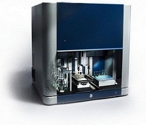
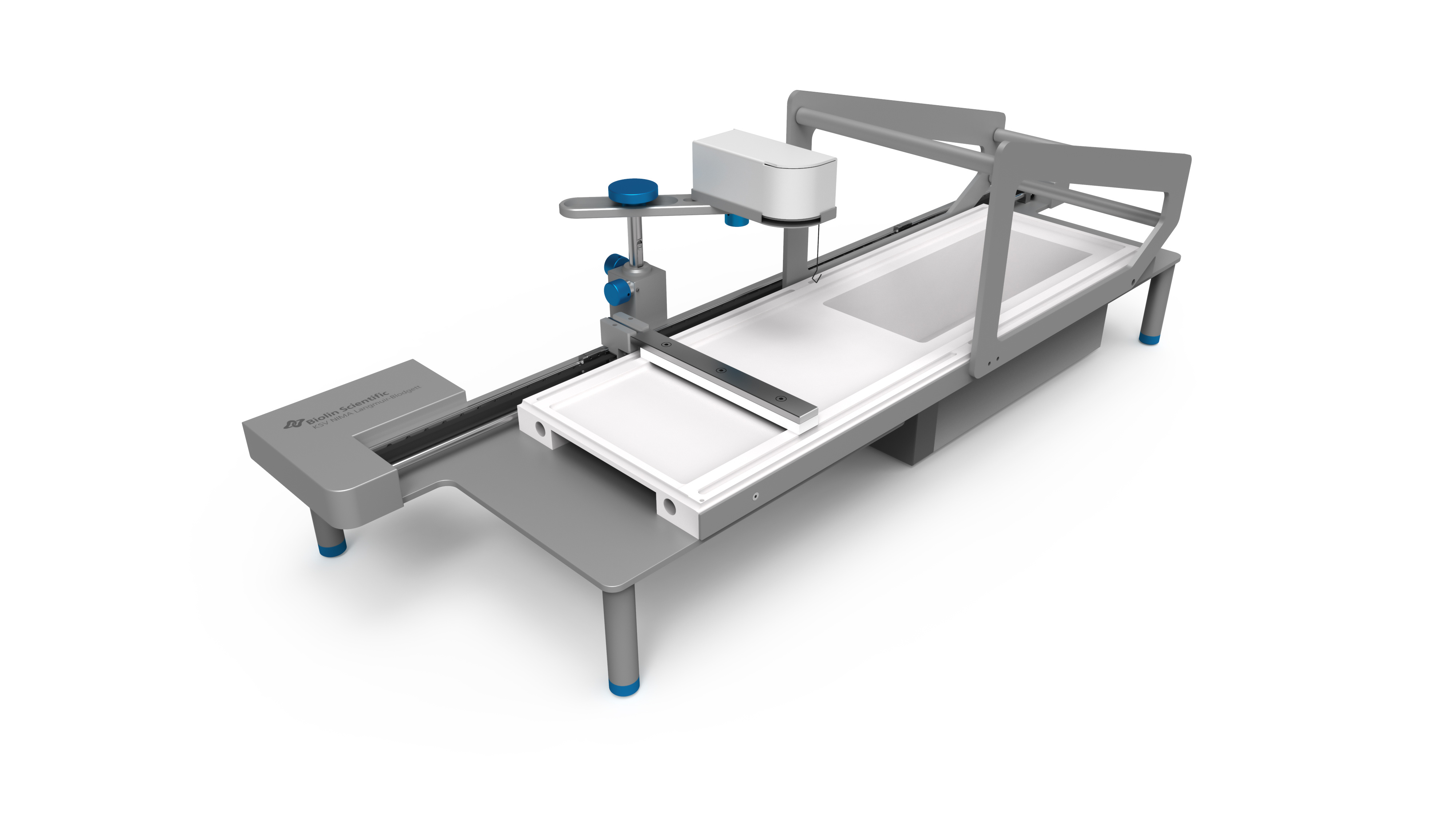
瑞典百欧林科技有限公司为您提供《纳米悬浮液中粒径检测方案(石英晶体天平)》,该方案主要用于其他中理化分析检测,参考标准《暂无》,《纳米悬浮液中粒径检测方案(石英晶体天平)》用到的仪器有QSense全自动八通道石英晶体微天平、KSV NIMA roll to roll 柔性LB膜制备系统。
我要纠错
相关方案


 咨询
咨询


