

方案详情文
智能文字提取功能测试中
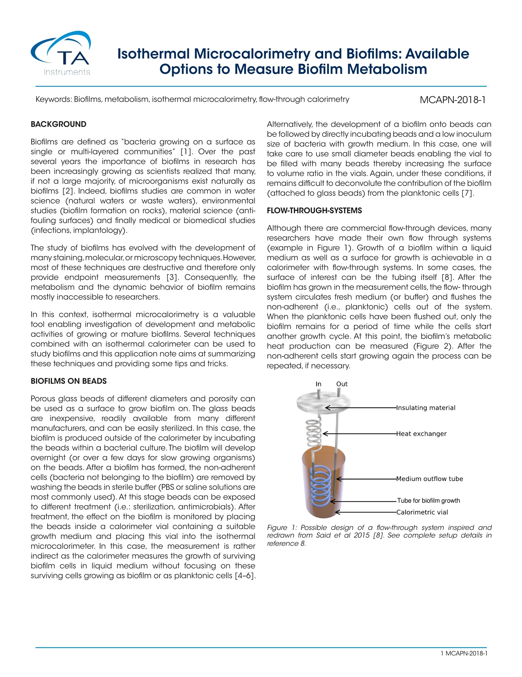
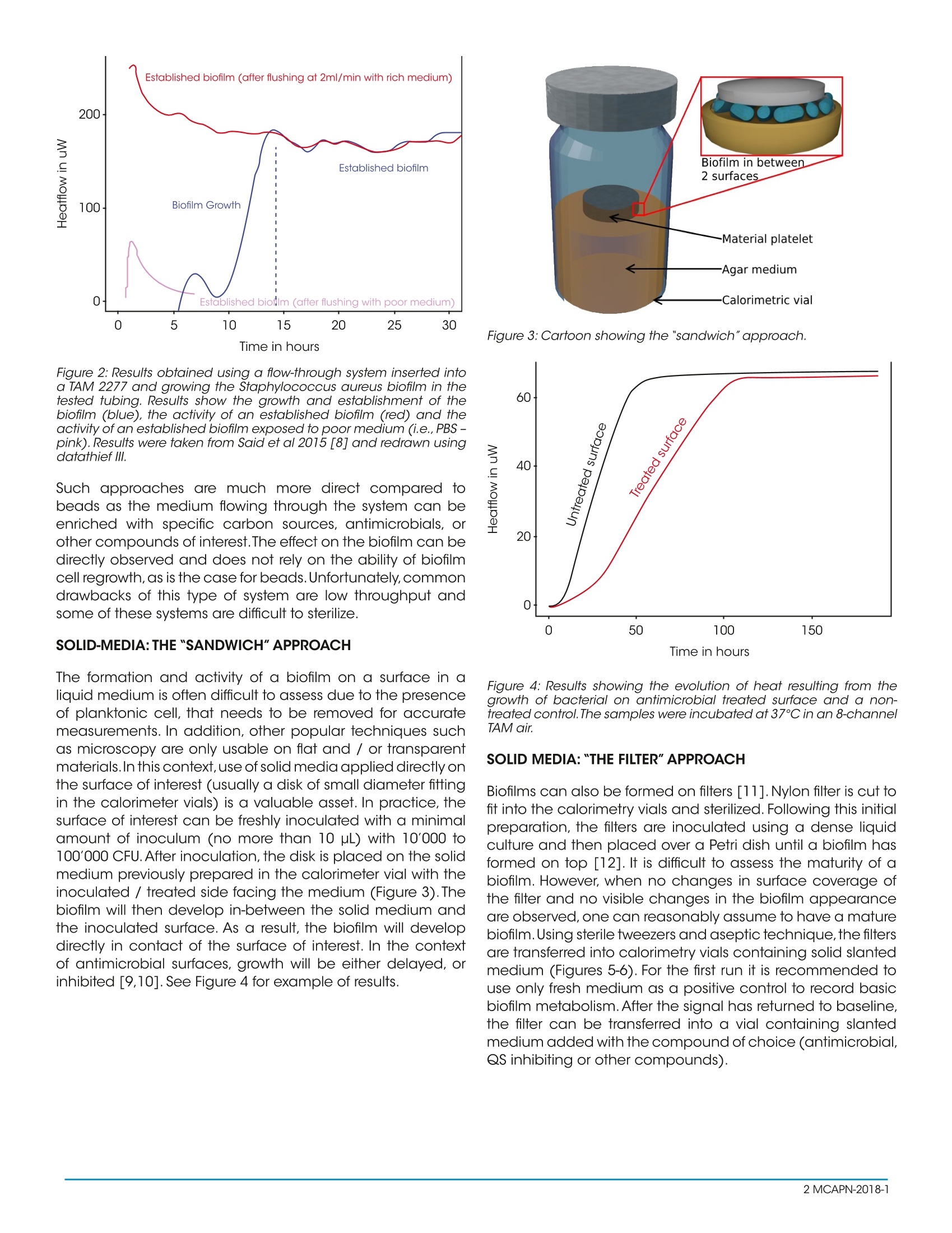
Isothermal Microcalorimetry and Biofilms: AvailableOptions to Measure Biofilm Metabolism Keywords: Biofilms, metabolism, isothermal microcalorimetry, flow-through calorimetry MCAPN-2018-1 BACKGROUND Biofilms are defined as "bacteria growing on a surface assingle or multi-ayered communities"[1]. Over the pastseveral years the importance of biofilms in research hasbeen increasingly growing as scientists realized that many,if not a large majority,of microorganisms exist naturally asbiofilms [2]. Indeed, biofilms studies are common in waterscience (natural waters or waste waters), environmentalstudies (biofilm formation on rocks), material science (anti-fouling surfaces) and finally medical or biomedical studies(infections, implantology). The study of biofilms has evolved with the development ofmany staining, molecular,or microscopy techniques.However,most of these techniques are destructive and therefore onlyprovide endpoint measurements[3]. Consequently, themetabolism and the dynamic behavior of biofilm remainsmostly inaccessible to researchers. In this context, isothermal microcalorimetry is a valuabletool enabling investigation of development and metabolicactivities of growing or mature biofilms. Several techniquescombined with an isothermal calorimeter can be used tostudy biofilms and this application note aims at summarizingthese techniques and providing some tips and tricks. BIOFILMS ON BEADS Porous glass beads of different diameters and porosity canbe used as a surface to grow biofilm on. The glass beadsare inexpensive, readily available from many differentmanufacturers, and can be easily sterilized. In this case, thebiofilm is produced outside of the calorimeter by incubatingthe beads within a bacterial culture.The biofilm will developovernight (or over a few days for slow growing organisms)on the beads. After a biofilm has formed, the non-adherentcells (bacteria not belonging to the biofilm) are removed bywashing the beads in sterile buffer (PBS or saline solutions aremost commonly used). At this stage beads can be exposedto different treatment (i.e.: sterilization, antimicrobials). Aftertreatment, the effect on the biofilm is monitored by placingthe beads inside a calorimeter vial containing a suitablegrowth medium and placing this vial into the isothermalmicrocalorimeter. In this case, the measurement is ratherindirect as the calorimeter measures the growth of survivingbiofilm cells in liquid medium without focusing on thesesurviving cells growing as biofilm or as planktonic cells [4-6]. Alternatively, the development of a biofilm onto beads canbe followed by directly incubating beads and a low inoculumsize of bacteria with growth medium. In this case, one willtake care to use small diameter beads enabling the vial tobe filled with many beads thereby increasing the surfaceto volume ratio in the vials. Again, under these conditions, itremains difficult to deconvolute the contribution of the biofilm(attached to glass beads) from the planktonic cells [7]. FLOW-THROUGH-SYSTEMS Although there are commercial flow-through devices, manyresearchers have made their own flow through systems(example in Figure 1). Growth of a biofilm within a liquidmedium as well as a surface for growth is achievable in acalorimeter with flow-through systems. In some cases, thesurface of interest can be the tubing itself [8]. After thebiofilm has grown in the measurement cells, the flow-throughsystem circulates fresh medium (or buffer) and flushes thenon-adherent (i.e., planktonic) cells out of the system.When the planktonic cells have been flushed out, only thebiofilm remains for a period of time while the cells startanother growth cycle. At this point, the biofilm’s metabolicheat production can be measured (Figure 2). After thenon-adherent cells start growing again the process can berepeated, if necessary. Figure 1: Possible design of a flow-through system inspired andredrawn from Said et al 2015 [8]. See complete setup details inreference 8. Figure 2: Results obtained using a flow-through system inserted intoa TAM 2277 and growing the Staphylococcus aureus biofilm in thetested tubing. Results show the growth and establishment of thebiofilm (blue), the activity of an established biofilm (red) and theactivity of an established biofilm exposed to poor medium (i.e., PBS-pink). Results were taken from Said et al 2015 [8] and redrawn usingdatathief lll. Such approaches are much more direct compared tobeads as the medium flowing through the system can beenriched with specific carbon sources, antimicrobials, orother compounds of interest.The effect on the biofilm can bedirectly observed and does not rely on the ability of biofilmcell regrowth, as is the case for beads.Unfortunately,commondrawbacks of this type of system are low throughput andsome of these systems are difficult to sterilize. SOLID-MEDIA:THE"SANDWICH"APPROACH The formation and activity of a biofilm on a surface in aliquid medium is often difficult to assess due to the presenceof planktonic cell, that needs to be removed for accuratemeasurements. In addition, other popular techniques suchas microscopy are only usable on flat and /or transparentmaterials.In this context, use of solid media applied directly onthe surface of interest (usually a disk of small diameter fittingin the calorimeter vials) is a valuable asset. In practice, thesurface of interest can be freshly inoculated with a minimalamount of inoculum (no more than 10 pl) with 10'000 to100'000 CFU. After inoculation,the disk is placed on the solidmedium previously prepared in the calorimeter vial with theinoculated / treated side facing the medium (Figure 3). Thebiofilm will then develop in-between the solid medium andthe inoculated surface. As a result, the biofilm will developdirectly in contact of the surface of interest. In the contextof antimicrobial surfaces, growth will be either delayed, orinhibited [9,10]. See Figure 4 for example of results. Figure 3: Cartoon showing the "sandwich"approach. Figure 4: Results showing the evolution of heat resulting from thegrowth of bacterial on antimicrobial treated surface and a non-treated control.The samples were incubated at 37℃ in an 8-channelTAM air. SOLID MEDIA:THE FILTER"APPROACH Biofilms can also be formed on filters [11].Nylon filter is cut tofit into the calorimetry vials and sterilized. Following this initialpreparation, the filters are inoculated using a dense liquidculture and then placed over a Petri dish until a biofilm hasformed on top [12]. It is difficult to assess the maturity of abiofilm. However, when no changes in surface coverage ofthe filter and no visible changes in the biofilm appearanceare observed, one can reasonably assume to have a maturebiofilm.Using sterile tweezers and aseptic technique,the filtersare transferred into calorimetry vials containing solid slantedmedium (Figures 5-6). For the first run it is recommended touse only fresh medium as a positive control to record basicbiofilm metabolism. After the signal has returned to baseline,the filter can be transferred into a vial containing slantedmedium added with the compound of choice (antimicrobial,QS inhibiting or other compounds). Figure 5: Sketch showing the filter approach. Note that in thisapproach filters can be re-used and transferred to other mediumwith different conditions. The advantage of such approach is that filters can be usedseveral times and transferred easily from one condition toanother. Except for the flow-through system described above,biofilms are usually used only once because they are difficultto handle without destroying their structure. Considering theheterogeneity of biofilm (and the potential variations resultingfrom such heterogeneity), it is very helpful to compare thevery same biofilm under different conditions. Figure 6: Results of 2 duplicates M.smegmatis biofilms on filters andincubated at 37℃ in an 8-channel TAM air. TOWARD BIOFILM ENERGETICS The metabolism and bio-energetic profile offbiofilmsis a complicated topic and calorimetry data can becomplemented by respiration data,O,consumption or CO,production. In sealed glass vials such measurements aremade practical using parallel samples that are prepared inthe same way but incubated in an oven. The O, and CO,concentration variations in the headspace over time canbe recorded using laser absorption spectroscopy (TDLAS)[13,14]. Alternatively,oxygen optrodes have also been usedsuccessfully [15,16]. CONCLUSION Isothermal microcalorimetry using 4 or 20 ml vials or aflow-through system offers several options to assess biofilmmetabolism. Although the filter and sandwich approachare more convenient in larger vials (20 ml option) they canbe used in 4 mL vials as well. The possibility to combinemicrocalorimetry with other techniques remains attractiveas isothermal microcalorimetry is non-destructive and offersfurther options to investigate biofilm bioenergetics. ACKNOWLEDGMENTS This application note was written by Dr. Olivier Braissant andedited by TA Instruments. ( 1 . H all-Stoodley, L.; Costerton, J.; Stoodley, P. Bacterial biofilms: from t he natural e nvironment to i n fectious diseases.Nat. Rev.Microbiol. 2004,2,95-1 0 8, doi:10.1038/nrmicro821. ) ( 2. Sutherland, I. W . Biofi l m exopolysaccharides: A stron g andsticky framework.Microbiology 2001,147,3-9. ) ( 3. F F lemming, H. C.; Wingender, J.Th e biofilm matrix. Nat. Rev. Microbiol.2010,8,623-633. ) ( 4. Tafin, U. F; Corvec, S.; Betrisey, B.; Zimmerli, W.; Trampuz, A.Role o f rifampin against Propionibacterium acnes biofilmin vitro and in an e xperimental f oreign-body infectionmodel. Antimicrob. Agents Chemother. 2 012, 56, 1885- 1891,doi:10.1128/AAC.05552-11. ) ( 5. C orvec, S . ; Tafin, U . E ; B etrisey, B.; Borens, O.; Trampuz,A. Activities of f osfomycin, tigecycline, colistin, andgentamicin against extended-spectrum-lactamase-producing escherichia coli in a f oreign-body infectionmodel. Antimicrob. Agents Chemother. 2 013, 57, 1421- 1427, doi:10.1128/AAC.01718-12. ) ( 6. M aiolo, E. M.; Tafin, U. F ; B orens, O.; Trampuz, A. Activities of fluconazole, , c aspofungin, anidulafungin, andamphotericin b on planktonic and b iofilm candidaspecies d etermined b y microcalorimetry. A ntimicrob.Agents C hemother. 2014, 58, 2 7 09-2717, d o i:10.1128/ AAC.00057-14. ) ( 7. Hauser-Gerspach,l.;Kulik,E . M.;Weiger,R.;Decker,E.-M.;VonOhle, C .; Meyer, J. Adhesion of Streptococcus sanguinisto dental implant and res t orative mat e rials in vit r o. Dent.Mater.J. 2007, 26, 361-366, doi:10.4012/dmj.26.361. ) ( 8. Said, J.; Walker, M.; Parsons, D.; Stapleton, P; Beezer,A. E.; Gaisford, S. Development o f a flow s y stem fo r studying biofilm formation on m edical d e vices withmicrocalorimetry. Methods 2015, 76,35-40, doi:10.1016/j. vmeth.2014.12.002 ) ( 9.E Braissant,O.;Chavanne,P; D e Wild,M.;Pieles,U.;Stevanovic, S.; Schumacher, R.; S traumann, L. ; Wirz, D.; G runer, P;Bachmann, A.; Bonkat,G. Novel microcalorimetric a s say for antibacterial activity o f impla n t coatings: The casesof silver-doped hydroxyapatite a nd c a lcium hydroxide.J. Biomed. Mater. Res. - Part B Appl. Biomater. 2 0 15, 103, doi:10.1002/jbm.b.33294. ) ( 10.Guimond-Lischer, S .; Ren,Q . ; Braissant, O.; Gruner, P ; Wampfler, B.; Maniura-Weber, K.Vacuum plasma sprayedcoatings using i ionic silver doped hydroxyapatitepowder t o p r event bacterial infection o f b o ne im p lants.Biointerphases 2016,11, doi:10.1116/1.4943225. ) ( 11.Merritt, J . H.; Kadouri , D . E . ; O'Toole, G. A. Growing andanalyzing s t atic b iofilms. C urr. Protoc. M icrobiol. 2 011 , doi:10.1002/9780471729259.mc01b01s22. ) ( 12.Solokhina, A.; Bruckner, D.; Bonkat, G.; Braissant, O. Metabolic activity of mature biofilms of Mycobacteriumtuberculosis and other n on-tuberculous mycobacteria. Sci. Rep.2017,7,doi:10.1038/s41598-017-10019-4. ) ( 13. B rueckner, D.; Solokhina,A.; K rahenbuhl, S.; B raissant,O.A combined application of tunable diode laser absorptionspectroscop y andisothermal micro-calorimetry forcalorespirometric analysis. J . Microbiol. Methods 2 017, 139,210-214, doi:10.1016/j.mimet.2017.06.012. ) ( 14.Brueckner, D. ; Roesti, D. ; Zuber , U. G. ; Schmidt, R .; Kraehenbuehl, S .; Bonkat, G. ; Braissant,O. Comparisonof Tunable Diode Laser Absorption Spectroscopy andIsothermal Micro-calorimetry for Non-invasive Detectionof Microbial Growth in Me d ia Fills. S c i. R e p. 2016, 6, doi:10 . 1038/srep27894. ) ( 15.Regan, M. D .; Gosline, J. M.; Richards, J. G. A simple andaffordable calorespirometer for assessing the metabolicrates of fishes. J .Exp. Biol.2013,216,4507 LP-4513. ) ( 16.ltoga,N.K.;Hansen,L.D.Gas-phase optrodemeasurementsof oxygen in calorimetric vessels.Thermochim. Acta 2 0 09, 490,78-81. ) MCAPN- Biofilms are defined as “bacteria growing on a surface as single or multi-layered communities”. Over the past several years the importance of biofilms in research has been increasingly growing as scientists realized that many, if not a large majority, of microorganisms exist naturally as biofilms. Indeed, biofilms studies are common in water science (natural waters or waste waters), environmental studies (biofilm formation on rocks), material science (antifouling surfaces) and finally medical or biomedical studies (infections, implantology).The study of biofilms has evolved with the development of many staining, molecular, or microscopy techniques. However, most of these techniques are destructive and therefore only provide endpoint measurements. Consequently, the metabolism and the dynamic behavior of biofilms remains mostly inaccessible to researchers.In this context, isothermal microcalorimetry is a valuable tool enabling investigation of development and metabolic activities of growing or mature biofilms. Several techniques combined with an isothermal calorimeter can be used to study biofilms and this application note aims at summarizing these techniques and providing some tips and tricks.
关闭-
1/4
-
2/4
还剩2页未读,是否继续阅读?
继续免费阅读全文产品配置单
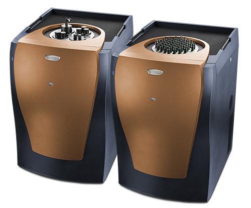
TA仪器为您提供《生物膜中生长代谢能量检测方案(差示扫描量热)》,该方案主要用于其他中生长代谢能量检测,参考标准《暂无》,《生物膜中生长代谢能量检测方案(差示扫描量热)》用到的仪器有TA仪器+TAM IV+微量热仪。
我要纠错
推荐专场
差示扫描量热仪(DSC/DTA)
更多
相关方案
 咨询
咨询