

方案详情文
智能文字提取功能测试中
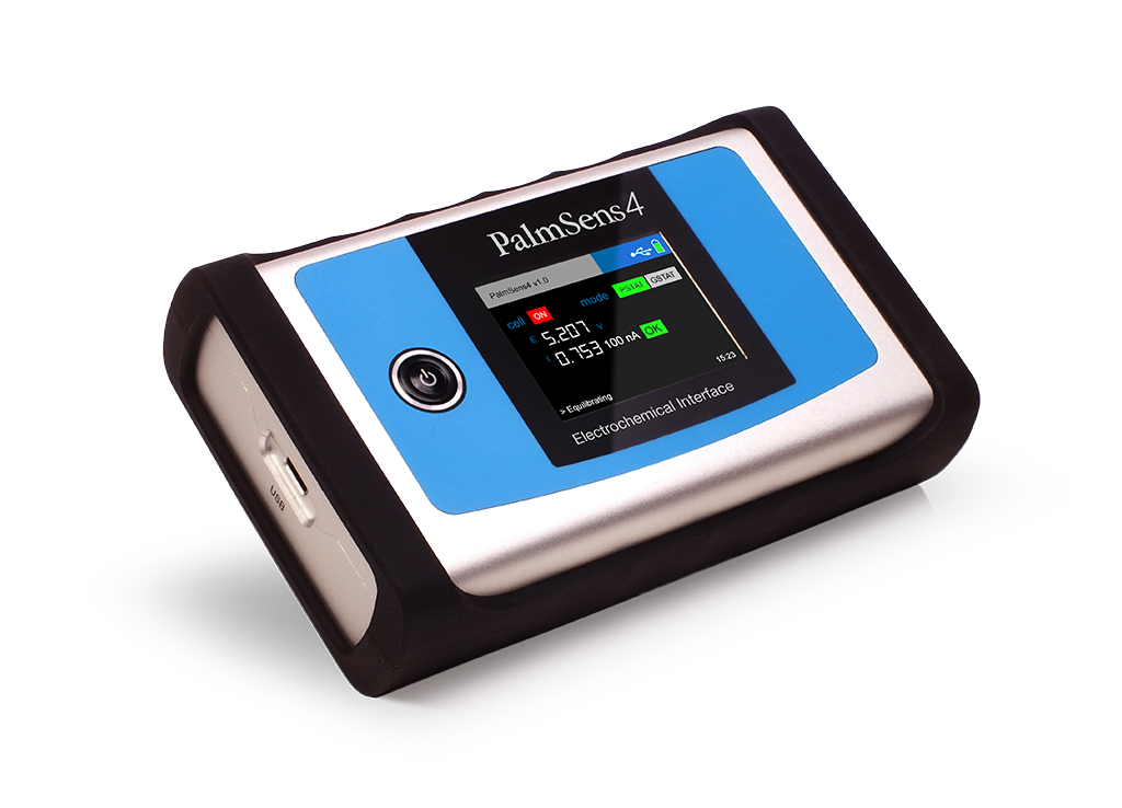
导致食物中毒的细菌对食物和饮用水的污染是一个关键的公共卫生问题。一般来说,在公共场合检测病原菌的应用场景,因为耗时,而且不利于在现场环境中进行实时监测。因此,本文研究中引入了一个全自动平台,一般分析人员能够在现场环境中轻松检查主要的食物中毒细菌,从预处理样本到最终检测。利用甘露糖的特异性结合力,成功地将四种不同细菌(即大肠杆菌和鼠伤寒沙门氏菌)中的两种肠道细菌固定在磁性表面,并且,磁性-电化学阻抗测量技术能够以高灵敏度检测细菌。大肠杆菌是典型的肠道细菌,在25分钟内被检测到,检测限为100CFU/mL。另外本文证明,与手动进行的实验相比,自动性能和实验一致性都得到大大的提高。本文中使用一次性抛弃式的碳丝印电极,避免电极交叉污染;利用磁性增强检测物质的富集能力,检测系统中嵌入PalmSens便携式电化学分析仪进行循环伏安法和交流阻抗的电化学测试。--摘自BioChip Journal (2021) 15:233–242,https://doi.org/10.1007/s13206-021-00024-1--自动细菌富集和检测系统的示意图BioChip Journal (2021) 15:233–242https://doi.org/10.1007/s13206-021-00024-1ORIGINAL ARTICLE BioChip Journal (2021) 15:233–242234 Fully Automated System for Rapid Enrichment and Precise Detection of Enterobacteria Using Magneto-Electrochemical Impedance Measurements Kirok Kwon1.· Taehee Yoon1·· Hogyeong Gwak1 · Kyungyeon Lee1l· Kyung-A. Hyun1·· Hyo-Il Jung1 Received: 7 January 2021 / Revised: 30 March 2021 / Accepted: 5 April 2021 / Published online: 20 May 2021© The Korean BioChip Society 2021 Abstract The contamination of food and drinking water by bacteria that cause food poisoning is a critical public health issue. In gen-eral, the procedures for testing the presence of pathogenic bacteria in public is not very efefctive, because it is time consuming and does not facilitate real-time monitoring in a feild setting. Therefore, this study introduced a fully automated platform that would allow non-experts to easily examine the major food poisoning bacteria in a feild setting, from pretreatment sam-ples to fnial detection. Two enterobacteria species, of four difefrent bacteria species (i.e., Escherichia coli and Salmonella typhimurium) were successfully immobilized on the surface of magnetic beads by exploiting the specifci binding force with mannose. Subsequently, magneto-electrochemical impedance measurement technology allowed bacterial detection with a high sensitivity. E. coli, typical enterobacteria, was detected in 25 min with a detection limit of 100 CFU/mL. Moreover, we demonstrated that the automatic performance and the experimental consistency was greatly improved in comparison with those of manually conducted experiments. Keywords Enterobacteria·· Food poisoning·· Fully automated bacterial detection·· Magneto-electrochemical and mannose 1Introduction The development of technology that enables detection of very low concentrations of pathogenic bacteria in a feild setting is vital for the prevention of various diseases caused by bacterial contamination of food. However, it is dif cfulit to simplify the equipment or procedure for non-expert per-sonnel, because ordinary and conventional detection equip-ment are based on complicated and sequential molecular reactions such as sample leading, biomarker extraction, and the polymerase chain reaction (PCR) [1, 2]. Therefore, the need for automated equipment that allows non-experts to easily detect infectious pathogens in the feild has risen stead-ily [3–7]. Moreover, these systems should be miniaturized and be automated for easy use by non-experts in the feild, and they should be designed to detect infectious bacteria * Hyo-Il Jung uridle7@yonsei.ac.kr 1 School of Mechanical Engineering, Yonsei University,50 Yonsei-ro, Seodaemun-gu, Seoul 120-749, Republic of Korea without employing expensive and complicated laboratory equipment [8–11]. Traditional detection and monitoring methods entail cul-turing the infectious bacteria on plates to detect the presence or type of specifci bacteria by comparing their growth rate, specifci nutritional needs, and inoculum size [12, 13]. How-ever, this method requires about 24–48 h to fully cultivate and detect bacteria, so the use of automated systems is not suitable in feild settings. Another method involves isolat-ing the bacteria present in food and extracting their DNA to identify them through a conventional or real-time PCR; however, this method requires complex chemical sample processing to extract bacterial DNA [14, 15]. The disad-vantage of this method is that a treatment time of several hours is required, including a sample treatment, although it is less time-consuming compared to the culture method. A series of sample processing steps are necessary for the efcfieint enrichment and lysis of the bacteria using physical or chemical methods, to extract adequate amount bacterial DNA for PCR. Therefore, PCR is not suitable for developing simple and fast automatic equipment that can be used easily by non-experts. Studies have focused on the development of equipment for concentrating and detecting pathogenic bacteria in food samples. The advantages of automated devices include the use of a continuous method to obtain accurate results with fewer variations compared to those obtained with laboratory procedures. Through previous research, we have developed a rapid and efcfieint bacteria pretreatment technology using microfulidic chips [16, 17]. In addition, it was confrimed that successful PCR analysis was possible through the extracted DNAs. However, there are some limitations to manufacture such complex automated devices for pretreatment and detec-tion of bacteria using microfluidic chips. To continuously enrich and extract DNAs using the microfulidic devices, the automated process requires folw control systems and mas-sive syringe pumps for multiple inlets and outlets. Therefore, to overcome these limitations and simplify the devices, we developed a cartridge-based sample processing technology to concentrate the sample loaded within a cartridge-type chamber using gravity and magnetic force. Moreover, we developed a cartridge-based automated bacterial detection system based on electrochemical impedance measurement without additional chemical modifciation of the sensor sur-face (Fig. 1). To enrich and detect bacteria, the magnetic beads were bound to the bacteria using a bacterial adhesive surface structure to bind specifci bacteria, and then the sample pre-treatments were performed by magnetic force. Enterobac-teria, such as E. coli and Salmonella, express a structure known as type 1 pili on their cell membrane, which contains a lectin called FimH at the structural end of the pili [18–21]. Essentially, the type 1 pili detect the mannose residues on the surface of the host cell to allow the bacteria to recog-nize the host cells [22]. Using this rationale, mannose was coated on the magnetic beads using chemical surface modi-fciations, so that the magnetic particles could combine with the bacteria through the binding force between type 1 pili and mannose. In this study, we detected E. coli and S. typhimurium using electrochemical impedance spectroscopy (EIS) after capturing them with mannose-modifeid magnetic beads (Man-MBs). The bacteria captured by the magnetic parti-cles were loaded on the electrochemical sensor using a mag-netic force and the bacteria were detected by measuring the change in the impedance of the magnetic particles using EIS. 2 Materials and Methods 2.1 Materials and Bacterial Preparation Ethylenediamine, methanol, D-mannose, concanavalin A (Con A), nitric acid, sulfuric acid, 1-ethyl-3-(3-dimeth-ylaminopropyl) carbodiimide (EDC), K3Fe(CN)6, and K4Fe(CN)6·3H20 were purchased from Sigma Aldrich. The MagnaBind™ Carboxyl derivatized magnetic beads, SYTO 9 green fulorescent stain, and BupH™ MES Bufefred Saline were purchased from Thermo Scientifci. Polydimethylsilox-ane (PDMS) (Dow Corning, USA), a biocompatible mate-rial, was used to fabricate the disposable electrochemical chamber. We employed four bacterial samples (E. coli, S. typhimu-rium, Staphylococcus aureus and Bacillus cereus), which were cultured overnight in a shaking incubator (37°°C,200 rpm) in Luria–Bertani broth to evaluate the selectivity and sensitivity of the automated bacterial pre-treatment and detection chip. All bacteria species were obtained from the Korean Collection for Type Culture. The bacterial samples were diluted to a concentration of 106°CFU/mL by meas-uring the optical density at 600 nm. Subsequently, each diluted bacterial sample was stained with a SYTO 9 solu-tion, a green fulorescent nucleic-acid stain (excitation/emis-sion maxima: 480/500 nm), and followed by measuring the intensity of fulorescence after binding to the magnetic beads. The results of fulorescence intensity measurement were used to determine the time and binding afnfitiy difefrence required for the bacteria to bind the magnetic beads sufcfieintly. 2.2 Preparation of Mannose-Modified Magnetic Beads The mannose-modifeid magnetic beads (Man-MBs) were synthesized using D-mannose to prepare mannosylated eth-ylenediamine in accordance with a previously reported and established method [23, 24]. Carboxyl derivatized magnetic microparticles (size: 1–4 um, magnetization: 25–35 Electric Multiple Unit/g) (Thermo Scientifci, USA) were completely dispersed in MES bufefr (pH 4.7, 0.1 M). EDC (5 mg/mL) was added and the mixture was stirred at room temperature for 30 min. The remaining EDC was removed after capturing the magnetic particles with a magnet. The magnetic beads were washed three times with deionized water and dispersed in MES bufefr again. Finally, mannosylated ethylenediamine (20 mg/mL) was added to the mixture, and the reaction was allowed to occur for 24 h at 25 °C. The residual bufefr was removed, and the products were washed three times with deionized water. 2.3 Electrochemical Detection The screen-printed carbon electrodes (SPCEs) (Dropsens Inc., Spain) were frist washed with sulfuric acid, and then washed lightly with deionized water to prepare clean elec-trodes, which were placed on a three-dimensionally (3D)-printed holder. Electrochemical experiments were performed using the CHI-650E (CH Instruments, US) electrochemi-cal analyzer. Magnetic beads were added to the equimolar Fig. 1 a Schematic workfolw of the conventional bacteria detection procedures. b Schematic drawing and workfolw of our automated bacteria detection procedures ferricyanide/ferrocyanide solution (5 × 10⁻³-M) containing 0.1 M KCl. When magnetic particles were loaded on the sensor at a low concentration, they formed a thin flim on the working electrode of the sensor by a magnetic force. Subsequently, EIS experiments were conducted in a redox probe solution. Nyquist plots were recorded at open circuit and an alternating current potential of 0.005 V over a fre-quency range of 10 kHz–0.1 Hz. Cyclic voltammetry (CV) was performed at a scan rate of 0.1 V/s. Additionally, a com-pact electrochemical analyzer (PalmSens, Netherlands) was further utilized with the automated system to increase the portability and compatibility of our system. 2.4 Automated System An automated system that could enrich and subsequently detect specifci bacteria was constructed. We designed a fully automated system capable of quantifying bacterial enrich-ment to make it simple for anyone working in a feild set-ting. Polylactic acid (XYZ printing, Republic of Korea) was used to print the entire frame and connecting parts using 3D printer (Sindoh Co., Republic of Korea). The sample was designed to folw from top to bottom under gravity without a pump, to overcome sample loss and complexity issues (in the connection process) that could arise as a result of the use of liquid pumps. The 3-way valves (Hyupsung Medical, Republic of Korea) and its connected servo motors (OEM, China) were positioned to control the folw and direction of the fulid. Moreover, a polypropylene enrichment chamber and neodymium magnet holder capable of moving up and down were used to enrich the bacteria bound to the magnetic beads. Thereafter, a sensor holder consisting of a neodym-ium magnet and PDMS chamber was fabricated to fix the magnetic beads on the surface of the electrochemical sensor. The folw control and sequential operation of the whole pro-cess were controlled via Arduino, which is an open-source hardware and software microcontroller. 3 Results and Discussion 3.1 Characterization of Man-MBs to Capture Bacteria Mannose can bind with type 1 pili in the bacterial cell’s membrane through lectin–carbohydrate interaction. Man-nose can also bind to a lectin known as Con A [25]; there-fore, the (synthesized) Man-MBs were characterized by the reaction with bacteria and Con A. First, the absorb-ance was measured at 280 nm to determine the change in absorbance during the reaction of the magnetic particles with the Con A protein, because a protein such as Con A concentration exhibits a linear relationship with absorbance at 280 nm (Fig. 2a). Normal carboxylated magnetic beads were incubated with Con A (1 mg/mL) for 30 min, and the magnetic particles were separated using a magnetic force and the absorbance of unbound Con A was measured. It was observed that about 200 µg/cm2 of Con A was bound to the surface of the magnetic beads after reaction with plain magnetic beads MB, since the non-specifci binding capacity of Con A allows it to adhere to various surfaces. On the other hand, the reaction of the same concentration of ConA with Man-MBs resulted in an adherence of approximately 400 µg/cm2 of ConA to the surface, i.e., approximately 200 µg/cm2more than that with plain magnetic beads. Our results prove that the surface of the prepared Man-MBs was sufcfieintly modifeid with mannose, which specifcially binds ConA (Fig.22b). Moreover, the formation of mannose layer on the sur-face of magnetic beads and binding afnfitiy with bacteria were characterized using EIS techniques. EIS is one of the most efcfieint methods for probing the features of surface and biosensing assays. The semicircle of the EIS response determines the interface layer resistance occurring on the surface of working electrode. A larger curve semicircle shows a higher impedance, which means that charge trans-fer is reduced. In this case, it refers to Man-MBs and con-jugated bacterial cells in contact with the electrode surface through magnetic force. The inset in Fig. 2c shows Randles equivalent circuit model for EIS analysis and curve ftiting in this research. The model consists of an active electrolyte resistance (Rs) in series with the parallel connection of the double-layer capacitance (Cdl) and impedance of a faradaic reaction. The total faradaic impedance corresponds to the electron transfer resistance (Rct) and Warburg impedance (Zw). The electron transfer of (1) a bare SPCE, (2) an electrode with Man-MBs bonded, and (3) an electrode with E. coli Fig. 2 a Linear relationship between absorbance at 280 nm and concentration of Con A. b Specifci binding due to the interaction between mannose and type 1 pili of the bacteria. c Nyquist plot of electrochemical impedance spectroscopy showing modifciation in mannose and capture of E. coli. ((i) a bare SPCE, (ii) an electrode with Man-MBs bonded, (iii) an electrode with E. coli conjugated to Man-MBs bonded) conjugated to Man-MBs bonded was measured in 0.1 M KCl solution including 5 mM [Fe(CN)6]3−/4− (Fig. 2c). Man-MBs (0.1 mg/mL) were added to phosphate-bufefred saline (PBS) (0.1 M, pH 7.4) containing E. coli (106°CFU/mL) and allowed to react at 25 °C for 30 min. As Man-MBs and bacteria were added to the unmodifeid electrode, it can be observed that modifeid electrodes have a larger curve semicircle, which means that electron transfer is disturbed. This means that mannose was successfully modifeid on the surface of the magnetic beads. Furthermore, to determine the time required for binding between bacteria and Man-MBs, the bacteria underwent fluorescence staining with SYTO 9, and the fluorescence intensities of the bound bacteria were quantifeid using Image J software (Fig. 3a). Moreover, the binding force for each bacterial species was compared with each other (Fig. 3b). The fulorescence intensity increased, as the reaction time between the bacteria and Man-MBs increased, and reached the maximum value approximately 30 min after the initiation of the reaction. Moreover, it has been commonly reported that S. aureus and B. cereus do not express type 1 pili [26,27]. To verify these previous reports, the same experiment was further conducted using four types of bacteria including E. coli, S. typhymurium, S. aureus, and B. cereus. As a result, two strains of E. coli and S. typhimurium showed a marked increase in fulorescence intensity, but no signifciant change was observed in S. aureus and B. cereus, reconfriming that these two bacterial species do not express type 1 pili. 3.2 Evaluation of Bacterial Enrichment Neodymium magnets were placed outside the chamber to capture the magnetic beads and concentrate the bacteria con-jugated with the magnetic beads within a small volume, to enrich the bacteria at high concentrations. This activity was controlled by opening the valve leading to the condensing chamber located below it (without the use of a fulid pump). At this time, the capture efcfieincy of the magnetic particles was calculated according to the enrichment time by deter-mining the concentration of the unbound magnetic particles through absorbance measurements. The capture efcfieincy of the magnetic particles increased signifciantly with an increase in the enrichment time. Approximately 4 min or longer were required to capture most of the magnetic par-ticles without loss with highly consistent results (Fig. 3c). After enrichment of the bacteria conjugated with Man-MBs, 5 mL of PBS was introduced into the chamber to wash out and remove the unbound bacteria, and the amount of time required for the washing procedure, which aimed to remove the bacteria that were not bound to the magnetic Fig. 3 a Fluorescence intensity of stained bacteria increases with incubation time. b Selec-tivity test with four types of bacteria. c Changes of capture efcfieincy according to the enrichment time. d Changes of recovery according to washing time particles, was measured. The longer the washing time, the higher the recovery rate of the magnetic particles, which tended to limit the magnetic particles lost through the wash-ing process. No loss of magnetic particles was observed, because the recovery yield was 100% if the reaction was performed for about 3 min or longer (Fig. 3d). 3.3 Electrochemical Detection of Bacteria E. coli and S. typhimurium express type 1 pili on the bacte-rial cell membrane and, therefore, possess a specifci bind-ing afnfitiy for mannose. Therefore, the impedance of the magnetic beads conjugated with bacteria increases as the bacteria start binding to the synthesized Man-MBs, resulting in signal changes in EIS and CV. These signal changes can be used to detect and quantify the concentration of bacteria bound to the magnetic particles. After varying the bacterial concentration, the reac-tion was performed with magnetic beads (0.1 mg/mL) for 30 min, concentrated, and loaded onto a sensor to meas-ure EIS and CV response. The assays were performed at difefrent concentrations of bacteria varying from 101to 108°CFU/mL. First, as shown in Fig. 4a, c, both bac-terial strains of E. coli and S. typhimurium were moni-tored by cyclic voltammograms. The faradaic peak cur-rent (Ip) decreases along with the increasing bacterial Fig. 4 a CV curves for difefrent concentrations of E. coli b EIS Nyquist diagrams (Z im vs Z) re for Man-MBs that correspond to difefrent concentrations of E. coli c CV curves for difefrent concentrations of S. typhimu-rium d EIS Nyquist diagrams (Zfor Man-MB’MM im vs Z) re s that correspond to difefrent concentrations of S. typhimu-rium, and the calibration curve for the change in electron transfer resistance (R ) with the ct logarithm of e E. coli and f S. typhimurium concentration (CV cyclic voltammetry, EIS electro-chemical impedance spectros-copy, Man-MBs mannose-modi-feid magnetic beads) concentration, which proves that the successful conjuga-tion between Man-MBs and enterobacteria through the lectin–carbohydrate binding forces. Then, the quantifcia-tion of each bacteria was performed on the impedance change due to the attachment of Man-MBs–bacteria com-plex on the working electrode. In Nyquist plots, EIS data with difefrent concentration of bacteria was analyzed by ftiting it to an equivalent circuit model. (Fig. 4b, d) Thus, the values corresponding to each elements of the equiva-lent circuit model can be calculated. Especially, the value of charge transfer resistance (Rct) at various bacteria con-centration can be obtained. Both bacterial strains of E. coli and S. typhimurium tended to increase Rct values of the EIS response with an increase in the bacterial concentration. Moreover, when the logarithm value of each concentration and EIS response was plotted, the linear relationship obtained could be used to quantify the bacterial concentration (Fig. 4e, f). In case of E. coli, the linear range is from 102 to 107'CFU/mL, and the slope of the linear regression equation through the calibra-tion plot is 93.32 (R2 = 0.95). The limit of detection (LOD) was estimated as 41 CFU/mL based on three times the stand-ard deviation of the blank. In case of S. typhimurium, the linear range is from 104 to 108 CFU/mL, and the slope of the linear regression equation through the calibration plot is 493.52 (R2 = 0.98). The LOD was calculated to be about 1.2 × 10⁴ CFU/mL based on three times the standard devia-tion of the blank. The same analysis was performed at con-centrations outside the linear range, but data were excluded, since there was no linear relationship with more than 95%confidence. The reason that the LOD for S. typhimurium is worse than that of E. coli is because the species used in this study has a lower binding efcfieincy with the modifeid mannose used in the experiment compared to E. coli, or the impedance per individual cell is lower than that of E. coli. In addition, a comparison of the introduced methods with other reported electrochemical methods for the detection of enterobacteria species has also been performed, and the results are summarized in Table 1 [28–30]. Compared to other electrochemical methods, our sensor demonstrated a shorter total assay time, a wide linear response range, and a lower LOD (in case of E. coli). Moreover, our proposed sen-sor can be applied to detect most enterobacteria species that cause similar diseases, unlike most electrochemical sensors that can detect only one species of bacteria. 3.4 Performance of Automated System We developed an automated system to simplify complex steps ranging from enrichment of the bacteria using Man-MBs to the detection of the captured bacteria using electro-chemical impedance measurements (Fig.55). The Man-MBs were allowed to react with bacteria for 30 min, and the con-jugated bacteria and magnetic beads were injected into inlet 1 that had a cartridge-type chamber. Both inlet 2 and inlet 3were loaded with the washing bufefr (PBS, 0.1 M, pH: 7.4) and redox probe bufefr. When the normally closed valve 1, which is connected to inlet 1, was opened, the sample was injected along the tube into the enrichment chamber using gravitational force. Subsequently, the magnetic beads were captured along the inner wall of the enrichment chamber using a neodymium magnet placed around the chamber. After enrichment of the magnetic beads using magnetic force for 4 min, valve 4, which is a 3-way valve, opened to eject the unbound bacteria through the waste outlet. Next, the normally closed valve 2, which is connected to inlet 2, opened to fulsh the washing bufefr into the enrichment chamber and removed the residual bacteria. Finally, the normally closed valve 3, which is connected to inlet 3, was opened and the magnets connected to the enrichment chamber were removed to inject the redox probe bufefr into the electrochemical sensor located at the bottom of the system. Subsequently, the magnetic beads conjugated with the bacteria formed a thin flim on the surface of the sensor because of the magnet under it and the concentration of captured bacteria was measured using EIS. The sequence described above was coded using Arduino so that it could be processed and controlled sequentially with a single click at the initial state. Meanwhile, the same experimental sequence was applied in the manual procedure without utilizing any programmed mechanical modules. In the manual procedure, bufefr injection/extraction and mag-netic bead enrichment were substituted for hand pipetting and extraction kit (STEMCELL Technologies, Canada). The automated and manual procedures were then compared to evaluate the enrichment efcfieincy, washing recovery, and Table 1 Comparison of the developed methods for the detection of E. coli and S. typhimurium Target bacteria Total assay time Limit of detection (CFU/ml) Linear range References E. Coli S. Typhimurium < 0.5hh 41 1.2 × 10⁴ 101–107 This work E. Coli < 3hh 2 101–104 [28] S. Typhimurium < 2hh 80 102–106 [29] E. Coli S. typhimurium < 1.5hh 3.90 × 10² 102–106 [30] 1.66 × 10³ Fig. 5 Schematic illustration of the automated bacterial enrichment and detection system and the simplifeid algorithm for the bacterial enrich-ment and detection system sensitivity for E. coli (106°CFU/mL), as shown (Fig. 6). The capture efcfieincy of the automated system increased signifciantly from 93.8 to 99.0% and the standard devia-tion decreased considerably from 5.2 to 0.03. The average recovery which was used to determine the loss of magnetic beads after washing the unbound bacteria after enrichment, increased from approximately 98.9 to 99.9% for the auto-mated system compared with the manual procedure and the standard deviation reduced from 0.67 to 0.13. Hence, we may conclude that the reliability of the process was higher for the automated procedure. Finally, the standard deviation decreased greatly and the uniform results could be obtained, with a considerable increase in the enrichment efcfieincy with the automated system, while the loss of magnetic beads during the washing process could also be prevented. Fig. 6 Comparison of the a capture efcfieincy and b post-washing recovery performance between the automated system and manually operated method 4 Conclusion This study introduced the concept of a simple automated system that can specifcially enrich and detect enterobacte-ria-expressing type 1 pili on their surfaces, such as E. coli and S. typhimurium. We have demonstrated the potential use of this automated system by presenting frim experi-mental data. The magnetic particles that can specifcially capture E. coli and S. typhimurium in liquid samples based on the lectin–carbohydrate bond between type 1 pili and mannose were synthesized. Secondly, the bacteria conju-gated with the modifeid magnetic beads captured by mag-netic force, and the unbound bacteria were removed using the washing bufefr, to concentrate the bacteria within a small volume. Subsequently, the concentrated magnetic beads were loaded on to a clean SPCE and a magnet was placed under the sensor so that the magnetic particles could successfully form a thin flim on the working elec-trode surface. Thereafter, the concentration of the bacteria bound to the magnetic particles was measured using elec-trochemical impedance measurement method. Our novel system can detect most of the enterobacte-ria, since the bacteria were isolated using mannose, which targets lectin that are expressed in most of enterobacte-ria species, not binding through specifci antibodies. The enterobacteria species with variation in less-expression of type 1 pili are excluded; however, in this case, the bacterial infectivity is expected to be declined, since the lectin on type 1 pili acts as an important material to combine with the animal cell’s receptor. Moreover, mannose is more cost-efefctive and stable at room temperature than anti-bodies, making it suitable for developing precise, simple and easy-to-use automated system in the real feild. After enriching bacteria combined with Man-MBs, MB–bacteria were directly contacted to unmodifeid sensor surface by only magnetic force, resulting signifciant reduction of the total assay time. Besides, our system automatically accom-panies the enrichment process, enabling our platform to be fast and sensitive automation equipment that detects the current even from low concentrations of enterobacteria. As a result, compared with other bacteria detection methods, E. coli and S. typhimurium were rapidly detected within 30 min, and the LODs were 41 and 1.2 × 10⁴ CFU/mL, respectively. Our automated system would allow non-experts to use the equipment in a precise and simple manner, resulting in a considerable reduction in standard deviation and con-tamination, compared to those observed upon using man-ual operation. Although the experiments were conducted only in LB condition in this research, additional research on applying real bacterial samples in this system could be extended as a prerequisite to deal with any infectious disease in real time, as very urgent and rapid detection of pathogens is required in places with widespread prolifera-tion of infectious microbes. AcknowledgementsThis research was supported by the National Research Foundation of Korea(NRF) Grant funded by the Korea Gov-ernment (MSIT) (No. 2020R1A5A1018052), Korea Environment Industry & Technology Institute (KEITI) through Aquatic Ecosystem Conversion Research Program, funded by Korea Ministry of Environ-ment (MOE) (2020003030007) and Korea Institute of Planning and Evaluation for Technology in Food, Agriculture and Forestry (IPET) through Crop Viruses and Pests Response Industry Technology Devel-opment Program, funded by Ministry of Agriculture, Food and Rural Afafirs (MAFRA) (320035031HD030). Declarations Ethical ConsiderationEthical approval was not required for this study, as it was not performed on animals or humans. Conflict of Interests There are no confilcts to declare. References 1. Mobed, A., Baradaran, B., Guardia, M., Agazadeh, M., Hasanza-deh, M., Rezaee, M.A., Mosafer, J., Mokhtarzadeh, A., Hamblin, M.R.: Advances in detection of fastidious bacteria: from micro-scopic observation to molecular biosensors. Trends Anal. Chem.113, 157–171 (2019) 2. Gracias, K.S., McKillip, J.L.: A review of conventional detection and enumeration methods for pathogenic bacteria in food. Can. J. Microbiol. 50, 883–890 (2004) 3. Ma, F., Rehman, A., Liu, H., Zhang, J., Zhu, S., Zeng, X.: Gly-cosylation of quinone-fused polythiophene for reagentless and label-free detection of E. coli. Anal. Chem. 87, 1560–1568 (2015) 4. Liu, D., Zhu, Y., Li, N., Lu, Y., Cheng, J., Xu, Y.: A portable microfulidic analyzer for integrated bacterial detection using vis-ible loop-mediated amplification. Sens. Actuator B Chem. 310,127834 (2020) 5. Nguyen, V.D., Nguyen, H.V., Bui, K.H., Seo, T.S.: Smart phone-powered capillary electrophoresis on a chip for foodborne bacteria detection. Sens. Actuator B Chem. 301, 127108 (2019) 6. Yoon, T., Moon, H.S., Song, J.W., Hyun, K.A., Jung, H.I.: Auto-matically controlled microfulidic system for continuous separation of rare bacteria from blood. CYTOM PART A 95, 1135–1144(2019) 7. Liu, Q., Zhang, X., Li, X., Liu, S., Sui, G.: A semi-quantitative method for point-of-care assessments of specifci pathogenic bio-aerosols using a portable microfulidics-based device. J. Aerosol Sci. 115, 173–180 (2018) 8. Lee, W.I., Park, Y., Park, J., Shrivastava, S., Son, Y.M., Choi, H.J., Lee, J., Jeon, B., Lee, H., Lee, N.E.: A smartphone fulorescence imaging-based mobile biosensing system integrated with a passive fulidic control cartridge for minimal user intervention and high accuracy. Lab Chip. 19, 1502–1511 (2019) 9. Schulz, M., Calabrese, S., Hausladen, F., Wurn, H., Drossart, D., Stock, K., Sobieraj, A.M., Eichenseher, F., Loessner, M.J., Schmelcher, M., Gerhardts, A., Goetz, U., Handel, M., Serr, A., Haecker, G., Li, J., Specht, M., Koch, P., Meyer, M., Tepper, P., Rother, R., Jehle, M., Wadle, S., Zengerle, R., Stetten, F., Paust, N., Borst, N.: Point-of-care testing system for digital single cell detection of MRSA directly from nasal swabs. Lab Chip. 20,2549–2561 (2020) 10. Geng, Z., Gu, Y., Li, S., Lin, B., Liu, P.: A Fully integrated in vitro diagnostic microsystem for pathogen detection developed using a “3D Extensible” microfulidic design paradigm. Micromachines.10, 873 (2019) 11. Stumpf, F., Schwemmer, F., Hutzenlaub, T., Baumann, D., Strohmeier, O., Dingemanns, G., Simons, G., Sager, C., Plobner, L., Stetten, F., Zengerle, R., Mark, D.: LabDisk with complete rea-gent prestorage for sample-to-answer nucleic acid-based detection of respiratory pathogens verifeid with infulenza A H3N2 virus. Lab Chip. 16, 199–207 (2016) 12. Pignato, S., Marino, A.M., Emanuele, M.C., Lannotta, V., Cara-cappa, S., Giammanco, G.: Evaluation of new culture media for rapid detection and isolation of salmonellae in foods. Appl. Envi-ron. Microbiol. 61, 1996–1999 (1995) 13. Fu, C.J., Carter, J.N., Li, Y., Porter, J.H., Kerley, M.S.: Compari-son of agar plate and real-time PCR on enumeration of Lactobacil-lus, Clostridium perfringens and total anaerobic bacteria in dog faeces. Lett. Appl. Microbiol. 42, 490–494 (2006) 14. Clifford, R.J., Milillo, M., Prestwood, J., Quintero, R., Zuraw-ski, D.V., Kwak, Y.I., Waterman, P.E., Lesho, E.P., Mc Gann, P.:Detection of bacterial 16S rRNA and identifciation of four clini-cally important bacteria by real-time PCR. PLoS ONE 7, e48558(2012) 15. Yang, J., Zhang, N., Lv, J., Zhu, P., Pan, X., Hu, J., Wu, W., Li, S., Li, H.: Comparing the performance of conventional PCR, RTQ-PCR, and droplet digital PCR assays in detection of Shigella. Mol. Cell. Probes. 51, 101531 (2020) 16. Kwon, K., Park, J.W., Hyun, K.A., Kwak, B.S., Yong, D.E., Hwang, J., Jung, H.I.: Continuous adsorption and photothermal lysis of airborne bacteria using a gold-nanoparticle-embedded-geometrically activated surface interaction (gold-GASI) chip. Sens. Actuator B Chem. 248, 580–588 (2017) 17. Kwon, K., Gwak, H., Hyun, K.A., Kwak, B.S., Jung, H.I.: High-throughput microfulidic chip for magnetic enrichment and photo-thermal DNA extraction of foodborne bacteria. Sens. Actuator B Chem. 294, 62–68 (2019) 18. Kisiela, D., Laskowska, A., Sapeta, A., Kuczkowski, M., Wiel-iczko, A., Ugorski, M.: Functional characterization of the FimH adhesin from Salmonella enterica serovar Enteritidis. Microbiol-ogy 152, 1337–1346 (2006) 19. Kline, K.A., Fälker, S., Dahlberg, S., Normark, S., Henriques-Normark, B.: Bacterial adhesins in host-microbe interactions. Cell Host Microbe. 5, 580–592 (2009) 20. Wang, Q., Kaminska, I., Niedziolka-Jonsson, J., Opallo, M., Li, M., Boukherroub, R., Szunerits, S.: Sensitive sugar detection using 4-aminophenylboronic acid modifeid graphene. Biosens. Bioelectron. 50, 331–337 (2013) 21. Wang, B., Anzai, J.: Recent progress in lectin-based biosensors. Materials. 8, 8590–8607 (2015) 22. Raetz, C.R.H., Whitfield, C.: Lipopolysaccharide endotoxins. Annu. Rev. Biochem. 71, 635–700 (2002) 23. Abellan-Flos, M., Timmer, B.J.J., Altun, S., Aastrup, T., Vin-cent, S.P., Ramstrom, O.: QCM sensing of multivalent interactions between lectins and well-defnied glycosylated nanoplatforms. Bio-sens. Bioelectron. 139, 111328 (2019) 24. Sousa, M.D., Martinez, D.S.T., Alves, O.L.: Alternative man-nosylation method for nanomaterials: application to oxidized debris-free multiwalled carbon nanotubes. J. Nanopart. Res. 18,143 (2016) 25. Yu, L., Hou, Y., Cheng, C., Schlaich, C., Noeske, P.M., Wei, Q., Haag, R.: High-antifouling polymer brush coatings on nonpolar surfaces via adsorption-cross-linking strategy. ACS Appl. Mater. Interfaces. 9, 44281–44292 (2017) 26. Telford, J., Barocchi, M., Margarit, I., et al.: Pili in Gram-positive pathogens. Nat Rev Microbiol 4, 509–519 (2006) 27. Spaulding, C. N., Schreiber IV, H. L., Zheng, W., Dodson, K. W., Hazen, J. E., Conover, M. S., Wang, F., Svenmarker, P., Luna-Rico, A., Francetic, O., Andersson, M., Hultgren, S., Egelman, E. H. Functional role of the type 1 pilus rod structure in mediating host–pathogen interactions. eLife. 7, 1–25 (2018) 28. Santos, M.B., Agusil, J.P., Prieto-Simon, B., Sporer, C., Teixeira, V., Samitier, J.: Highly sensitive detection of pathogen Escheri-chia coli O157:H7 by electrochemical impedance spectroscopy. Biosens. Bioelectron. 45, 174–180 (2013) 29. Wang, L., Huo, X., Qi, W., Xia, Z., Li, Y., Lin, J.: Rapid and sensi-tive detection of Salmonella Typhimurium using nickel nanowire bridge for electrochemical impedance amplifciation. Talanta 211,120715 (2020) 30. Xu, M., Wang, R., Li, Y.: Rapid detection of Escherichia coli O157:H7 and Salmonella Typhimurium in foods using an elec-trochemical immunosensor based on screen-printed interdigitated microelectrode and immunomagnetic separation. Talanta 148,200–208 (2016) Publisher’sNote Springer Nature remains neutral with regard to jurisdictional claims in published maps and institutional aflfiatiions.
关闭-
1/10
-
2/10
还剩8页未读,是否继续阅读?
继续免费阅读全文产品配置单
雷迪美特中国有限公司为您提供《【PalmSens4电化学应用】全自动肠道细菌快速富集和精确检测系统--磁性电化学阻抗测量》,该方案主要用于饮用水中大肠杆菌和鼠伤寒沙门氏菌检测,参考标准《暂无》,《【PalmSens4电化学应用】全自动肠道细菌快速富集和精确检测系统--磁性电化学阻抗测量》用到的仪器有PalmSens4便携式电化学工作站。
我要纠错
推荐专场
相关方案
 咨询
咨询