蛋白中双栖小泡蛋白和N-WASP调节肌蛋白组装的相互作用动力学检测方案(CCD相机)
检测样品 其他
检测项目 双栖小泡蛋白和N-WASP调节肌蛋白组装的相互作用动力学

 金牌会员
752 篇解决方案
金牌会员
752 篇解决方案
方案详情文
智能文字提取功能测试中
Supplemental Material can be found at:http://www.jbc.org/content/suppl/2009/09/16/M109.064204.DC1.htmlTHE JOURNAL OF BIOLOGICAL CHEMISTRY VOL.284,NO.49,pp.34244-34256,December 4, 2009O 2009 by The American Society for Biochemistry and Molecular Biology,Inc. FPrinted in the U.S.A. Supplemental Material can be found at:http://www.jbc.org/content/suppl/2009/09/16/M109.064204.DC1.htmlAmphiphysin 1 in Actin Dynamics Dynamic Interaction of Amphiphysin with N-WASP RegulatesActin Assembly**S Received for publication, September 8, 2009, and in revised form, September 8,2009 Published, JBC Papers in Press, September 16, 2009, DOI 10.1074/jbc.M109.064204 Hiroshi Yamada, Sergi Padilla-Parra, Sun-Joo Park,Toshiki Itoh, Mathilde Chaineau,b.h llaria Monaldi,9hOttavio Cremona,Fabio Benfenati,9.h2 Pietro De Camilli, Maite Coppey-Moisan,Marc Tramier,Thierry Galli,and Kohji Takei From the‘Department of Neuroscience, Okayama University Graduate School of Medicine,Dentistry,and PharmaceuticalSciences,2-5-1 Shikata-cho, Kita-ku, Okayama 700-8558,Japan,lnstitut Jacques Monod, UMR 7592, CNRS, Universite Paris-Diderot and Universite Pierre et Marie Curie, 75205 Paris Cedex 13, France, Division of LipidBiochemistry, Kobe UniversityGraduate School of Medicine, 7-5-1 Kusunoki-cho, Chuo-ku, Kobe 650-0017,Japan, the"Department of Chemistry, PukyongNational University, 599-1 Daeyeon 3-Dong, Busan 608-737, Korea,Division of Membrane Biology, Kobe University GraduateSchool of Medicine, 7-5-1 Kusunoki-cho, Chuo-ku, Kobe650-0017,Japan,'Membrane Traffic in Neuronal and EpithelialMorphogenesis, INSERM U950, F-75013 Paris, France, the 9Department of Neuroscience and Brain Technologies, Italian Institute ofTechnology, Via Morego 30, 16163 Genova, Italy, the "Department of Experimental Medicine, University of Genova and IstitutoNazionale di Neuroscienze, Viale Benedetto XV, 3, 16132 Genova,Italy,'Fondazione Italiana Ricerca sul Cancro Institute ofMolecular Oncology, Universitá Vita-Salute San Raffaele and Istituto Nazionale di Neuroscienze, 20141 Milano, Italy, and theDepartment of Cell Biology and Neurobiology, Howard Hughes Medical Institute, Yale University School of Medicine, New Haven, Connecticut 06510 Amphiphysin 1, an endocytic adaptor concentrated at synapses The dynamic nature of the actin cytoskeleton is crucial for athat couples clathrin-mediated endocytosis to dynamin-depend- variety of cellular events, including cell morphogenesis, cellentfission, was also shown to have a regulatory role in actin dynam- migration, and intracellular membrane traffic (1). Actin polym-ics. Here, we report that amphiphysin 1 interacts with N-WASP erization is stimulated by a variety of actin regulatory proteins,and stimulates N-WASP-and Arp2/3-dependent actin polymeri- prominent among which are WASP family proteins that func-zation. Both the Src homology 3 and the N-BAR domains are tion by triggering Arp2/3-mediated actin nucleation (2). Acti-requiredforthis stimulation.Acidicliposome-triggered,N-WASP- vation of WASP family proteins, in turn, is controlled by factorsdependent actin polymerization is strongly impaired in brain that bind these proteins and release an autoinhibitory intramo-cytosol of amphiphysin1 knock-out mice.FRET-FLIM analysis of lecular interaction that prevents their VCA domain from inter-Sertoli cells, where endogenously expressed amphiphysin 1 co- acting with the Arp2/3 complex. As extensively shown forlocalizes with N-WASP in peripheral ruffles, confirmed the N-WASP, many such factors are proteins that bind to theassociation between the two proteins in vivo. This association N-WASP proline-rich region via the SH34 domain.undergoes regulation and is enhanced by stimulating phos-phatidylserine receptors on the cell surface with phosphatidyl-serine-containing liposomes that trigger ruffle formation.These results indicate that actin regulation is a key function ofamphiphysin 1 and that such function cooperates with the endo-cytic adaptor role and membrane shaping/curvature sensingproperties of the protein during the endocytic reaction. ( *This work was s u pported, in w h ole or in part, by a N a tional I n stitutes ofHealth grant (to P.D. C.). This work was also supported in par t by grantsfrom the Ministry o f Education, Science, Sports, and Culture of Japan, agrant from the United S t ates-Japan Brain Research Collaborative (t o K.T.),grants from INSERM (to T. G.), an INSERM-Japan Society for the Promotionof Science grant (to K. T. and T. G.), grants from Foundation pour la Recher- c he M edicale and f r om CNRS (to M. C. M.), E uropean U n ion-Marie-Curie F ellowship MRTN-CT-2005-019481 (to S . P.P.), grants from Ministerodell'lstruzione, d ell'Universita e d e lla Ri c erca (Italy) (t o F. B . and O.C. ) , grants from Compagnia di San Pao l o, Torino, Italy (t o F. B.), and b y Basic Science Research P rogram through the National Research Foundation ofKorea funded by the Ministry of Education, Science, and Technology Grant 2009-0065739 (to S.P.). ) ( The on-line version of this article (available at http://www.jbc.org) contains su pp lemental F i g s. S1 and S2 and Movie S1. ) ( 1 Supported by the Association pour la Recherche sur le Cancer. ‘Supported by Telethon (Italy). ) ( To whom correspondence should be a ddressed.Tel.: 81-86-235-7120; Fax: ) ( 81-86-235-7126; E-mail: kohji@md.okayama-u.ac.jp. ) Recently, we have found that the SH3 domain containing pro-tein amphiphysin 1 stimulates actin polymerization during phag-ocytosis in testicular Sertoli cells, and this effect requires interac-tions of its C-terminal SH3 domain (3). Amphiphysin 1 is anendocytic adaptor present at high levels in brain at neuronal syn-apses but is also expressed at significant levels in Sertoli cells (4,5).In addition to a C-terminal SH3 domain, known to bind theGTPase dynamin and the phosphoinositide phosphatase synapto-janin (6), amphiphysin 1 contains an N-terminal BAR domain, acurved protein module that binds lipid bilayers and generates andsenses curvature (7,8). It also contains binding motifs for clathrinand for the clathrin adaptor AP-2 (9). Hence, amphiphysin 1 wasprimarily studied as an endocytic protein capable of assembling atthe neck of endocytic pits and of coupling clathrin-mediated bud-ding to dynamin-mediated fission (10, 11). However, regulatoryroles of amphiphysin 1 in actin cytoskeleton have also been sug-gested by studies of neuronal growth cones (12) and by the func-tion of the amphiphysin homologue in yeast, Rvs167 (13-15). ( The abbreviations used are: S H3, S rc homology 3; GFP, green fluorescentprotein; GST, g lutathione S-transferase; PS, p hosphatidylserine; PI, p hos- p hatidylinositol; PI(4,5)P2, phosphatidylinositol 4 , 5-bisphosphate; PC,phosphatidylcholine; PBS, phosphate-buffered saline; F RET-FLIM, fluores- cence resonance energy transfer-fluorescence lifetime imaging microsco- py; Ni-NTA, nickel-nitrilotriacetic acid; WT, wild type. ) We have searched for a mechanistic explanation of the stim-ulatory action of amphiphysin 1 on actin dynamics. We nowshow that amphiphysin 1 acts as an activator of N-WASP, andwe provide evidence for the occurrence of this activation inlivingcells. EXPERIMENTAL PROCEDURES Reagents and Antibodies-Phosphatidylinositol (4,5)-bisphos-3phate (PI(4,5)P,) from bovine brain was purchased from Cal-1biochem (Darmstadt, Germany). PS, phosphatidylethanol-1amine, phosphatidic acid, phosphatidylinositol (PI), andphosphatidylcholine (PC) were from Avanti Polar Lipids (Ala-baster,AL). Glutathione-Sepharose 4B beads,pGEX-6P vector,PreScission protease, and thrombin were from GE Healthcare.Rabbit anti-Myc antibodies were purchased from Santa CruzBiotechnology (Santa Cruz, CA). Monoclonal anti-amphiphy-sin 1 antibodies (monoclonal antibody 3) were as described pre-viously (3). Rabbit anti-PAN-WAVE antibody was a kind giftfrom Dr. Scita (IFOM Istituto Fondazione Italiana Ricerca sulCancro di Oncologia Molecolare Via Adamello 16, Italy). Rabbitanti-N-WASP antibody was a kind gift from Dr. Kirschner (Har-vard Medical School, Boston, MA). Anti-synaptophysin and anti-dynamin 1 antibodies were purchased from Sigma and SynapticSystems (Goettingen, Germany), respectively. Wiskostatin waspurchased from EMD Biosciences, Inc. (Darmstadt,Germany). Animals and Cell Culture-Wild type mice were purchasedfrom Shimizu Laboratory Supplies Co. (Kyoto, Japan). Am-phiphysin 1 knock-out mice (amphiphysin 1-/-) were gener-ated by gene targeting in embryonic stem cells as describedpreviously (16). Ten-week-old wild type or amphiphysin 1-/-brains were used for cytosol preparation. All animals weremaintained in clean conditions with free access to food andwater. They were allowed to adapt to their environment formore than 1 week before initiating the experiments. Ser-W3cells, a rat Sertoli cell line, were cultured with Dulbecco's mod-ified Eagle’s medium containing 10% fetal bovine serum at37C under 5% CO,(3). Protein Purification-N-WASP was expressed in Sf9 cells by aBac-to-Bac baculovirus expression system (Invitrogen) with a His,tag. Recombinant virus-infected Sf9 cells were lysed, and His,-N-j-WASP was purified with Ni-NTA-agarose (Qiagen). GST fusionVCA was purified as described previously (17,18).Arp2/3 com-plex was purified as described previously (19). Actin was puri-fied from rabbit skeletal muscle, and monomeric actin (G-ac-tin) was isolated by gel filtration on Superdex 200 (GEHealthcare) in G buffer (2 mM Tris-HCl, pH8.0, 0.2 mM CaCl,,0.5 mM dithiothreitol, 0.2 mM ATP). Full-length amphiphysin 1,1-306 amino acids (N-BAR-PRS), 1-626 amino acids (ASH3),226-695 amino acids (AN-BAR), and △248-601 amino acids(N-BAR-SH3) were subcloned into the plasmid pGEX-6P asdescribed previously (8). The SH3 domain was subcloned intothe pGEX-2T vector. The nucleotide sequences of the con-.structs were verified using a DNA sequence analyzer. Theexpression of GST fusion proteins was induced by 0.1 mM iso-propyl 1-thio-D-galactopyranoside at 37℃ for 3-6 h in LBmedium supplemented with 100 ug/ml ampicillin atA600=0.8The purification of GST fusion proteins was performed asdescribed previously (9), and the cleavage of the GST with Pre- Scission protease was carried out according to the manufactur-er’s instruction. Finally, the proteins were purified on Mono Qcolumn equilibrated in 20 mM Tris-HCl, pH 7.7, and 0.2 MNaCl. The protein solution (1 mg/ml protein) was stored at-80℃ and thawed at 37℃ before use. The purity of theseproteins was confirmed by SDS-PAGE. cDNA Constructs and Transfection-Full-length amphiphy-sin 1 or 1-626 amino acids (ASH3) containing the BamHI andEcoRI restriction sites were subcloned into a pEGFP-C1 vector(Clontech) or mCherry-C1 kindly gifted by Dr. Tsien (HowardHughes Medical Institute, University of California, San Diego).In another case, amphiphysin 1 containing XhoI and EcoRIrestriction sites was subcloned into mCherry-N1. The plasmidspEF-BOS-myc-N-WASPandpEGFP-N-WASP were describedpreviously (20). Full-length N-WASPand 265-391 amino acids(PRD ofN-WASP) containing XhoI and EcoRI restriction siteswere subcloned into mCherry-C1. The nucleotide sequences ofthe constructs were verified using DNA sequence analysis. Theconstructs were transfected into Ser-W3 cells using a Lipofectamine 2000 transfection system (Invitrogen). For FRET-FLIM analysis, Ser-W3 cells (7 × 10⁴ cells per well) wereco-transfected with 3 pg of plasmids containing cDNA forGFP-tagged protein and 1 ug of cDNA for mCherry-taggedprotein. Four ug of GFP-amphiphysin 1 were transfected as anegative control. Twenty four hours after transfection, cellswere subjected to FRET analysis. Preparation of Liposomes-For stimulation of Ser-W3 cells,small unilamellar liposomes containing 70% PC and 30% PSwere prepared by sonication as described previously (3). Foractin polymerization assay, small liposomes of uniform size,~100 nm in diameter, composed of 50% PS/50% PC, 50%phosphatidylethanolamine/50% PC, 50% PI/50% PC, 50%phosphatidic acid/50% PC, or 10% PI(4,5)P,/90% PC wereused. For this purpose, large unilamellar liposomes in 1 mMEDTA, 20 mM Hepes, pH 7.4, were prepared as describedpreviously (21) and then extruded in a syringe-type extruder(Avanti Polar Lipids) six times through polycarbonate mem-branes (pore size, 100 nm). Quantification of Membrane Ruffle Formation-Ser-W3cells (1×10⁴ cells/coverslip) in serum-free Dulbecco’s modi-fied Eagle’s medium were stimulated with 0.25 mM PS-contain-ing liposomes and incubated at 37℃ for 10 min. The cells werethen washed with PBS containing 1.5 mM CaCl, and 1 mMMgCl, (PBS(+)) three times, fixed with 4% paraformaldehyde,permeabilized, and stained with Alexa 488-phalloidin or anti-c-Myc antibodies. Formed ruffles were characterized as thickactin filament accumulation at the cell periphery (3). To quan-tify ruffle formation, cells that had no ruffles were scored asnegative, whereas cells that had one or more ruffles were con-sidered to be positive. The number of ruffle-positive cells werecounted and expressed as a percentage of total number of cellsanalyzed. At least 100 cells in different areas of the wells werecounted in each experiment. Microscopy-Ser-W3 cells (1×10+ cells/coverslip) were fixedwith 4% paraformaldehyde in PBS(+) at room temperature,per-meabilized with 0.1% Triton X-100, and double stained by immu-nofluorescence as described previously (22). The samples wereexamined using a spinning disk confocal microscope system Supplemental Material can be found at:http://www.jbc.org/content/suppl/2009/09/16/M109.064204.DC1.html (CSU10, Yokogawa Electric Co.,Japan) combined with an invertedmicroscope (IX-71, Olympus Opti-cal Co., Ltd., Japan) and a Cool-SNAP-Pro camera (Roper Indus-tries, Sarasota, FL). The system wassteered by Metamorph software(Molecular Devices). When neces-sary, images were further processedusing Adobe Photoshop and Illus-trator software. Preparation of Brain Cytosol-Brain cytosolwasprepared asdescribed previously (23). Briefly,20brains of wild type or amphiphysin1knock-out mice were homogenizedin 5 ml of XB buffer (10 mM Hepes,100 mM KCl, 2 mM MgCl,, 0.1 mMCaCl,,5mM EGTA,50 mM sucrose,1 mM dithiothreitol, 1 ug/ml leu-peptin, 5 ug/ml pepstatin, and 0.4mg/ml phenylmethylsulfonyl fluo-ride), pH 7.4. The homogenate wascentrifuged at 3,000 × g for 20 minand 10,000 × g for 20 min. Theresultant supernatant was dilutedwith XB buffer up to 4-fold and cen-trifuged at 400,000×g for 1 h. Theclear supernatant was carefullycollected and re-concentrated toone-fourth the volume using Cen-triprep-10 concentrators (AmiconCorp.). A final concentration ofthe cytosol was 40-50 mg/ml.Amounts of actin in wild typeoramphiphysin 1--brain cytosolwere estimated to be equal byWestern blotting. Preparation of Synaptosomes-Synaptosomes from adult mousecortices were purified from P2fractions by centrifugation on dis-continuous Percoll gradients as de-scribed previously (24) with 0.5mM EGTA in the initial homogeni-zation buffer. The final synapto-somal pellet was resuspended inresting buffer (20 mM Hepes, 145mM NaCl, 5 mM KCl, 1 mM MgCl,,0.1 mM EGTA, 10 mM glucose,pH 7.4) to yield a synaptosomesuspension with an OD7500.75-0.85. Determination of Actin Levels inSynaptosomes-G-actin/F-actin cy-cling was evaluated using a proce-dure describedpreviously (25)with some modifications. One-ml A 120 FIGURE 1—continued tosomes were sedimentedandsubsequentlyypermeabilized in0.1%Triton X-100 (v/v) and lmg/ml NaBH in resting buffer for2-3 min. The buffer was removed,and rhodamine-phalloidin or Ore.gon Green-DNase I (Invitrogen) in5 mm KCl, 145 mM NaCl, 2 mmCaCl, was added (final volume 100ul). Staining for 20 min in the darkat room temperature was followedby 1-2 washes with 500 ul of rest-ing buffer. Labeled synaptosomeswereresuspendedin0.32 Msucrose and stored in the dark at4°C. The fluorescence associatedwith the samples was measured24 h later using an LS50 spec-trofluorometer (PerkinElmer LifeSciences) at the excitation andemission wavelengths of 540 and566 nm for rhodamine-phalloidinand 497 and 524 nm for OregonGreen-DNase I, respectively (with 2.5 and 5 nm excitation/emission slits). In Vitro Actin Assembly Assay-For quantitative analysis of actinassembly using cytosol, pyrene-ac-tin assay was carried out accordingto Ma et al. (26). Briefly, dilutedcytosol (8 mg/ml) with XB buffersupplemented wit 0.4mg/mlpyrene-actin (Cytoskeleton Inc.),1.3 mM MgCl2,0.1 mM EGTA, andATP-generating system (11mMATP, 8 mM creatine phosphate, 8units/ml phosphocreatine kinase) aliquots of the synaptosome stock were preincubated at30 ℃ for 20 min and collected by centrifugation at 10,000 ×g for 20 s. Synaptosomes were resuspended in 70 ul of depo-larizing buffer (20 mM Hepes, 75 mM NaCl, 75 mM KCl,2 mMCaCl,, 1 mM MgCl2, 10 mM glucose, pH 7.4) and fixed by theaddition of 120 ul of 2.5% glutaraldehyde at various times ofdepolarization (from 3 to 120 s). For the zero time point,depolarizing buffer and glutaraldehyde were combinedbefore the resuspension of the synaptosomal pellet. Synap-D- was incubated in a quartz cuvette at room temperature for10 min. Lipid membrane at the indicated concentration wasadded, and pyrene fluorescence was then measured at 407nm with excitation at 365 nm in an F-2500 fluorescencespectrophotometer (Hitachi Co. Ltd.,Japan) with a 10-nmslit width. Pyrene-actin assays for N-WASP-dependentArp2/3 activation were performed as described previously(20). ( FIGURE 1. Actin dynamics is reduced in amphiphysin 1-/-in synapse and brain cytosol.A, liposome-induced actin polymerization measured by pyrene fluores-cence. Wild type brain cytosols were treated with 100 uM liposomes composed of 50% PS and 50% PC (+PS -l ip o .) or 90% PC and 10% PI(4,5)Pz(+PI( 4 ,5)P-lipo.). Ascontrols, the incubation was carried out in the presence of 100 uM 100% PC (+PC-lipo.) or in the absence of liposomes (+Buffer). Time 0 indicates the time when theliposomes were added. B, inhibition of PS-induced actin polymerization by wiskostatin. Brain cytosol was pretreated with 5 or 10 uM wiskostatin (Wisko.) for 10 min . T hen, the cytosol was stimulated with PS- l iposomes or PC-liposomes. C, red u ction of actin polymerization in amphiphysin 1-- brain c y t o s o l ((-/-) + PS-lipo.). The reduction was recovered in the p resence of PS by adding back1 or 2 juM of recombinant full-length amphiphysin to the amphiphysin1-/-cytosol.Theincubation was c arried out as in B.D, actin,but not dynamin 1 and synaptophysin, was decreased in amphiphysin 1-/- s yn a p tosomes.Quantitative comparison of actin l evels i n tota l brain cytosol ( left panel,n =8 for both genotypes) and s y naptosomes (right panel,n = 4 fo r both genotypes) from WT or amphiphysin 1-- mic e . T w e nty ug of each fraction per lane were analyzed. R epresentative Western blots using antibody directed against actin, synaptophysin, or dynamin 1 we r e shown (upper panel). Theamount of actin was measured by d e nsitometry. Statistical significance was determined by Student’s t tests (**,p<0.01). E, cycling of actin assembly in depolarized s ynaptosomes.Synaptosomes from of WT (clos e d circles) and amphiphysin1-- mice (open circles) were depolarized with hig h K* for t he indicated times.Amounts of F-actin (left panel) an d G-actin (right panel) were fluorometrically measured by rhodamine-phalloidin and Oregon Green-DNase l, r e spectively. Each data point represents the mean ± S .E. of the fluorometric readings performed from 10 independent experiments. ) Supplemental Material can be found at:http://www.jbc.org/content/suppl/2009/09/16/M109.064204.DC1.html Amphiphysin 1 in Actin Dynamics FIGURE 2. Amphiphysin 1 directly interacts with N-WASP.SH3-mediated binding of amphiphysin 1 (Amph)with N-WASP in synaptosomes (A) and Sertoli cells (B). A, Western blot analysis of a representative pulldownexperiment fromRIPA extracts of rat brain synaptosomes (2 mg of protein/sample) with GST or GST fused to theSH3 domains of amphiphysin 1 (5 nmol of fusion protein/sample preadsorbed to 60 ul of GSH-Sepharose).Wave was used as a negative control. Proteins that bound to GSH beads were probed with antibodies recog-N-Ynizing both endogenous N-WASP and WAVE. The 1st lane (Input) shows the total amount of N-WASP and WAVEin the starting material. The experiments were performed five times with similar results.B, 300 ug of GST-Amph(WT), GST-Amph (ASH3), GST-Amph (SH3), GST-Amph (AN-BAR), or GST bound to GSH beads were incubatedwith 0.7 mg of Ser-W3 cell lysates transfected with myc-N-WASP proteins solubilized by 1%Triton X-100 for 2h.N-WASP bound to the beads was analyzed by Western blotting with antibodies against Myc. The same sampleswere analyzed using anti-dynamin 2 antibodies. C, pulldown experiments using purified proteins. His -N-WASP at 100 ug was incubated with 50 ul of Ni-NTA beads for 1 h at 4℃. After gentle washing, 20 ul ofHis-N-WASP-bound Ni-NTA beads or Ni-NTA beads alone were incubated with 50 ug of amphiphysin WT inPBS (pH 8.0) containing 0.01% Tween 20 at 4℃ for 2 h.N-WASP and amphiphysin WT (Amph WT) bound to thebeads were separated by SDS-PAGE and stained with Coomassie Brilliant Blue. Five hundred ng of His-N-WASPor amphiphysin WT were loaded as standards. LaVision Biotec. Analysis of thedata was done by using Image-J.Quantitative analysis was carriedout as described previously (27).Briefly, the images from a timegated stack are first smoothed by a3 × 3 mask to decrease the noise.After that, Equation 1 was appliedon the resulting background-sub-tracted time-gated images to re-cover mean lifetime pixel by pixel. Finally, Equation 2 was applied onmean lifetime images using fixedlifetime donor values (Tp) to recovermfp. The quantity mfp stands forthe minimal percentage of donorengaged in FRET. mfp=(1 -((T)/TD))/(((T)/2Tp)-1)2‘ (Eq.2) The mfp was an interesting param-eter because it retrieves informationabout a known threshold of inter-acting donor protein. In this study,mfstands for the minimal percent-age of amphiphysin 1/amphiphysin1 interaction and the minimal per-centage of amphiphysin 1/N-WASPinteraction. RESULTS Amphiphysin 1 Is Implicated inN-WASP-dependent Actin Assem-bly-Incubation of liposomes con-taining acidic phospholipids withcytosol in the presence of ATPresults in a powerful polymerizationof actin that can be monitored bypyrene fluorescence assay (supple Mutifocal Multiphoton Fluorescence Lifetime Imaging Mi-croscopy and Data Analysis-The FRET-FLIM system wasdescribed previously (27). Briefly, the FRET-FLIM apparatuscombines multifocal multiphoton excitation(TriMscope,LaVision Biotec, Bielefeld,Germany) and a fast-gated CCDcamera (Picostar, LaVision Biotec, Bielefeld, Germany). Two-photon multifocal excitation was carried out using the Tri-MScope connected to an inverted microscope (IX 71, Olympus,TokyoJapan). A mode-locked Ti:Sa laser at 950 nm for theexcitation of GFP (Spectra Physics, France) was split into 2-64beams by utilizing a 50/50 beam splitter and mirrors. A line offocus was then created at the focal plane, which can be scannedacross the sample. A filter wheel of spectral filters (535AF45 forGFP) was used to select the fluorescence imaged onto a fast-gated light intensifier connected to a CCD camera. All instru-mentation was controlled by IMSpector software developed by mental Fig. S1) (3, 28). Time courses of the PS- or PI(4,5)P,induced actin polymerization are shown in Fig. 1A. The actirpolymerization in the presence of PS, but not PC, was inhibitedby wiskostatin, an N-WASP inhibitor (Fig. 1B)(29). To deter-mine the impact of amphiphysin 1 on this lipid bilayer-inducedactin polymerization, we compared the effect of brain cytosolobtained fromWT or amphiphysin 11knock-out mice(amphiphysin1-/-). Amphiphysin 1-cytosol is almost com-pletely devoid of not only amphiphysin 1 but also amphiphysir2, a major brain-specific isoform that forms a heterodimer withamphiphysin 1 (16). PS-dependent actin polymerization inamphiphysin 1--cytosol was reduced by ~60% comparedwith wild type cytosol at the 1500-s time point (Fig.1C). Thedecrease was rescued by supplementing the amphiphysin1--cytosol with recombinant amphiphysin 1 (Fig. 1C). In the pres- Supplemental Material can be found at:http://www.jbc.org/content/suppl/2009/09/16/M109.064204.DC1.html B C FIGURE 3.Amphiphysin 1 stimulates N-WASP-dependent actin assembly. A, actin polymerization with 60nM Arp2/3 complex and 100 nM N-WASP in XB buffer was examined in the presence of various concentrationsof recombinant amphiphysin 1 (+Amph WT). One hundred nM GST-VCA was used as positive control asdescribed previously (20,44). B, domain structures of the amphiphysin 1 constructs used in C. N-BAR, N-termi-nal amphipathic helix preceding the consensus BAR (BIN/amphiphysin/Rvs) domain; PRS, proline-rich stretch;CLAP, clathrin-AP2-binding domain; SH3, Src homology 3. C, both N-BAR and SH3 domain are required forstimulating N-WASP-dependent actin assembly. Actin polymerization with Arp2/3 complex and N-WASP pro-teins was examined as described in A using 5 uM of mutant proteins. amphiphysin is implicated in PS-de-pendent actin polymerization, andN-WASP is likely to be involved inthis process. Because both amphiphysin 1 andN-WASP are concentrated at thesynapse (30, 31), we investigatedwhether the lack of amphiphysinaffects the formation of F-actin insynaptosomes. The amount of actinin total brain cytosol was essentiallythe same in WT or amphiphysin1-/-nmice (Fig. 1D, upper leftpanel). In synaptosomes, however,actin was selectively reduced inamphiphysin 1--(Fig. 1D, upperright panel). Despite the reducedlevel of actin, the activity-depend-ent cycling of actin assembly wasnot changed in amphiphysin 1--synaptosomes (Fig. 1E). Indeed, inboth WT and amphiphysin 1--synaptosomes, F-actin levelsshowed an early peak (10 s afterdepolarization), a drop, and a laterpeak (30-40 s after depolarization),although the fluorescence levelswere constantly and significantlylower in amphiphysin 1-:synap-tosomes than in WT ones (Fig. 1E,left panel). The parallel assay of thecycling of DNase I-labeled G-actinrevealed, in bothgenotypes,,anopposite pattern of cycling andlower G-actin levels in amphiphysir1-- synaptosomes (Fig. 1E, rightpanel).These data strongly supportan implication of amphiphysin 1 inactin assembly and dynamics in thesynapse. Amphiphysin 1 Directly Stimu-lates N-WASP-dependent Arp2/3Actin Nucleation-We next ex-plored a potential interaction ofamphiphysin 1 with N-WASP usingpulldown assays from neurons andtesticular Sertoli cells. Amphiphy-sin 1 pulled down N-WASP bothfrom extracts of rat brain synapto-somes (Fig. 2A) and from extracts ofSer-W3 cells,a rat Sertoli cell line,expressing Myc-tagged N-WASP8M(Fig.2B). The interaction was medi-ated by amphiphysin 1 SH3 domain, Amphiphysin 1 in Actin Dynamics F-ActinMyc-N-WASP Overlay FIGURE 4. Amphiphysin 1 and N-WASP accumulate at ruffles. Ser-W3 cellswere transfected with myc-N-WASP. After 24 h of transfection, ruffle forma-tion was induced by stimulating PS receptors on the cell surface with 0.25 mmPS-liposomes at 37 ℃ for 10 min. Cells were fixed,permeabilized, and stainedwith anti-Myc antibodies and/or anti-amphiphysin 1 (Amph 1) antibodies(monoclonal antibody 3). Actin filaments were visualized by Alexa 568-phal-loidin. Arrowheads in the upper panel indicate ruffles. Areas enclosed withrectangles in the middle panel were shown at higher magnification in thelower panel. Note that the co-localization of amphiphysin 1 and N-WASP isrestricted to ruffles (arrowheads).Bar,20 um. GST-Amph-ASH3 with endogenous amphiphysin 1. Directinteraction between N-WASP and amphiphysin 1 was demon-strated by pulldown assay using purified proteins (Fig. 2C). To determine whether amphiphysin 1 directly regulatesN-WASP-dependent Arp2/3 actin nucleation, formation ofF-actin was quantified in an in vitro assay, in which pyrene-conjugated actin was incubated withN-WASP,G-actin, Arp2/3complex, and amphiphysin. The N-WASP-triggered actinassembly was enhanced by amphiphysin 1 in a dose-dependentmanner (Fig.3A). In absence of N-WASP, amphiphysin 1 hadno stimulatory effect on the actin assembly (Fig.3A). Further-more, in vitro actin polymerization assay using amphiphysin1-deletion mutants revealed that both the N-BAR and SH3domain of amphiphysin 1 were required for the stimulation ofactin assembly. Neither the SH3 alone nor an amphiphysin 1construct lacking the N-BAR domain (AN-BAR) stimulatedactin assembly (Fig. 3C). Amphiphysin 1/N-WASP Interaction Takes Place at CellPeriphery-To elucidate physiological significance of am-phiphysin 1/N-WASP interaction, in vivo interaction ofthese molecules was examined in Ser-W3 cells. Previously,we have reported that amphiphysin 1 stimulates actin polym-erization, which in turn supports ruffle formation during phag-ocytosis. Such ruffle formation can be induced in Sertoli cells bystimulating surface PS receptors with PS-containing liposomes(3). In PS-stimulated Ser-W3 cells, amphiphysin 1 localized atruffles (Fig. 4) as reported previously (3).Double immunofluo-rescence staining of the cell revealed co-localization ofamphiphysin 1 and myc-N-WASP at ruffles in particular attheir very edge(Fig. 4). Next, physiological relevance of the amphiphysin 1-N-J-WASP complex was examined in PS-stimulated Ser-W3 cells. Dysfunction ofN-WASP by wiskostatin drastically reduced PS-dependent ruffle formation (Fig. 5A). Expression of mCherry-PRD ofN-WASP inhibited recruitment of amphiphysin 1 to thecell periphery and reduced the ruffle formation more than 50%(Fig.5B). Expression of AN-BAR ofamphiphysin 1 also stronglyinhibited the ruffle formation (Fig.5C). These results suggestthat amphiphysin 1 and N-WASP, at least partially, contributeto PS-dependent ruffle formation. I association ofamphiphysin 1 with N-WASP in Ser-W3cells,FRET-FLIM wasperformed (27,32). First, we proved sensitivity ofthe method bydemonstrating the homotypic interaction of amphiphysin 1 toform a homodimer. Homodimerization of amphiphysin 1 hasbeen shown by in vivo and in vitro experiments (33). This assayallows monitoring the spatio-temporal decrease of the meanfluorescence lifetime of GFP-tagged amphiphysin 1 because ofthe interaction of this protein with an mCherry-tagged partner.GFP-amphiphysin 1 and amphiphysin 1-mCherry were co-ex-pressed in Ser-W3 cells,and the fluorescence lifetime of GFP.amphiphysin 1 was acquired. The average mean lifetime ofGFP-amphiphysin 1 decreased from 2.48 ± 0.01 ns in cellsexpressing GFP-amphiphysin 1 alone (n= 10) to 2.40 ±0.03 nsin co-transfected cells (n =14) (Fig.6). This decrease in lifetimeis characteristic of FRET between GFP and mCherry. Consid-ering the fact that amphiphysin 1 acts as both donor and accep-tor, the decrease of the mean lifetime of GFP could be under-estimated more than the actual homotypic interaction. Thehigh resolution map of the homotypic interaction can be displayed by using the minimal fraction of donor protein involvedin FRET (mfp) (Fig. 6) (27) (see"Experimental Procedures").The mf map represents the spatial distribution of the relativevariation of the effective amount of interaction. In this case,thequantification is underestimated, and the three-dimensionalrepresentation highlights the differences from the control(Fig.6A,right panel). Next, Ser-W3 cells expressing GFP-amphiphysin 1 andmCherry-N-WASP were imaged using the FRET-FLIM assay.In the absence of PS, only one co-expressing cell showed FRET(n=4). The average mean lifetime decreased from 2.48±0.01ns in cells expressing GFP-amphiphysin alone to 2.44±0.01 nsin the FRET-positive cell (S.D. calculated from the spatialdistribution within the single cell). In the presence of PS, ninecells expressing GFP-amphiphysin 1 and mCherry-N-WASPshowed FRET (n =10), and the average mean lifetimedecreased to 2.42 ± 0.02 ns (S.D. calculated from the spatialdistribution within the nine cells). A representative FRET-positive cell in the presence of PSis shown in Fig. 7A (arrowhead in right bottom panel). Thisdemonstrates that the interaction is localized at a restrictedarea ofthe cell periphery. The specificity ofthis interaction isrevealed by the fact that cells co-expressing GFP-amphiphy-sinn with N-WASP-mCherry (position of the acceptorchanged) did not show FRET either in the absence or in thepresence of PS. In this configuration, when mCherry isplaced C-terminal to the N-WASP, the acceptor is likely toofar away from GFP-amphiphysin or in a wrong orientationrespective to the donor to allow FRET. The fact that we didnot observe a significant change in the lifetime upon PS seMe Wisko.(uM) B C mCherry-PRD FIGURE 5.Amphiphysin 1-N-WASP complex is involved in PS-dependent ruffle formation. A, dysfunction of N-WASP causes decrease of PS-depend-ent ruffle formation in Ser-W3 cells. Ser-W3 cells were preincubated with or without wiskostatin (Wisko.) at the indicated concentrations at 37℃ for 30min, and then cells were stimulated, and the ruffle formation was analyzed. All results represent the mean ± S.E. from the three experiments. Statisticalsignificance was determined by Student's t tests (**,p<0.01;*,p<0.05). Bar, 20 um. B, expression of mCherry-tagged proline-rich domain of N-WASPinhibited PS-dependent recruitment of amphiphysin 1 (Amph) to cell periphery in Ser-W3 cells (B, left two columns) and ruffle formation (B, right twocolumns). Cells were stimulated with PS-liposomes at 37℃ for 10 min. Then, endogenous amphiphysin 1 or F-actin was stained with anti-amphiphysin1 antibodies (monoclonal antibody 3) or Alexa 488-phalloidin. Bar, 10 um. C, effect of Myc-taggedAN-BAR of amphiphysin 1 on PS-dependent ruffleformation was shown. Quantitative analysis for the results in B and C represents the mean ± S.E. from the three experiments. Statistical significance wasdetermined by Student's t tests (**, p<0.01). Bar, 10 um. All the samples were examined by fluorescent confocal microscopy, and representativemicrographs are shown.Arrowheads indicate ruffles. addition (2.48 ± 0.01, n =5) using GFP-amphiphysin/N-WASP-mCherry shows the specificity of PS addition uponthe FRET signal occurring between GFP-amphiphysin andmCherry-N-WASP. Specificity of this method was further strengthened, byconfirming that the FRET signal was absent in other negativecontrol cells as follows. In Sertoli cells co-expressing GFP-amphiphysin and mCherry, the average mean lifetime after suppementc.atferallensepel2005/09/16/M109.064204.DC1.html FIGURE 6. Dimerization of amphiphysin 1 in Ser-W3 cell by FRET-FLIM. A, dimerization of amphiphysin 1 in Ser-W3 cells. FLIM and minimal fraction of donorengaged in FRET (mf )images of GFP-amphiphysin expressed alone as control (upperpanel) or with amphiphysin 1-mCherry (lower panel) were obtained usingTriMscope as described under "Experimental Procedures."Three-dimensional representation of mfp images was obtained using a threshold limit given by thecontrol (0.2).GFP-lifetime decreased in the presence of amphiphysin 1-mCherry.Bar, 10 um.B, lifetime (/eft panel) andmf histograms(rightpanel) of the control(green) and the co-transfection (red) cells show a decrease of the GFP lifetime from 2.48 ns (control) to 2.41 ns (co-expression) and the mean value of mffrom0 (control) to 0.11 (co-expression). PS stimulation was 2.47 ± 0.01 ns (n =5). Thus, it was verylikely that the FRET signal occurring between GFP-am-1-phiphysin and mCherry-N-WASP represents the interactionbetween amphiphysin and N-WASP in living cells. Using this approach, we were able to quantify and follow theinteraction in time in the presence of PS. For each cell analyzed,20 FRET-FLIM images were sequentially acquired every 12 s,the time required for one image acquisition. From these FRET-FLIM time lapses, the spatio-temporal variations of the amountof the interaction were imaged by mf time lapses (for oneexample see supplemental Movie S1). Snapshots of the interac-tion are displayed in Fig. 7C for sequential time lag numbers5-7 over the 20 acquisitions. These mf images reveal the local-ized and transient interaction between GFP-amphiphysinlandmCherry-N-WASP. Even if we measure the minimal fraction ofdonor engaged in FRET, because the GFP/mCherry ratio likelydoes not change during time lapse course (4 min), the spatio-temporal changes of mf within single cells represent the dynamic changes of the amount of interacting donor. The timeaxis profile of one representative time-lapse acquisition of aco-transfected cell in the presence of PS are shown in Fig. 7D. This profile shows that the mean amount of interactingdonor increases by a factor of 2 within 1 min (Fig. 7D, 1st peak)followed by a decrease in 12 s. After a stable period for 1 min,rapid increase to the second peak is observed (Fig. 7D). Therough correlation of this stimulated interaction with the timecourse of ruffle formation, within 10 min, suggests a role in thecontrol of the actin nucleation that underlies their generationand dynamics. Thus, these findings provide a mechanisticexplanation for our previous observation that PS-stimulatedruffling and phagocytosis was strongly impaired in Sertoli cellsof amphiphysin 1--mice. DISCUSSION Until this study, although a functional link between am-phiphysin and actin had been identified, the underlying mech- suppementc.ateraltennspel200o9/16/M109.064204.DC1.html A Intensity FLIM 2.9 mfD 0.5 0.5 AsB 34253 FIGURE 8. Possible role of amphiphysin 1-N-WASP complex in actin assembly. N-WASP is activated upon binding to the amphiphysin dimer, and thecomplex induces Arp2/3-dependent actin assembly. This interaction occurs in proximity to the plasma membrane where polymerized actin networkmay assist dynamin-dependent endocytosis or support ruffle formation. For simplification, coat components and other actin related proteins areomitted. anistic link had remained elusive (12-15). Our results provethat amphiphysin, like other SH3 domain-containing proteins,functions as an activator ofN-WASP, although additional moreindirect connections to actin function are also possible. Severalother SH3 domain-containing proteins that, like amphiphysin,also contain modules of the BAR domain superfamily were pre-P-viously shown to stimulate actin nucleation via N-WASP andArp2/3 in vitro (34-37). Furthermore, we have used FRET-FLIM to provide evidence that amphiphysin 1/N-WASPinter-action occurs in a living cell and that it is enhanced at the cellperiphery by a stimulus that induces ruffles formation. Themultiplicity of factors that regulate the activation of N-WASP isexplained by the many different contexts in which N-WASPmust function. The profound impact of amphiphysin 1 on actin nucleationin brain and Sertoli cells, as genetically shown by studies in synaptosomes (this study) or in Sertoli cells (3) of amphiphysin1--mice, likely reflects the abundance of amphiphysin 1 inthese cells. Amphiphysin 1--mice exhibit cognitive defects(16). It is therefore of interest to note that mutations in severalgenes encoding actin-regulatory proteins, including the BARand SH3 domain-containing protein oligophrenin 1 (7,38,39),are responsible for inherited cases of mental retardation inhumans. Stimulation of N-WASP-dependent actin assembly re-quired, surprisingly, not only the SH3 domain but also theN-BAR domain (Fig. 3C). The BAR domain is responsibleboth for homo- or heterodimerization of amphiphysin (33and for lipid bilayer binding(7, 8). The property ofamphiphysin1 stimulates N-WASP-dependent actin assem-bly even in the absence of liposomes (Fig. 3, A and C). Theseresults strongly suggest that dimerization but not membrane ( FIGURE 7. Spatio-temporal association of amphiphysin 1 with N-WASP. A, amphiphysin 1/N-WASP interaction. FLIM and mfimages of GFP-amphiphysin 1 expressed alone as control (upperpanel)or with mCherry-N-WASP (lowerpanel) present a localized decrease at the periphery of the cell (shown by arrowhead).Three-dimensional mf ima g e (threshold limi t of 0.1 gi v en by the control) highlights the localization a n d its extent (up to 0.30 for this example). Bar, 10 um.B, lifetime (left panel) and mf histograms (right panel) of co n trol (green) and co-transfected (red) cells show a lifetime and mf variation from 2.48 ns (control)to 2.43 ns (co-expression) and from 0 (control) to 0.09 (co-expression), respectively. C, time-lapse of amphiphysin 1/N-WASP interaction.GFP-amphiphysin 1 expressed with mCherry-N-WASP in Ser-W3 cell was imaged and captured every 12 s during 240 s using the TriM - FLIM system. Time-lapse of the 20 mf images is presented i n su p pl e mental Movie S 1. H ere, three-dimensional images of mfp values at time 60, 72, and 84 s are shown (threshold limit of 0.1 given by thecontrol), exhibiting the increase of transient localized amphiphysin 1/N-WASP interaction at the periphery of the cell. D, time profile o f the region of interest shown in C during all the time-lapse experiments presents two consecutive peaks for amphiphysin 1/N-WASP transient interaction (time lag 7 (84 s) and time lag 15 ( 180 s)) at the same region of the cell. ) binding may be crucial for the activation. One potentialexplanation of this finding is that dimerization may in turnpromote the clustering and dimerization of N-WASP. Arecent study has shown that full activation of N-WASPrequires its dimerization following its allosteric relief ofautoinhibition by regulatory factors such as SH3 domainsand that dimeric SH3 domains are much more powerful acti-vators than monomeric SH3 domains (40). In any case,because full N-WASP activation requires its binding toPI(4,5)P, in the plasma membrane, the physiological sites ofthese interactions is the cell cortex. Several previous studiesof amphiphysin 1 have focused on its role in endocytosisThe current model is that the curvature-generating and cur-vature-sensingproperties of theN-BARdomain1OI. amphiphysin mediate its accumulation at the neck of endo-,cytic pits. In endocytosis, amphiphysin 1 couples clathrin-mediated budding to fission and clathrin uncoating. In theformer process, amphiphysin 1 binds to clathrin and AP-2,the clathrin adaptor, and in the latter process it binds todynamin and to the PI(4,5)P2phosphatase synaptojanin viaits SH3 domain (6,11). However, it is now clear that actinand endocytosis, including clathrin-mediated endocytosisare intimately interconnected and that actin also assemblesat the neck of at least a subset of clathrin-coated pits (Fig.8)(15, 41). Thus, amphiphysin may function in cooperationwith other BAR superfamily proteins that also contain SH3domains to induce curvature-dependent actin polymeriza-tion at the neck of endocytic pits (34-37). Evidence for a roleof amphiphysin in ruffles and the occurrence of isoforms ofamphiphysin that lack the clathrin and AP-2-binding motifsindicate that endocytosis-independent actions may occur(42, 43). Our identification of a role for the BAR domain ofamphiphysin independent from bilayer binding further sup-ports this possibility. In conclusion, we have shown here thatamphiphysin 1 is an important regulator of actin polymeri-zation through its interaction with N-WASP and that thisinteraction is regulated in time and space. Acknowledgments-We thank Dr. Tsien (Howard Hughes MedicalInstitute, University of California, San Diego) for mCherry constructs,Dr. Scita (Istituto Fondazione Italiana Ricerca sul Cancro di Oncolo-gia Molecolare Via Adamello 16, Italy) for anti-PAN-WAVE anti-bodies, and Dr. Kirschner (Harvard Medical School, Boston) for anti-N-WASP antibodies. We also thank the Imaging Facilities of theInstitut Jacques Monod. REFERENCES ( 1. P ollard, T . D., and Borisy,G. G. (2003) Cell 112, 4 5 3-465 ) ( 2. T akenawa, T., and Suetsugu, S. (2007) Nat.Rev. Mol. Cell Biol. 8,37-48 ) ( 3. Y amada, H., O h ashi, E. , A b e, T., Kusumi, N., Li, S . A., Y oshida, Y ., Wa-tanabe, M., Tomizawa, K., Kashiwakura, Y.,Kumon, H., Matsui, H., andTakei, K. (2007) Mol. Biol . Cell 18, 4669-4680 ) ( 4. D e Camilli, P. , T h o mas, A., C ofi e ll, R., Folli, F., Li c hte, B., Pi c colo, G., M einck, H. M. , Austoni, M. , Fassetta, G., Bottazzo, G., Bates, D., Cart l idge,N., Solimena, M., and Kilimann, M. W. (1993) J. Exp. Med. 178, 2219 - 2223 ) ( 5. Watanabe, M., Ts utsui, K ., Hosoya, O., Tsutsui, K., Kumon, H., and To-kunaga, A. (2001) Biochem.Biophys. Res. Commun. 287,739-745 ) ( 6. C estra, G., Castagnoli, L ., Dente, L., Minenkova,O., Petrelli, A., Migone, ) N., Hoffmuller,U., Schneider-Mergener,J.,and Cesareni, G.(1999)J. Biol.Chem.274, 32001-32007 7. Peter, B. J., Kent, H. M., Mills, I. G., Vallis, Y., Butler, P. J., Evans, P. R., andMcMahon, H. T. (2004) Science 303, 495-499 8..YYoshida, Y., Kinuta, M., Abe, T., Liang, S., Araki, K., Cremona, O., DiPaolo, G., Moriyama, Y., Yasuda, T., De Camilli, P., and Takei, K. (2004)EMBOJ. 23,3483-3491 ( 9.8 S lepnev, V. I., Ochoa, G. C., B utler, M. H ., and De Camilli,P. (2000) J. Biol. Chem.275,17583-17589 ) 10. Schmid, S. L.,McNiven, M. A., and De Camilli,P. (1998) Curr. Opin. CellBiol. 10,504-512 11. Takei, K., Slepnev, V.I., Haucke, V., and De Camilli, P. (1999) Nat. CellBiol. 1,33-39 12. Mundigl, O.,Ochoa, G. C., David, C., Slepnev, V. I., Kabanov, A., and DeCamilli, P. (1998) J. Neurosci. 18,93-103 13. SSivadon, P., Bauer, F., Aigle, M., and Crouzet, M. (1995) Mol. Gen. Genet.246,485-495 ( 1 4. Munn, A. L.,Stevenson, B. J., G eli, M . I., and Riezman,H. (1995) Mol. Biol. Cell 6, 1721 - 1742 ) 15. Kaksonen,M., Toret, C. P., and Drubin,D. G. (2005) Cell 123,305-320 16. Di Paolo,G., Sankaranarayanan, S.,Wenk, M. R.,Daniell, L., Perucco, E.,Caldarone,B. J., Flavell, R., Picciotto, M. R., Ryan, T. A., Cremona,O., andDe Camilli, P. (2002) Neuron 33, 789-804 ( 17. Miki, H., Miura , K., a n d Takenawa,T. (1996) EMBO J . 15,5326-5335 ) ( 18. Suetsugu, S., Miki, H . , and Takenawa, T. (1998) EMBO J.17,6516-6526 ) ( 19. Egile, C., Loisel, T. P.,L a urent, V.,Li, R., Pa n taloni, D., Sansonetti,P. J., a nd C arlier, M. F. (1999) J . Cell Biol. 146, 1319-1332 ) ( 20. Park, S. J., Suetsugu , S., and Takenawa, T .(2005) EMBO J. 24,1557-1570 ) ( 21. Kinuta, M . , Y a mada, H., Abe, T., Watanabe, M., Li, S. A . , Kam i tani, A.,Yasuda, T ., Matsukawa, T., K umon, H., and Takei, K. (2002) Proc. Natl.Acad. Sci. U.S.A. 99,2842-2847 ) ( 2 2. Kamitani,A.,Yamada, H., Kinuta,M.,Watanabe,M., L i , S . A., Matsukawa, T .,McNiven, M., Kumon, H., and Takei, K. (2002) Biochem. Biophys. Res. Commun. 294, 261-267 ) ( 23. L I ebensohn,A.M.,Ma, L., Ho, H. Y.,and Kirschner,M. W.(2006) Methods Enzymol.406,156-173 ) ( 24.I D unkley, P. R., Heath, J. W., H arrison, S. M. , Jarvie, P. E., Glenfield, P . J. ,a nd Rostas, J. A. (1988) Brain Res.441, 59 - 71 ) ( 25. B ernstein, B. W., and Bamburg, J. R .(1989) N euron 3, 257-265 ) ( 2 6. Ma, L., C antley, L. C., Janmey, P. A., and Kirschner, M. W . (1998)J. CellBiol. 140, 1125-1136 ) ( 27. Padilla-Parra,S., Auduge,N., Coppey-Moisan,M., and Tramier,M.(2008)Biophys. J . 95,2976- 2 988 ) ( 2 8. H o , H. Y. , R o hatgi, R. , Le b ensohn, A. M., L e M a , L i , J. , G y gi , S. P., a nd K irschner, M. W. (2004) Cell 118, 2 03-216 ) ( 29. Peterson,J.R., Bickford,L. C.,Morgan,D., Kim, A. S., Ouerfelli,O., K irsch- n er, M. W. , and R osen, M . K. (2004)Nat. Struct. Mol. Biol. 11,747-755 ) 30. BFauerfeind, R., Takei, K., and De Camilli, P. (1997) J. Biol. Chem. 272,30984-30992 31.\Wegner, A. M., Nebhan, C. A., Hu,L., Majumdar,D., Meier, K. M.,Weaver, A. M., and Webb, D. J. (2008) J. Biol. Chem. 283,15912-15920 32. Tramier, M., Gautier, I., Piolot, T., Ravalet, S., Kemnitz, K., Coppey, J.,Durieux, C., Mignotte, V., and Coppey-Moisan, M. (2002) Biophys. J. 83,3570-3577 33. Wigge, P., Kohler, K., Vallis, Y., Doyle, C. A., Owen, D., Hunt, S. P., andMcMahon, H. T. (1997) Mol. Biol.Cell 8, 2003-2015 ( 3 4. Otsuki, M ., I t oh, T . , a nd T a kenawa, T. (2 003) J. Bi o l. C he m. 278, 6461 - 6469 ) ( 35. Kessels, M. M., and Qualmann, B. (2004)J. C ell Sci . 117,3077-3086 ) ( 3 6. Yarar,D., Waterman-Storer, C. M., and Schmid, S. L. ( 2 0 07) De v . Cell 13, 43-56 ) ( 37. T akano,K., Takano, K . , Toyooka, K.,and Suetsugu, S. (2008)EMBOJ. 27,2817-2828 ) 38. BEilluart, P.,Bienvenu, T.,Ronce,N., des Portes, V., Vinet, M. C., Zemni, R.,Roest Crollius, H., Carrie, A., Fauchereau, F., Cherry, M., Briault, S.,Hamel, B., Fryns, J. P., Beldjord, C., Kahn, A., Moraine, C., and Chelly, J.(1998) Nature 392, 923-926 39. Itoh, T., and De Camilli, P. (2006) Biochim. Biophys. Acta 1761, ( 897-912 ) ( 40. P adrick, S . B., Cheng, H. C., Ismail, A. M., Panchal, S. C., Do o little, L. K . ,Kim, S., Skehan, B . M., Umetani, J . ,Brautigam, C. A., L e ong, J. M., andRosen, M. K. (2008) Mol . Cell 32,426-438 ) ( 41. M errifield, C. J., Perrais, D. , and Zenisek, D. (2005) Cell 121,593-60642. Go ld, E. S., Morrissette, N. S., Underhill, D. M.,Guo, J., B assetti, M.,and ) ( Aderem, A. (2000) Immunity 12,285-292 ) ( 43. L ee, E ., Marcucci, M., Daniell, L ., Pypaert, M., Weisz, O. A ., Ochoa,G. C ., F arsad, K ., Wenk, M. R., a n d De Camilli, P. (2002) Science 297, 1193-1196 ) ( 44. Rohatgi, R . , Ma, L . , Miki, H., Lopez, M . , K i rchhausen, T., Ta k e n awa, T., a nd Kirschner, M. W. (1999) Cell 97, 221 - 231 ) 342566 OURNAL OF BIOLOGICAL CHEMISTRYVOLUME NUMBERDECEMBER , OURNAL OF BIOLOGICAL CHEMISTRYDECEMBER, VOLUME NUMBER
关闭-
1/13
-
2/13
还剩11页未读,是否继续阅读?
继续免费阅读全文产品配置单

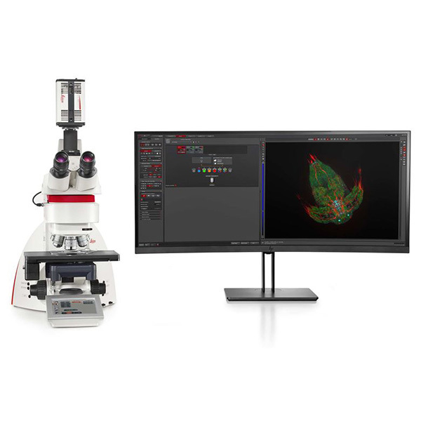
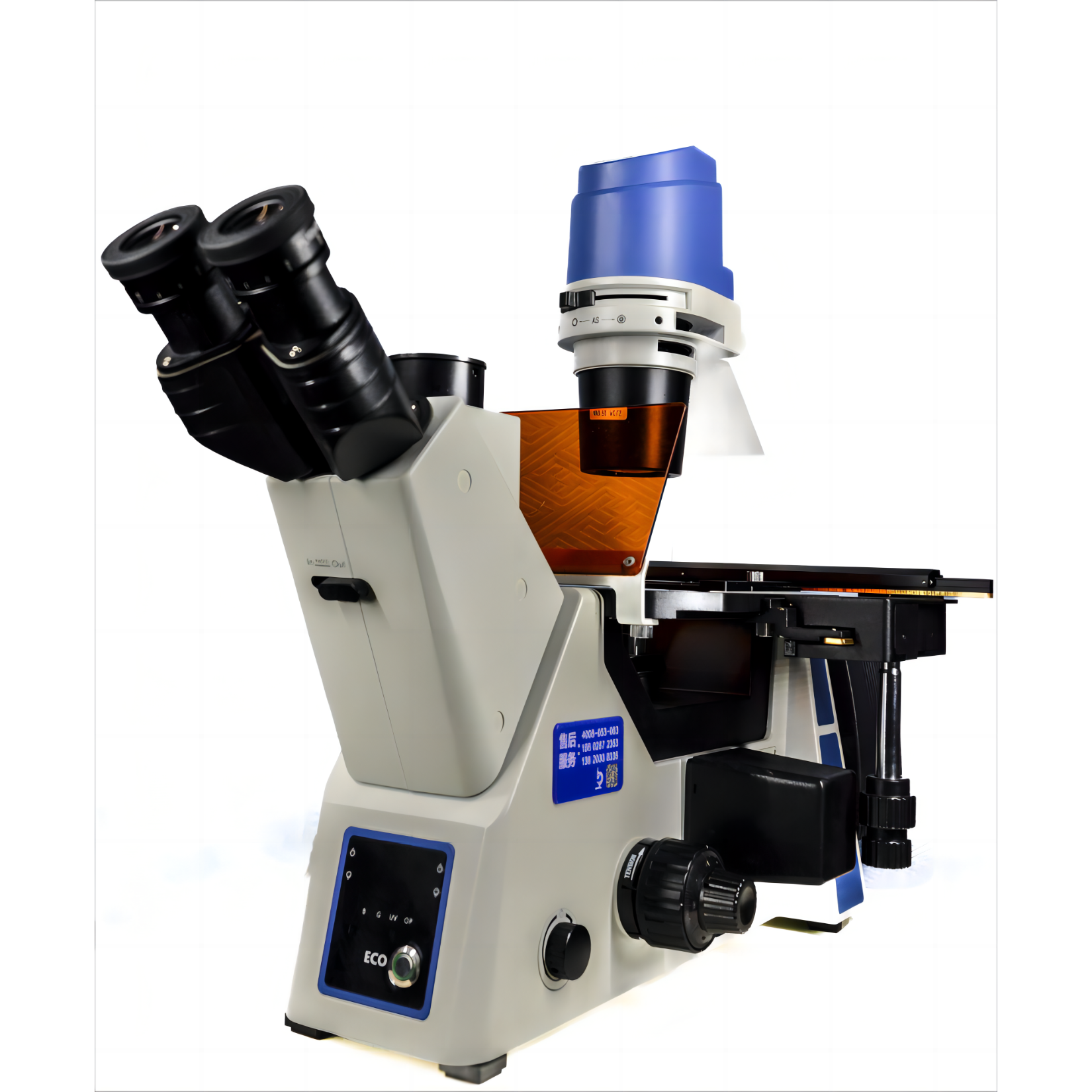
北京欧兰科技发展有限公司为您提供《蛋白中双栖小泡蛋白和N-WASP调节肌蛋白组装的相互作用动力学检测方案(CCD相机)》,该方案主要用于其他中双栖小泡蛋白和N-WASP调节肌蛋白组装的相互作用动力学检测,参考标准《暂无》,《蛋白中双栖小泡蛋白和N-WASP调节肌蛋白组装的相互作用动力学检测方案(CCD相机)》用到的仪器有Ekspla CARS 相干反斯托克斯拉曼显微光谱仪、MCL单分子成像RM21™显微镜平台、荧光寿命成像显微镜。
我要纠错
推荐专场
激光拉曼光谱(RAMAN)
更多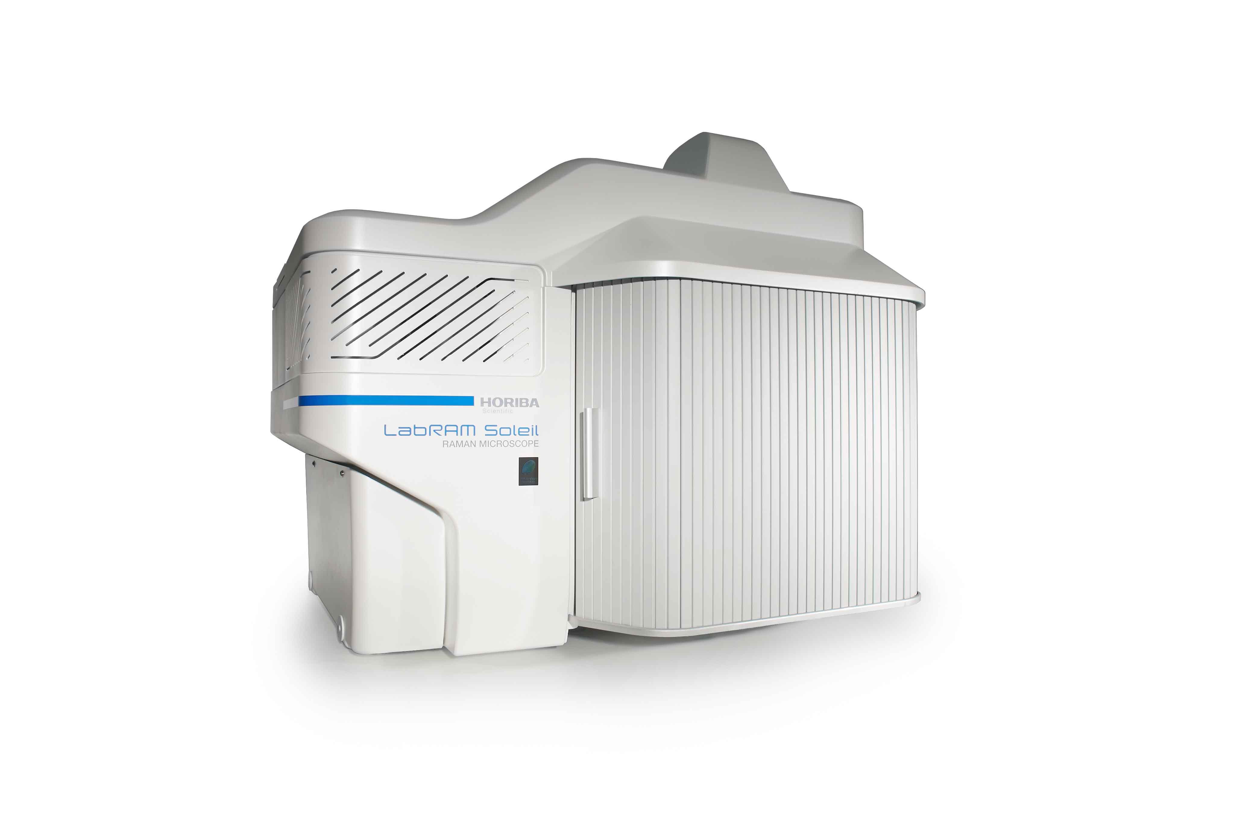

相关方案
 咨询
咨询