方案详情文
智能文字提取功能测试中
The official journal ofINTERNATIONAL FEDERATION OF PIGMENT CELL SOCIETIES·SOCIETY FOR MELANOMA RESEARupdates Received:27 April 2018 Revised: 15 October 2018Accepted: 1 November 2018DOI: 10.1111/pcmr.12752 1O 2018 John Wiley & Sons A/S.Pigment Cell Melanoma Res. 2018;1-14.wileyonlinelibrary.com/journal/pcmrPublished by John Wiley & Sons Ltd PIGMENT CELL & MELANOMA If you wish to order reprints of this article,please see the guidelines here Supporting Information for this article is freely available here EMAIL ALERTS Receive free email alerts and stay up-to-date on what is publishedin Pigment Cell & Melanoma Research -click here Submit your next paper to PCMR online at http://mc.manuscriptcentral.com/pcmr Subscribe to PCMR and stay up-to-date with the only journal committed to publishingbasic research in melanoma and pigment cell biology As a member of the IFPCS or the SMR you automatically get online access to PCMR. Sign up asa member today at www.ifpcs.org or at www.societymelanomaresarch.org ORIGINALARTICLE WILEY Photobleaching of pheomelanin increases its phototoxicpotential: Physicochemical studies of synthetic pheomelaninsubjected to aerobic photolysis Andrzej Zadlo1|Grzegorz Szewczyk1|Michal SarnaD| Theodore G. Camenisch21Jason W. Sidabras²| Shosuke ltoiD Kazumasa Wakamatsu°D| Filip SaganIMariusz Mitoraj1 Tadeusz SarnaliD ‘Department of Biophysics, Facultyof Biochemistry, Biophysics andBiotechnology,Jagiellonian University,Krakow, Poland ‘Department of Biophysics, Medical Collegeof Wisconsin, Milwaukee, Wisconsin Department of Chemistry, Fujita HealthUniversity School of Health Sciences,Toyoake, Japan “Department of Computational Methods inChemistry, Faculty of Chemistry, JagiellonianUniversity, Krakow, Poland Correspondence Tadeusz Sarna,Department of Biophysics, Faculty of Biochemistry, Biophysics andBiotechnology, Jagiellonian University,Krakow, Poland. Email: tadeusz.sarna@uj.edu.pl Funding information Poland National Science Centre, Grant/Award Number: Maestro-2013/08/A/NZ1/00194, Symfonia-2013/08/W/NZ3/00700 and Sonata-2015/19/D/ST4/01964; Japan Society for the Promotionof Science,Grant/Award Number:23591659,26461705 and 15K09794:National Institute of Biomedical Imaging andBioengineering of the National Institutes ofHealth, Grant/Award Number: EB001980;Polish Ministry of Science and Education “T-subsidy" for young researchers Abstract Although melanin is a photoprotective pigment, its elevated photochemical reactiv-ity could lead to various phototoxic processes.Photoreactivity of synthetic pheomela-nin, derived from 5-S-cysteinyldopa (5SCD-M) and its photodegradation productsobtained by subjecting the melanin to aerobic irradiation with UV-visible light, wasexamined employing an array of advanced physicochemical methods. Extensive pho-tolysis of 5SCD-M was accompanied by partial bleaching of the melanin, modificationof its paramagnetic properties, and significant increase in the ability to photogen-erate singlet oxygen. The changes correlated with a substantial decrease in the mela-nin content of benzothiazine (BT) units and increase of modified benzothiazole (BZ)units. Synthetically prepared BZ exhibited higher efficiency to photogenerate singletoxygen than the synthetic BT, and the free radical form of BZ, unlike that of BT, didnot show measurable spin density on nitrogen atom, which was confirmed by quan-tum chemical calculations. Formation of modified BZ units in the photobleached5SCD-M is responsible for the paramagnetic and photochemical changes of the mel-anin and its elevated phototoxic potential. Given a relatively constant pheomelanin-eumelanin ratio, such undesirable changes could occur in individual of all skin types. KEYWORDSoxidative modifications, pheomelanin, photobleaching, photoreactivity, singlet oxygen Melanin, one of the most common pigments of the animal king-dom, is synthesized in specialized cells such as melanocytes orpigment epithelial cells via non-enzymatic and enzyme-catalyzedredox and tautomerization reactions (d'lschia et al., 2013,2015).Although it is generally accepted that the most important posi-tive regulator of melanogenesis is the melanocortin 1 receptor (MC1R), with its ligands melanocortins and ACTH, the biosyn-thesis of melanin is regulated by many cellular and extracellu-lar factors including hormones (Slominski, Tobin, Shibahara, &Wortsman, 2004). Melanin is typically viewed as a natural sunscreen and efficientantioxidant that protects pigmented tissues against photochem-ical damage induced by solar radiation (Brenner & Hearing, 2008;Hu, Simon, & Sarna, 2008; Sarna, 1992). However, growing body of experimental evidence suggests involvement of melanin in cer-tain phototoxic reactions and its role in the development of mela-noma, one of the most aggressive types of cancer (Noonan et al.,2012; Wood et al., 2006). Interestingly, chemiexcitation of melaninderivatives was reported to induce DNA damage in cells (Premi etal., 2015). The apparent dichotomy in the behavior of melanin pig-ments may result from different photochemical properties of vari-ous melanins and diverse experimental conditions employed in theirstudy.Indeed, it has been postulated long ago that while eumela-nins, originating from oxidative polymerization of L-dopa, are rela-tively stable, and exhibit photoprotective and antioxidant activity(Brenner & Hearing, 2008; Rozanowska, Sarna, Land, & Truscott,1999; Tadokoro et al., 2003; Zareba, Skumatz, Sarna, & Burke,2014), pheomelanins, derived from the oxidation of cysteinyldopaprecursors, are prone to photodegradation and exhibit higher photo-reactivity (Koch & Chedekel, 1987; Kvam & Dahle, 2004; Pilas, Felix,Sarna, & Kalyanaraman, 1986; Takeuchi et al., 2004; Ye et al., 2006).However, action spectra and quantum yields of photoinduced oxy-gen consumption were found to be rather similar for both melaninpigments (Sarna, Menon, & Sealy, 1984; Sarna & Sealy,1984). Whilephotochemical lability of pheomelanin is of considerable mechanis-tic interest, it raises an intriguing question about photoreactivity ofthe melanin oxidative degradation products that could potentiallybe formed in the human skin under excessive exposure to solar radi-ation. This is an important issue considering that human epidermis,regardless of the degree of pigmentation,almost always containspheomelanin, comprising approximately 3/4 of eumelanin and 1/4 ofpheomelanin (Del Bino et al.,2015). To address this issue, we examined chemical and photochemicalproperties of a synthetic pheomelanin, derived from enzymatic ox-idation of 5-S-cysteinyldopa, subjected to aerobic irradiation withintense UV-visible light. Results of our study show that the pheom-elanin undergoes distinct photochemical modifications affecting itsoptical and paramagnetic properties, and photoreactivity. Partiallyphotobleached pheomelanin becomes a much more efficient gen-erator of singlet oxygen when excited with UV or short-wavelengthvisible light. Since photobleaching of pheomelanin is also accompa-nied by a gradual decrease in its efficiency to quench singlet oxy-gen, biological ramifications of such photochemical changes of theskin pheomelanin, if occurred in vivo, could be severe. 2 MATERIALS AND METHODS Sodium hydroxide,99.5% acetic acid, and potassium hexacyano-ferrate (III) were from Polish Chemical reagents (POCH), Gliwice,Poland. Sodium acetate was from Chempur (Piekary Slaskie,Poland).Sodium chloride,potassium chloride, disodium hydrogen phosphatedodecahydrate, potassium dihydrogen phosphate, and zinc acetatewere from Standard Co. (Lublin, Poland). Mushroom tyrosinase,5,5-dimethyl-1-pyrroline N-oxide (DMPO), chelex-100, and dialysisbags were from Sigma-Aldrich Chemie GmbH (Steinheim,Germany).Dimethyl sulfoxide (DMSO) was from Acros Organics (Geel, Significance Although pheomelanin, compared to eumelanin, is consideredto be more photoreactive and potentially phototoxic, thephysicochemical reasons of these differences remain obscure.We have demonstrated in this study that partial photobleach-ing of a synthetic pheomelanin is accompanied by a dramaticincrease in its ability to photogenerate singlet oxygen and insubstantial reduction in the melanin efficiency to quench thisreactive oxygen species. The results suggest that exposure tointense solar radiation, regardless of the skin type, could ele-vate cytotoxic potential of the skin melanin that results fromphotochemical modifications of the pigment. Belgium). 4-Protio-3-carbamoyl-2,2,5,5-tetraperdeuteromethyl-3-pyrroline-1-oxol(mHCTPO) was a generous gift from ProfessorH.J. Halpern, University of Chicago, Chicago, IL, USA. All chemicalswere reagent grade or better and used as supplied except DMPO,which was additionally purified by extraction in water:toluene sys-tem (1:1), and then, the water fraction was shaken with activatedcharcoal and finally centrifuged and filtered. Buffer solutions,made of double-deionized water, were treated with Chelex-100to remove any adventitious metal ions. 5-S-cysteinyldopa-melanin(5SCD-M) was prepared by the tyrosinase-catalyzed oxidation of5-S-cysteinyldopa according to the method described elsewhere(d'lschia et al., 2013). 7-(2-amino-2-carboxyethyl)-5-hydroxy-3,4-dihydro-2H-1,4-benzothiazine-3-carboxylic acid (DHBTCA) wasprepared (98% purity by HPLC)by oxidizing 5-S-cysteinyldopa at pH9.0, 6-(2-amino-2-carboxylethyl)-4-hydroxybenzothiazole (BZ-AA)was prepared (>99% purity by HPLC) by oxidizing 5-S-cysteinyldopawith FeNH (SO), and (NH) S,, and 7-(2-amino-2-carboxye-thyl)-5-hydroxy-3-oxo-3,4-dihydro-2H-1,4-benzothiazine (ODHBT)was prepared (>99% purity by HPLC) by oxidizing 5-S-cysteinyldopawith 0.1 M NaOH, as described previously (Wakamatsu, Ohtara, &Ito, 2009). Preparation of 6-(2-amino-2-carboxylethyl)-4-hydroxy-2methyl benzothiazole (Me-BZ-AA) was also described previously(Wakamatsu et al.,2009). 2.1 Aerobic photolysis of 5-S-cysteinyldopa-melanin 5SCD-M in distilled water (2 mg/ml) at pH 7.4 was subjected to con-trolled photodegradation employing 1 kW high-pressure compactarc xenon lamp (Cermax, PerkinElmer) equipped with 5-cm-longwater filter and blue dichroic color filter (TFI Technologies, Inc.,Greenfield, MA, USA). The intensity of the UVA-blue visible light(326-502 nm) was 1,500 mW/cm². During photodegradation, themelanin solution (7.25 ml initial volume) was gently stirred and itstemperature was maintained at 4°C using a circulating water bath. Atselected time intervals, small aliquots were taken for electron para-magnetic resonance and UV-vis spectrophotometric measurements. 2.2| Preparation of radicals from selectedpheomelanin monomers DHBTCA and BZ-AA were dissolved in water at concentration0.01 M, and pH of the solutions was adjusted to 7.4 by additionof small amounts of 1 M NaOH. To 0.2 ml of the monomer solu-tions, 0.16 ml of 0.1 M acetate buffer pH 5.0 or 0.1 M zinc acetate,0.032 ml of water, and 0.008 ml of 0.2 MK [Fe(CN) ] were addedand the samples were vigorously stirred. After about 30 s, the sam-ples were frozen and kept in liquid nitrogen until EPR examination. 2.3|X-band EPR measurements EPR examination of 5SCD-M, its photodegradation products, andradical forms of BZ and BT in 50 mM acetate buffer (pH 5.0) or50 mM zinc acetate were carried out at 77 K using a quartz finger-type dewar, employing Bruker EMX-AA EPR spectrometer (BrukerBioSpin, Rheinstetten, Germany). The apparatus settings were asfollows: center field 336 mT, sweep width 7 mT, modulation ampli-tude 0.15 mT, microwave power 0.017 mW, time constant 0.33 s,and sweep time 83.9 s. 10 or 40 scans were averaged to improvesignal-to-noise ratio. 2.4W-band EPR measurements Spectra were obtained on a spectrometer system constructed atthe National Biomedical EPR Center, Medical College of Wisconsin,Milwaukee, WI, USA. Samples in 0.20 mm l.D. and 0.33 mm O.D.synthetic silica capillaries (Fiber Optic Center,Inc.,New Bedford,MAUSA) were placed in a cylindrical TE011 cavity resonator, speciallydesigned for aqueous samples, operating at 94.04 GHz (Sidabras etal.,2007). The sample and resonator were maintained at 25°C, witha temperature controlled water bath circulating through a clamp at-tached to the resonator assembly. Typically, the samples were re-corded at an incident microwave power of -7 dBm (50pW). Fieldmodulation frequency was 937 Hz, and modulation amplitude was0.4 mT p-p. The magnetic field was scanned 20 mT over a period oftwo minutes. The obtained spectra were the result of multiple scansaveraged over a period of 10-45 min in order to obtain adequatesignal-to-noise ratio. For selected samples, the pseudomodulationwas performed with homebuilt software that convolutes a sinusoi-dal modulation on the collected data (Hyde, Pasenkiewicz-Gierula,Jesmanowicz, & Antholine, 1990). The procedure precisely mimicshardware-based higher-order modulation detection. 2.5Chemical characterization of 5SCD-M and itsaerobic photolysis products Fifty microliter aliquots of 5SCD-M and its selected photodegrada-tion products (0.1 mg) were subjected to alkaline hydrogen perox-ide oxidation (Ito et al., 2011) and hydroiodic hydrolysis (Ito, & Rees,2002; Ito & Wakamatsu, 2003). The analyses were performed in du-plicate and the average values are reported. 2.66Oxygen consumption measurements Time-dependent changes in oxygen concentration induced by lightwere determined by EPR oximetry using mHCTPO at concentra-tion 0.1 mM as dissolved oxygen-sensitive spin probe according tothe method described elsewhere (Rozanowska et al., 1995;Zadlo,Burke, & Sarna,2009).Samples containing 1 mg/ml 5SCD-M or itsphotodegradation products in PBS (pH 7.4) were irradiated in EPRquartz flat cells in the resonant cavity with 402-508 nm (24mW/cm²). 2.77|Electron paramagnetic resonance spintrapping studies Samples containing 0.5 mg/ml 5SCD-M or its photodegradationproducts, 0.1 M DMPO, used a spin trap, in 70% DMSO/30% waterwere irradiated with blue light employing the same light source andfilters as those described for oxygen photoconsumption (vide supra).EPR samples were run using microwave power 10.6 mW, modulationamplitude 0.05 mT, center field 339.0 mT, scan width 8 mT, and scantime 84 s. 2.88|Time-resolved singlet oxygen detection 5SCD-M or its photodegradation products, dissolved in phosphate-buffered (pH 7.2) D,O, were examined for their ability to photogen-erate and quench singlet oxygen according to the method describedelsewhere (Szewczyk et al., 2016). Photoexcitation of melanins wasexamined in the spectral range 300-560 nm, employing an integrated nanosecond DSS Nd:YAG laser system equipped with a nar-row bandwidth optical parametric oscillator (NT242-1k-SH/SFG;Ekspla,Vilnius, Lithuania). 2.99 Computational details Calculations have been performed with the use of ADF 2016.102package (ADFn.d., n.d.). Presented structures were optimized at theDFT/BLYP-D3/TZP level of theory and verified by calculations withthe use of B3LYP, PW91xc, and mPW1PW correlation-exchangepotentials in TZP basis set (Guerra, Snijders, te Velde, & Baerends,1998; te Velde et al., 2001). Distribution of the spin density was alsocalculated with solvation effects taken into account by means ofCOSMO continuum solvation model. Spin densities remained mostlyunaffected. 2.10|Zetasizer Size and zeta potential analysis of selected melanin samples wasmade using Malvern's Zetasizer Nano ZSP particle size analyzer(Malvern Panalytical Ltd, Malvern, UK). For the analysis, mela-nin samples were diluted in deionized, distilled and filtered waterto a concentration of 0.1 mg/ml, and suspended in a volume of800 ul. FIGURE1. O(ptical changes of 5SCD-M induced by aerobic photolysis.(a) Absorption spectra of control (solid line) and irradiated melanin:30 hr (dash line), 120 hr (dash-dot line), and 175 hr (dotted line).(b) Integrated absorbance (in the spectral range 300-700 nm) of control andphotolyzed melanin. Data points are connected with a b-spline 2.11.I Atomic force microscopy For AFM analysis, a small droplet of the sample was placed on freshlycleaved mica surface and evaporated in a desiccator. Topographyimages of the selected samples were obtained in Tapping AC modeemploying the blueDrive photothermal excitation and Cypher ESAFM (Asylum Research, Santa Barbara, CA, USA). Olympus OMCL-AC55TS probes were used with a nominal tip radius of 7 nm, springconstant of 85 N/m, and resonant frequency of 1.6 MHz. Details onAFM analysis can be found elsewhere (Wytrwal et al., 2015). 3 RESULTS AND DISCUSSION 3.1 | Photolysis of 5-S-cysteinyldopa melanin(5SCD-M) Irradiation of aqueous solution of 2 mg/ml5-S-cysteinyldopa melanin(5SCD-M) with 326-502 nm intense light, under aerobic conditions,brings about qualitative and quantitative changes in the melanin UV-Vis absorption spectra (Figure 1). Relatively short photolysis ofthe sample (20 hr and less) is accompanied by a small increase in theintegrated absorbance, and longer photolysis (52 hr and more) modi-fies the absorption spectra by making them almost monotonic withwavelength. Thus, the weak but distinguishable band in the 400-600 nm range, clearly seen in control non-irradiated sample, disap-pears after 30 hr of irradiation being replaced by almost exponentialdependence of the absorbance with wavelength. Longer irradiationtimes (above 90 hr) induce detectable bleaching of the melanin, asevident by its reduced integrated absorbance (Figure 1b). Optical changes of 5SCD-M accompanying aerobic photolysisindicate significant photochemical modifications of key monomerunits of the pigment. We examined this by measuring EPR spec-tra of control and irradiated pheomelanin (d'lschia et al., 2013),and analyzing their characteristic degradation products (d'lschiaet al.,2015). It is important to emphasize that all melanins exhibitpersistent-free radicals, which can be viewed as intrinsic molecu-lar probes providing information about the origin of melanin pig-ments and their physicochemical state (Sarna & Swartz, 2006). FIGURE 2>X-band EPR spectra of 5SCD-M in acetate buffer before and after aerobic photolysis; control sample (a), samples irradiatedfor: 20 hr (b), 120 hr (c), and 175 hr (d). Arrow indicates the lower field component typical for pheomelanin pigments.(e) Changes of theintegrated EPR signal of melanin with photolysis time. Data points are connected with a b-spline FIGURE 3 Changes in the yields of characteristic degradation products of 5SCD-M with its aerobic photolysis time. (a)Markers ofpheomelanin obtained by hydrolysis with HI:4-AHP (an indicator of benzothiazine units) (black squares), BZ-AA (red dots), and Me-BZ-AA(indicators of benzothiazole units) (blue triangles). (b) Markers of pheomelanin and eumelanin obtained by oxidation with alkaline hydrogenperoxide: TTCA (black squares), TDCA (indicators of native and modified benzothiazole units) (red dots), PTCA (blue triangles), PDCA (indicatorsof minute quantities of eumelanin) (green inverted triangles). Some of PTCA and PDCA also arise from pheomelanin (Wakamatsu, Nakanishi,Miyazaki, Kolbe, & Ito, 2012). Data points in (a) and (b) are connected with a b-spline [Colour figure can be viewed at wileyonlinelibrary.com] Magnetic parameters of the observable EPR signals of the photo-lyzed melanin exhibit changes that correlate with optical changesof the melanin samples (Figure 2a-d). Thus, the EPR signal of thenon-irradiated 5SCD-M is characteristic for an immobilized rad-ical with hyperfine splitting to nitrogen 2Az~3.0 mT typical forpheomelanins (Sealy et al., 1982). On the other hand, photolysisof the melanin decreases the ratio of the intensity of the low-fieldcomponent of the EPR signal to its central component. At longestirradiation times, the observable EPR signal consists of a singlefeatureless line without any hyperfine splitting (Figure 2d). SimilarEPR spectra have been reported for eumelanin, including syn-thetic dopa-melanin (d'lschia et al., 2013). Figure S1 shows EPRspectra of non-irradiated and irradiated 5SCD-M after exposureof the samples to saturating concentration of zinc acetate. It isapparent that the intensities of the corresponding EPR spectra areenhanced 10- to 20-fold compared to those in Figure 2, facilitatinga better comparison of the signal parameters. The effect is dueto zinc-ion-induced shift in the comproportionation equilibriumbetween the fully reduced and fully oxidized melanin moieties,and melanin radicals (Felix, Hyde, Sarna, & Sealy, 1978). Changesof the integrated EPR signal intensity of 5SCD-M with photoly-sis are shown in Figure 2e. The integrated signal intensity showsa weak increase at short irradiation times followed by a steadydecrease at longer irradiation times, so that after 175 hr the in-tegrated signal intensity is about one-third of that of the controlmelanin. On the other hand, in the presence of saturating concen-tration of zinc ions, no transient increase in the signal intensity isobserved and after 175 hr of the melanin photolysis, the signal isreduced almost by a factor of five (Figure S1e). The combined dataindicate that while in the non-irradiated 5SCD-M, compropor-tionation equilibrium involves o-aminophenols, o-quinonimines,and o-semiquinonimines, derived from hydroxybenzothiazines,the key structural and functional units of pheomelanin (d'lschia et al., 2013; Ito & Wakamatsu, 2006), in extensively photolyzedsynthetic pheomelanin the comproportionation equilibrium is onlyweakly diminished but it is dominated by other type of moieties,which are responsible for the formation of free radicals withoutdetectable hyperfine splitting to nitrogen. Of course, o-semiqui-nones detected in eumelanin, where the comproportionationequilibrium involves o-quinones and o-hydroquinones, the build-ing blocks of the melanin (d'lschia et al., 2013; Felix et al., 1978;Mostert et al., 2013; Sealy et al., 1982), do not have nitrogen atomsthat could give measurable hyperfine splitting. However, chemicalanalysis of the synthetic pheomelanin clearly showed that neitherin non-irradiated 5SCD-M, nor in photolyzed samples, any sub-stantial amount of eumelanin moieties was present as evidencedby the very low yield of pyrrole-2,3,5-tricarboxylic acid (PTCA).The hydroiodic acid (HI) hydrolysis data (Figure 3a) show that aer-obic photolysis of 5SCD-M, results in extensive degradation of themelanin with significant decrease in both its benzothiazine and“intact”benzothiazole moieties, as indicated by decreases of 4-amino-3-hydroxyphenylalanine (4-AHP-marker of benzothiazineunits), and BZ-AA and Me-BZ-AA-markers of “intact”benzothi-azole units. Even though the H,O, oxidation products, namelythiazole-2,4,5-tricarboxylic acid (TTCA) and thiazole-4,5-dicar-boxylic acid (TDCA)-markers of benzothiazole units are not asspecific as BZ-AA, the threefold increase in the yields of TTCAand TDCA in samples photolyzed for 175 hr (Figure 3b) indicatesthat the photolyzed melanin accumulates“photomodified"benzo-thiazole units of unknown structure. Figure 4 summarizes the current knowledge on pheomelano-genesis as well as key results obtained in this study: (i) benzothi-azine moiety is produced following oxidation of cysteinyldopa andis converted to benzothiazole moiety during late stage of pheom-elanogenesis (Wakamatsu et al., 2009), (ii) 4-AHP and BZ-AA (andMe-BZ-AA) decrease in parallel during photolysis (Figure 3b), and Photodegraded PM FIGURE 4A scheme illustrating biosynthetic pathway from 5-S-cysteinyldopa to pheomelanin and its possible photochemical reactions.5-S-Cysteinyldopa is oxidized by tyrosinase to produce DHBTCA (Wakamatsu et al., 2009). DHBTCA constitutes BT moiety in pheomelanin(in the figure, only a reduced form of BT moiety is shown).BT moiety is oxidatively converted to BZ moiety (the maturation of pheomelanin,see Wakamatsu et al.,2009). These two moieties can be analyzed by hydroiodic acid (HI) hydrolysis to afford 4-AHP and BZ-AA. Uponirradiation with UV or visible light, BT and BZ moieties are converted to photomodified BZ moiety that does not give rise to BZ-AA becauseof the newly formed covalent bond (shown in the red arrow). During this process, BT and BZ moieties produce superoxide anion and singletoxygen, respectively, as suggested in this study. The photomodified BZ moiety produces additional singlet oxygen with photodegradation ofpheomelanin.Both photomodified BZ moiety and photobleached pheomelanin can be analyzed by alkaline H,O, oxidation to afford TTCA[Colour figure can be viewed at wileyonlinelibrary.com] (iii) TTCA continues to increase during photolysis. It is likely that“photomodified”benzothiazole moiety is produced by cross-linkingwith another moiety of pheomelanin so that the moiety would notgive rise to BZ-AAupon HI hydrolysis. This would increase molecularmass of pheomelanin, which is consistent with a previous observa-tion on size exclusion HPLC of UVA-irradiated synthetic pheomela-nin (Wakamatsu et al.,2012). A photochemical ring contraction of dihydro-1,4-benzothiazineto give 2-methylbenzothiazoles has been described over 20 yearsago (Costantini, Testa, Crescenzi, & d'lschia, 1994). Interestingly,a similar pattern of photochemical changes was determined in redhair melanin with photoaging and in synthetic pheomelanin afterirradiation with UVA and also in human retinal pigment epithe-lium melanin with aging (Ito et al.,2013; Wakamatsu et al., 2012).It should be emphasized that the concentration of 4-AHP, deter-mined in non-irradiated 5SCD-M and in the photolyzed melanin,correlates very well with the intensity of the low-field componentof the nitrogen hyperfine line in the observable EPR spectra(FigureS2). The correlation confirms that the nitrogen hyperfine splittingis mostly due to the presence of benzothiazine units. It also sug-gests that their content in the examined pheomelanin could bedetermined by measuring the intensity of the corresponding EPRsignal. Our EPR data demonstrate that the comproportionationequilibrium involving benzothiazine groups is virtually shut downby extensive oxidative photodegradation of the pheomelanin. It remains to be determined, which remaining functional groups ofthe pheomelanin, or what type of groups formed in the melaninby its aerobic photolysis, participate in the comproportionationequilibrium that yields radicals detected in strongly photolyzedpheomelanin. Although the formation of a two-electron oxidationproduct of benzothiazole seems thermodynamically unfavorable,the presence of fully reduced and fully oxidized photomodifiedbenzothiazole moieties in photolyzed pheomelanin could not beruled out. It is postulated that while comproportionation equilib-rium between such units would not yield an o-semiquinonimine,an oxygen-centered radical could be formed. Taking the advan-tage of higher spectral resolution of W-band EPR spectroscopy,we examined control and photolyzed 5SCD-M samples saturatedwith zinc ions (Figure 5a-d). All spectra show significant anisotropywith distinct axial character. For non-irradiated 5SCD-M, hyperfinesplitting to nitrogen, with A, value about 1.5 mT, is apparent in theg, region of the observable EPR spectrum, while two components,probably related to two different radical centers, can be seen in theg,,gregion. These spectral features become more prominent afterpseudomodulation of the detected EPR signals, which generatessecond-harmonic-like spectra (Figure 5e-h). The W-band EPR examination of the irradiated 5SCD-M confirmsthe chemical analysis data that even relatively short aerobic photoly-sis (20 hr) of 5SCD-M results in melanin with significantly degradedbenzothiazine moieties. EPR spectra of pheomelanin photodegraded for 100 hr and even for 52 hr are almost identical exhibiting practi-cally a single component with profound axial anisotropy, which is sig-nificantly greater than that of a synthetic dopa-melanin (Figure S3).The W-band EPR data provide additional evidence that the chemicalnature of radical centers in the photolyzed pheomelanin is differentfrom that of eumelanin, even though magnetic parameters of theirX-band ERP spectra are rather similar. 3.2Studies on pheomelanin monomers To validate the conclusions based on EPR analysis of control andphotobleached 5SCD-M, attempts were made to detect by EPR one-electron oxidation products of synthetically prepared amino acids:DHBTCA and BZ-AA, typical benzothiazine monomer (in a reduced,dihydro form), and benzothiazole monomer of pheomelanin, respec-tively. Addition of potassium hexacyanoferrate (III) to aqueous solu-tion of these compounds resulted in the development of a weak EPRsignal detectable at 77 K. The signal intensity significantly increasedin the presence of 40 mM zinc acetate (Figure S4), as a result of spinstabilization of the radicals due to the formation of chelate com-plexes with the metal ions (Felix et al.,1978). It is important to stressthat without oxidation, neither DHBTCA nor BZ-AA exhibited anydetectable EPR signals (data not shown). The spectral parametersof the observed EPR signals are quite remarkable in that a distinctnitrogen splitting (2A,~3.0 mT), typical for o-semiquinonimine radi-cals of the control 5SCD-M, is only seen in the case of DHBTCA. TheEPR signal of the oxidation product of BZ-AA is a single asymmet-ric line very similar to that of photobleached 5SCD-M. Evidently, infree radicals formed by oxidation of BZ-AA, the unpaired electron ismostly localized on oxygen atoms, even though the molecule func-tional groups that participate in the proposed comproportionationequilibrium contain nitrogen atom (Figure S5). We have confirmed diverse spin density distributions for radical forms of DHBTCA andBZ-AA (Figure 6) and their complexes with zinc ions (Figure S6). TheNalewajski-Mrozek bond orders of DHBTCA and BZ-AA radicals,their electrostatic potentials, and the resulting resonance structuresare shown in Figure S7. The calculations also show the effect of thecarboxyl group, attached to the thiazine ring of DHBTCA, on the dis-tribution of spin density of the molecule radical form. Interestingly,for the radical of the decarboxylated form of benzothiazine,ODHBT,no significant spin density was found on nitrogen atom and no cor-responding hyperfine splitting of the radical was detected by EPR(data not shown). 3.3 Physical changes of 5SCD-M The observed chemical changes of 5SCD-M, induced by aerobicphotolysis, are accompanied by physical changes of the melaninnanoaggregates. Dynamic light scattering (DLS) analysis revealsthat the hydrodynamic diameter of the melanin nanoaggregatesdecreases with irradiation time (Figure 7a). These results were con-firmed by atomic force microscopy(AFM) measurements of selectedsamples subjected to photolysis. Figure 7c,d shows representativetopography images of control 5SCD-M and of melanin irradiatedfor 175 hr, respectively. As evident, the size of individual melaninnanoaggregates decreases significantly after prolonged photolysis.The observed physical changes may result from decomposition ofthe melanin nanoaggregates induced by chemical modifications ofthe building monomers. Figure 7b shows significant heterogeneityin the values of the zeta potential in samples subjected to photolysis.While control 5SCD-M exhibits only one distinct peak with the aver-age value of the zeta potential of -18 mV, photolyzed samples showmultiple peaks with the melanin irradiated for 175 hr being the mostextreme with 4 different values of the zeta potential (Figure 7b). DHBTCA FIGURE 6 The ADF/DFT/BLYP-D3/TZP optimized structures of DHBTCAand BZ-AA radicals (a), spin densitydistributions of DHBTCA and BZ-AAradicals together with the Bader andMulliken populations (b), and the electrondensities of SOMO for DHBTCA andBZ-AA radicals (c) [Colour figure can beviewed at wileyonlinelibrary.com] These results indicate that during photolysis melanin nanoaggre-gates release smaller fractions with different physicochemical prop-erties. If such changes occur in photolyzed melanosomes, it couldlead to instability of the pigment granules and the release of highlyphotoreactive melanin nanoaggregates. 3.4| Changes in photoreactivity of 5SCD-M To determine how aerobic photolysis of pheomelanin modifiesits photoreactivity, we measured the ability of non-irradiated andphotolyzed 5SCD-M to generate superoxide anion and consumeoxygen during photoexcitation with short-wavelength visible light(404-510 nm). Representative data are shown in Figure 8. Theseresults reveal that with increasing photolysis time the syntheticpheomelanin gradually loses its efficiency to photoreduce oxygen tosuperoxideanion, which, in part, is responsible for the observed oxy-gen photoconsumption. The effect is consistent with the expecteddepletion of electron-donating groups in melanin subjected to ex-tensive photooxidation.5SCD-M has recently been shown to pho-togenerate singlet oxygen, albeit at very low yield (Szewczyk et al.,2016). Therefore, we examined the effect of photobleaching of thesynthetic pheomelanin on its efficiency to generate singlet oxygen atdifferent excitation wavelengths employing the direct time-resolved singlet oxygen phosphorescence method (Kozinska, Oles, & Sarna,2012). Figure 9a shows action spectra, plotted as semi-logarithmicgraphs, of the ability of non-irradiated 5SCD-M and melanin photol-yzed for selected times to generate singlet oxygen. Two points need to be stressed when discussing the obtainedsinglet oxygen data. First, it is the extraordinary increase of theefficiency of 5SCD-M to photogenerate singlet oxygen with theextent of melanin photolysis. At 300 and 320 nm, the efficiencyof the longest photolyzed 5SCD-M, compared to control melanin,increases over one order of magnitude.Remarkably, a substantialincrease in the efficiency of the melanin to photogenerate singletoxygen is also apparent in the visible part of the spectrum (aboutfivefold at 490 and 520 nm), even though such a melanin, beingpartially photobleached, exhibits substantially reduced optical ab-sorption. Changes of the efficiency of 5SCD-M to photogeneratesinglet oxygen, at selected wavelengths, with time of the melaninphotolysis exhibit biphasic behavior: A significant increase in theefficiency to photogenerate singlet oxygen proceeds a decreaseobserved during early photolysis times (Figure 9b). The effectcoincides with a transient increase of the integrated optical ab-sorption and integrated EPR intensity of the photolyzed mel-anin (Figures 1b and 2b). Therefore, the initial changes inducedby photolysis can be attributed to a reversible photooxidation of FIGURE 7 Effect of photolysis onthe size and zeta potential of 5SCD-Mparticles. DLS data showing size (a) andzeta potential (b) for: control sample(solid line), 20 hr of irradiation (dash line),120 hr of irradiation (dash-dot line), and175 hr of irradiation (dotted line). AFMtopography images of: control melanin (c)and melanin irradiated for 175 hr (d). Largeimages indicate scan areas of 500×500nm², whereas inset images show highmagnification of individual melaninnanoaggregates. Scale bars in inset imagesrepresent 10 nm [Colour figure can beviewed at wileyonlinelibrary.com] FIGURE 8 Changes of photoreactivityof 5SCD-M induced by aerobic photolysis:rate of oxygen photoconsumption (a);rate of accumulation of DMPO-OOHspin adduct (b). Inset in (b) showsrepresentative EPR spectrum of DMPO-OOH spin adduct. Data points in (a) and(b) are connected with a b-spline Size (nm) Zeta potential (mV) (c) (d) 100 (b) mwM w 50· 2mT 0 50 100 Photolysis time (hr) the melanin subunits. Indeed, similar physicochemical changes of5SCD-M were observed upon treatment of the melanin with theone-electron oxidizing agent potassium hexacyanoferrate (III).Interestingly, while lower concentration of the oxidant induced aconcerted increase of both the low-field and middle-field compo-nent of the EPR spectrum of 5SCD-M, at higher concentration ofthe oxidant, the enhancement of the middle-field component wassubstantially greater than that of the low-field component (FigureS8). The data suggest that the benzothiazine moieties of 5SCD-Mare more susceptible to oxidation by mild oxidants than the ben-zothiazole units, which are affected only at higher concentration of the oxidant. The results also indicate that both types of themoieties contribute to comproportionation equilibrium responsi-ble for the presence of nitrogen-centered and oxygen-centeredradicals even in“pure"pheomelanins. A differential effect of glu-tathione on both components of the EPR signal of pheomelanin,observed by Panzella et al. (2014), was attributed to an irrevers-ible conversion of the reduced benzothiazine melanin moiety toanother species unable to take part in the comproportionationequilibrium. Although the authors of the cited paper did not con-sider any interactions of glutathione with the free radical form ofbenzothiazole units, their explanation could be justified in view FIGURE9Photogeneration of singlet oxygen by 5SCD-M and its photolysis products. (a) Action spectra of singlet oxygenphotoformation for: control sample (solid line); sample irradiated for 20 hr (dash line), sample irradiated for 120 hr (dash-dot line), and sampleirradiated for 175 hr (dotted line). (b) Changes in singlet oxygen photoformation by melanin subjected to photolysis at indicated wavelengths:320 (magenta), 370 (violet), 430 (blue), and 520 (green). Y-axis in (a) and (b) represents initial intensity of singlet oxygen phosphorescencedivided by laser power at indicated wavelengths. Data points in (a) and (b) are connected with a b-spline [Colour figure can be viewed atwileyonlinelibrary.com] of the expected differences in redox properties of the two mainpheomelanin units. It can be postulated that the melanin benzothi-azole units are less susceptible to oxidation-reduction than ben-zothiazine units. The actual efficiency of a chromophore to photogenerate sin-glet oxygen is given by its quantum yield, which can be determinedby a comparative method using a photosensitizer with a knownquantum yield of singlet oxygen generation at wavelength wherethe two samples exhibit the same absorbance (Kozinska et al.,2012; Vakrat-Haglili et al., 2005; Wilkinson & Abdel-Shafi, 1999).Results of such measurements are shown in Figure S9, and thecorresponding quantum yields of singlet oxygen photogenerationfor 5SCD-M subjected to different photolysis times are assem-bled in Table 1. The table also shows rate constants for quench-ing of singlet oxygen by selected melanins. At 332 nm, quantumyield for the control 5SCD-M is only 1.30×10³, but it increasesto 1.63×10for the most photolyzed melanin sample. Similartrend is also found in the visible range, except the correspond-ing values are at least an order of magnitude smaller. Even thoughquantum yields of singlet oxygen photogeneration for commonphoto-sensitizing dyes are typically much higher than that of thephotobleached pheomelanin, its steady photoexcitation with UVAcould generate considerable fluxes of this reactive oxygen species,particularly if the photogenerated singlet oxygen is less efficientlydeactivated by the melanin. Indeed, the singlet oxygen quenchingability of 5SCD-M decreases with its photobleaching (Table 1). An important issue of our study is the chemical nature of thechromophore responsible for photogeneration of singlet oxygenby the photobleached pheomelanin. The efficiency of 5SCD-Mto photogenerate singlet oxygen after oxidation of the melaninwith increasing concentration of potassium hexacyanoferrate(IIl) markedly decreases, indicating that it is not the oxidized formof benzothiazine or other melanin moieties (data not shown).Analysis of the chemical data (discussed above) demonstrated thataerobic photolysis of 5SCD-M was accompanied by a substantial TABLE 1 Quantum yield of singlet oxygen formation and rateconstants of singlet oxygen quenching by 5SCD-M: dependence onthe photolysis time Photolysis Quenching rate constant time (hr) Quantum yield ([mg/ml]-1s-1) 0 1.30×10 1.49×10° 74 2.76×10~ 7.66×10* 96 4.00×10 120 5.80×10³ 5.77×10+ 141 8.09×10 155 1.17×10~2 175 1.63×10~ decrease in its content of benzothiazine moieties with possibleaccumulation of photomodified benzothiazole groups (Figure 4).However, their role as an endogenous photosensitizer of the pho-tobleached pheomelanin is unclear. Although benzothiazole wasfound to be susceptible to photooxidiation and photodegradationwhen irradiated with light derived from medium-pressure mercurylamp (Borowska, Felis, & Kalka,2016), photochemistry of thesecompounds seemed to be limited to the excited state proton trans-fer to solvent and excited state intramolecular proton transfer (ElNahhas et al., 2014). No evidence for the photoformation of tripletexcited state of 4-hydroxybenzothiazole was obtained in an ear-lier study (Lambert et al., 1984), even though the authors readilyobserved triplet transients for 4-methoxybenzothiazole. The sameauthors also reported that the 4-hydroxybenzothiazole chromo-phore underwent monophotonic photoionization to generate hy-drated electron with a quantum yield of 0.06. We analyzed the efficiency of the two main constituents of phe-omelanins, namely (dihydro)benzothiazine DHBTCA and benzothi-azole BZ-AA to photogenerate and quench singlet oxygen. FigureS10a shows that BZ-AA in D,O, when excited at 320 nm, generatessinglet oxygen with quantum yield approaching that of fluorescein used as a standard. On the other hand,the yield of DHBTCA to pho-togenerate singlet oxygen, under similar conditions, is at least fivefoldlower. Interestingly, the efficiency of the two compounds to quenchsinglet oxygen differs in reversed order; that is, while the bimolecu-lar rate of singlet oxygen quenching by DHBTCA is about 4.6×10'M1s1, it is only about 6×10M1s1 in case of BZ-AA (Figure S10b). The other point that needs to be emphasized is the observeddecrease in the efficiency of photolyzed 5SCD-M to quench singletoxygen (Table 1). Both effects, the enhanced efficiency of photo-bleached pheomelanin to generate singlet oxygen and the reducedefficacy of the melanin to quench this reactive oxygen species, couldhave significant biological implications, if such phenomena alsooccur in vivo in the human skin. It is important to stress that pho-toprotective action of a melanin pigment is not only due to efficientthermal dissipation of energy of the absorbed photons, but also toself-quenching of reactive oxygen species that could be generated bythe pigment residual photochemistry (d'lschia, Napolitano, Pezzella,Meredith, & Sarna, 2009; Meredith & Sarna, 2006). Consideringthe high rate constants of the interaction of unbleached melaninwith singlet oxygen (Szewczyk et al., 2016) and with oxidizing rad-icals (Rozanowska et al., 1999), the probability of reactive oxygenspecies to escape from native melanin granules into cell compart-ments, where they could induce damage of critical cell constituents,is rather low. This could change, if the pigment granule morphologyis substantially modified by oxidative degradation and if the oxidizedmelanin constituents lose their efficiency to quench/scavenge reac-tive oxygen species. We recently demonstrated that melanosomesisolated from retinal pigment epithelium of bovine eyes, subjectedto experimental photobleaching, and exhibited significantly alteredmorphology with loose structure allowing for easier penetration ofthe granules by different reagents (Zadlo et al., 2016). In addition,such partially photobleached pigment granules showed higher pho-toreactivity, reduced efficiency to quench singlet oxygen and lowerantioxidant capacity (Zareba et al.,2006; Zadlo et al., 2016). The re-duced efficacy of photobleached synthetic pheomelanin to quenchsinglet oxygen is consistent with our postulate that the photoreac-tivity and phototoxicity of the skin pheomelanin could significantlyincrease with its in situ aerobic photolysis. 3.5 Photochemical and biological relevance An important issue of this study is the relevance of the observedchanges induced by photolysis of the synthetic 5SCD-M to possiblechanges in natural pheomelanin pigments. Previous studies havedemonstrated that 5SCD-M is a reliable model of natural pheomela-nins (Sarna & Swartz, 2006; Zadlo, Rozanowska, Burke, & Sarna,2007).We have used this synthetic pheomelanin to avoid ambiguityin interpreting results that could arise in experiments with naturalpheomelanins, which are always a different mixture of pheomela-nin and eumelanin (Ito & Wakamatsu, 2003). In addition, syntheticmelanins are devoid of components that are present in melano-somes such as proteins, lipids,carbohydrates, and metal ions. Thesecomponents could affect the observable photoreactivity of the examined melanin and modify its efficiency to quench reactive oxy-gen species. Finally, unlike natural pheomelanin pigments, the syn-thetic 5SCD-M is soluble in aqueous media at neutral pH, which isan important consideration for quantitative photochemical studies. 4 CONCLUSION The observed physicochemical changes of the pheomelanin modelinduced by aerobic exposure to intense UV-visible radiation suggestphotochemical instability of natural pheomelanin present in the skinepidermis. It should be emphasized that even though eumelanin isthe dominant type of pigmentation in Caucasians, the human skinepidermis contains 25% of pheomelanin, which is mostly the ben-zothiazole type, regardless the degree of pigmentation (Del Binoet al., 2015). An excessive exposure of the human skin to solar ra-diation could result in pheomelanin-mediated phototoxic reactions,leading to oxidative stress with severe biological implications if notefficiently counter-balanced by appropriate antioxidant responsesof the pigmented cells (Ito & Wakamatsu,2008). The biological im-pact of photooxidation of pheomelanin that may occur under in vivoconditions, as a result of an exposure of the human skin to intense/prolong solar radiation is unknown and should be verified in prop-erly designed animal studies. Nevertheless, the remarkable physico-chemical changes of the synthetic pheomelanin induced by aerobicphotolysis, reported in this work, give reasons for the postulate thatphototoxic reactions in the human skin could also be mediated bypheomelanin photodegradation products. The results described in this study may also be relevant for thephotobiology of ocular melanin, particularly the retinal pigment ep-ithelium (RPE).Although the human RPE is practically inaccessibleto ambient UV radiation, these post-mitotic cells are exposed tosignificant doses of short-wavelength visible light, which could in-duce age-related photobleaching of RPE melanin granules (Sarna etal., 2003). Indeed, distinct photoaging of human RPE melanin hasbeen reported by us in an earlier study (Ito et al.,2013), which alsodemonstrated for the first time that the melanin was a mixture ofboth eumelanin and pheomelanin. ACKNOWLEDGEMENTS This work was supported in part by Poland National ScienceCentre (Maestro-2013/08/A/NZ1/00194; Symfonia-2013/08/W/NZ3/00700:Sonata-2015/19/D/ST4/01964),Japan Societyfor the Promotion of Science (JSPS) (23591659;26461705;15K09794), and National Institute of Biomedical Imaging andBioengineering of the National Institutes of Health (EB001980).Faculty of Biochemistry, Biophysics and Biotechnology of theJagiellonian University is a partner of the Leading NationalResearch Center (KNOW) supported by the Ministry of Scienceand Higher Education. DFT calculations were partially performedusing the PL-Grid Infrastructure and resources provided by theACC Cyfronet AGH (Cracow, Poland). F.S. acknowledges financial support from the Polish Ministry of Science and Education"T-sub-sidy" for young researchers. CONFLICT OF INTEREST The authors have no conflict of interest to declare. ORCID Michal SarnaIDhttps://orcid.org/0000-0001-9306-7965Shosuke Ito https://orcid.org/0000-0001-9182-5144Kazumasa Wakamatsu https://orcid.org/0000-0003-1748-9001Tadeusz Sarna https://orcid.org/0000-0002-7693-5756 REFERENCES ( ADF2016.102. (n.d.) . A DF modeling suite, SCM, theoretical chemistry.Amsterdam: V r ije U n iversiteit. Retrieved f rom h t tps://www.scm. com ) ( Borowska,E., F elis, E., & Kalka,J. (2016).Oxidation of benzotriazole andbenzothiazole in photochemical p rocesses: K inetics and formationof transformation p roducts. Chemical E ngineering Journal , 304,852-863.https://doi.org/10.1016/j.cej.2016.06.123 ) ( Brenner, M . , & Hearing, V . J. ( 2008). T he p r otective role of melaninagainst UV damage in human skin. Photochemistry and Photobiology,84,539-549.https://doi.org/10.1111/j.1751-1097.2007.00226.x ) ( Costantini,C.,Testa , G.,Crescenzi,O.,&d'lschia,M.(1994).Photochemicalring contraction of dihydro-1,4-benzothiazines.T e trahedron L e tters,35,3365-3366. https://doi.org/10.1016/S0040-4039(00)76909-7 ) ( d'lschia, M., Napolitano, A., Pezzella, A., Meredith, P., & Sarna, T.(2009). Chemical and s t ructural diversity in eumelanins: Unexplored bio-op-toelectronic m aterials. A ngewandte Chemie International Edi t ion inEnglish, 48,3914-3921. https://doi.org/10.1002/anie.200803786 ) ( d'lschia, M., Wakamatsu, K., Napolitano, A., Briganti, S., Ga r cia-Bor r on, J . C., Kovacs, D. , .. . Ito, S . (2013). Melanin s and melanogenesis: Methods, s t andards, protocols. P i gment Cell and Melanoma Research,26,616-633. https://doi.org/10.1111/pcmr.12121 ) ( Del B ino, S., I to, S., Sok, J., Nakanishi, Y., Bastien, P., Wakamatsu, K . , &Bernerd, F. ( 2015). C hemical a n alysis o f c o nstitutive p i gmentationof human epidermis r eveals constant eumelanin to pheomelanin ratio. Pigment Cell and Melanoma Research, 28, 707-717. https://doi. org/10.1111/pcmr.12410 ) ( d'lschia, M., Wakamatsu, K., C i coira, F., Di Mauro, E., Garcia-Borron, J. C.,Commo, S., ... Ito,S . (2015). Melanins and melanogenesis: From pig- ment cells to human h e alth a n d technological applications. P i gmentCell and Melanoma Research, 2 8, 520-544.https://doi.org/10.1111/ pcmr.12393 ) ( El Nahhas, A . , P a scher, T . , L e one, L. , P a nzella,L. , Na p olitano, A., & Sundstrom, V. (2014). Photochemistry of pheomelanin building blocks and m odel c h romophores: Exc i ted-state intra- an d intermo-lecular proton transfer. Journal of Physical Chemistry Letters,5, 2094-2100. https://doi.org/10.1021/jz500720g ) ( Felix, C. C . , Hyde,J.S.,Sarna, T., & Sealy, R. C . (1978). Interactions of mela- nin with metal ions. E l ectron s pin r esonance evidence for chelate co m - p lexes of metal ions with f r ee r a dicals. Journal of the American C h emical Society, 100 , 3922-3926. https://doi.org/10.1021/ja00480a044 ) ( Guerra, C . F., Snijders, J. G., te Velde, G., & Baerends, E. J. ( 1998). T owards an order-N DFT method. Theoretical Chemistry Accounts,99,391-403.https://doi.org/10.1007/s002140050353 ) ( Hu, D . N ., Simon, J . D., & Sarna, T.(2008).Role of ocular melanin in oph- thalmic p hysiology and p a thology. Photochemistry and Photobiology,84,639-644. https://doi.org/10.1111/j.1751-1097.2008.00316.x ) ( H yde, J. S. , Pasenkiewicz-Gierula, M ., J esmanowicz, A . , & Antholine,W. E. (1990). Pseudo field modulation in EPR s pectrosocpy.Applied M agnetic Resonance, 1 , 483-496. https://doi.org/10.1007/ BF03166028 ) ( Ito, S., Nakanishi, Y., Valenzuela, R . K., Brilliant, M. H ., K olbe, L . , &Wakamatsu, K. ( 2 0 11). Usefulness of al k aline hydrogen peroxide oxidation to analyze e umelanin a n d pheomelanin in v arious tis - sue samples: Application t o c hemical analysis of human hair mela-nins. Pigment Cell and Melanoma Research,24 , 60 5 -613. https://doi.org/10.1111/j.1755-148X.2011.00864.x ) ( I to, S., Pilat , A. , Gerwat, W., Skumatz, C . M . , Ito, M . , Kiyono, A. , .. . Wakamatsu, K . ( 2 013). Photoaging o f h u man re t inal p i gment epi- thelium is accompanied by o x idative modifications of its eumela- n in. Pigment Cell and M elanoma Re s earch, 26 , 35 7 -366. https://doi.org/10.1111/pcmr.12078 ) ( Ito, S . , & Wakamatsu, K. ( 2 003). Q u antitative analysis o f eumelanin and p heomelanin i n humans, mice, and other animals : A com-parative review. Pigment C ell Research, 16, 5 23-531. https://doi.org/10.1034/j . 1600-0749.2003.00072.x ) ( Ito, S.,& Wakamatsu, K.(2006).Chemistry of melanins. In J. J. N ordlund, R. E. Boissy, V. J. Hearing, R. A. King,W. S . O e tting, & J. P. Ortonne (Eds.), The pigmentary system: Physiology and pathophysiology,2nd ed. (pp. 282-310). Oxford: Blackwell Publishing Ltd. ) ( Ito, S., & Wakamatsu, K. (2008). Chemistry of mixed melanogenesis - p ivotal roles of dopaquinone. Photochemistry and Photobiology, 84,582-592. https://doi.org/10.1111/j.1751-1097.2007.00238.x ) ( Koch, W. H., & Chedekel, M. R. (1987). Photochemistry and p ho-tobiology of melanogenic metabolites: Formation o f f ree r adi-cals. Photochemistry a nd P h otobiology, 46, 2 2 9-238. htt p s://doi.org/10.1111/j.1751-1097.1987.tb04761.x ) ( Kozinska,A.,Oles, T., & Sarna,T.(2012). Photoactivation and detection ofphotoexcited molecules and photochemical products. I s rael Journalof Chemistry,52,745-756.https://doi.org/10.1002/ijch.201200019 ) ( Kvam, E., & D a hle, J. ( 2004). M elanin s y nthesis m a y se n sitize m e - lanocytes to oxidative DNA d a mage by ultraviolet A ra d iationand protect melanocytes f rom direct D NA damage by ultravio- l et B r adiation. Pigment Cell R esearch, 1 7, 5 49-550. ht t ps://doi.org/10.1111/j.1600-0749.2004.00168.x ) ( L ambert, C., Sinclair, R. S., Truscott, T . G.,Land, E .J . , Chedekel, R. M., &Liu, C. T. (1984). Photochemistry of benzothiazole m o dels of ph e -omelanin. P hotochemistry a n d Photobiology, 3 9 , 5 - 10. ht t ps://doi.org/10.1111/j.1751-1097.1984.tb03396.x ) ( Meredith, P., & Sarna, T.(2006). The physical and chemical proper- ties of eumelanin. Pigment C e ll R e search, 1 9 ,57 2 -594. https://doi.org/10.1111/j.1600-0749.2006.00345.x ) ( M ostert, A . B., H a nson, G. R., Sar n a, T., Gen t le, I. R., Pow e ll, B. J ., &Meredith, P. (2013).Hydration-controlled X-band EPR spectroscopy:A tool f or u nravelling t he c omplexities o f the s o lid-state free rad- i cal in eumelanin. Journal o f Physical Chemistry B , 117, 4965-4972. https://doi.org/10.1021/jp401615e ) ( N oonan, F. P . , Zaidi, M . R., W olnicka-Glubisz,A., Anver, M. R ., Bah n , J., Wielgus,A.,... De Fabo,E. C.(2012).Melanoma inductionby ultravio-let A but not ultraviolet B radiation requires melanin pigment.Nature Communications, 3, 884.https://doi.org/10.1038/ncomms1893 ) ( Panzella, L ., Leone, L . , Greco, G., V i tiello, G., D ' Errico, G . , N a politano,A., & d'lschia, M . (2014). Re d human hair pheomelanin is a potentpro-oxidant mediating UV-independent contributory mechanisms ofmelanomagenesis. Pigment Cell and Melanoma Research, 27,244-252.https://doi.org/10.1111/pcmr.12199 ) ( Pilas, B., Felix, C. C., Sarna, T . , & Kalyanaraman, B. (1986). Photolysis of pheomelanin precursors: An ESR-spin t rapping study. ) ( Photochemistry :andd Photobiology, 44. 689-696. https://doi.org/10.1111/j.1751-1097.1986.tb05525.x ) ( P remi, S. , Wallisch, S., Mano, C . M ., W einer, A . B . , Bacchiocchi, A . , Wakamatsu,K., .. . Brash, D. E . ( 2015). Chemiexcitation o f melaninderivatives induces DNA p hotoproducts long after UV exposure.Science, 347,842-847. https://doi.org/10.1126/science.1256022 ) ( R ozanowska, M., Jarvis-E v ans,J., K o rytowski, W., Boulton, M. E. , Burke,J. M., & Sarna, T. (1995). Blue light-induced r eactivity of retinal agepigment. In v itro g eneration o f o x ygen-reactive sp e cies. Journal ofBiological Chemistry, 270, 18825-18830. ht t ps://doi.org/10.1074/ jbc.270.32.18825 ) ( R ozanowska, M., S arna, T . , Land, E . J., & T ruscott, T. G. (1999). Freeradical scavenging properties of m e lanin in t eraction o f eu - a n dpheo-melanin models wi t h re d ucing and oxidising rad i cals. Fr e eRadical Biology and Medicine, 26, 518-525. ht t ps://doi.org/10.1016/S0891-5849(98)00234-2 ) ( Sarna, T ., & Swartz, H. A. (2006). The p h ysical p r operties of melanins. InJ.J.Nordlund,R. E. Boissy, V.J. H earing, R. A. King,W. S. Oetting & J.P. Ortonne (Eds), The pigmentary system: Physiology and pathophysiol-ogy (2nd ed, pp. 311-341). Oxford: B l ackwell Publishing Ltd. ) ( Sarna, T. (1992) . New trends in photobiology: Properties and func-tion of the ocular melanin - a photobiophysical view. Journal of Photochemistry and Photobiology B, 12, 215-258. https://doi.org/10.1016/1011-1344(92)85027-R ) ( Sarna, T., Burke, J. M., Korytowski, W., Rozanowska, M., Sk u matz, C. M. B ., Zareba, A., & Zareba, M. ( 2003). L oss o f m elanin fromhuman R P E wi t h ag i ng: Po s sible ro l e of melanin ph o tooxidation. Experimental E ye Research, 76,8 9- 98. htt p s://doi.org/10.1016/ S0014-4835(02)00247-6 ) ( Sarna, T., Menon, I . A., & Sealy, R. C. (1984). Photoinduced oxygen con- sumption i n melanin s y stems -ⅡI. Action spectra and quantum yie l dsfor pheomelanins. P hotochemistry a nd Photobiology,39, 8 05-809. https://doi.org/10.1111/j.1751-1097.1984.tb08863.x ) ( Sarna, T. , & Sealy, R. C. (1984). Photoinduced oxygen consumption in melanin systems. Action spectra and quantum yields f or eumelaninand synthetic melanin. P h otochemistry an d Photobiology, 39, 69 - 74. https://doi.org/10.1111/j.1751-1097.1984.tb03406.x ) ( S ealy, R . C . , Hyde, J . S., F elix, C. C., Menon, I. A., Prota, G., Swartz, H.A., .. . Haberman,H . F . . ( 1982).Novel free radicals i n synthetic a n dnatural pheomelanins: Distinction between dopa m elanins and cys- t einyldopa melanins by ESR s p ectroscopy. Proceedings of the National Academy of Sciences U. S. A., 79 , 2885-2889. https://doi.org/10.1073/ pnas.79.9.2885 ) ( Sidabras,J.W.,Mett, R. R., Froncisz, W.,Camenisch, T. G.,Anderson,J.R.,& H yde,J. S.(2007).Multipurpose EPR loop-gap resonator and cylin-dricalTE011 cavity for aqueous samples at 94 GHz.Review of ScientificInstruments, 78,034701.https://doi.org/10.1063/1.2709746 ) ( Slominski, A., Tobin, D. J., Shibahara, S., & Wortsman,J. (2004). MelaninPigmentation i n Mammalian Skin and Its Hormonal Reg u lation. Physiological Reviews , 84, 1155-1228. h t tps://doi.org/10.1152/physrev.00044.2003 ) ( Szewczyk, G., Z a dlo, A., S a rna, M . , Ito, S . , Wakamatsu, K., & Sarna, T .(2016). Aerobic photoreactivity of synthetic eumelanins and p h eom-elanins: Generation of s inglet oxygen and superoxide anion. PigmentCell and Melanoma R esearch, 29, 6 69-678.https://doi.org/10.1111/ pcmr.12514 ) ( T adokoro, T ., Kobayashi, N. , Zmudzka, B. Z., I to, S., Wakamatsu, K.,Yamaguchi, Y .,... Hearing, V. J . (2003). UV-induced DNA damageand melanin content in hu m an skin differing in rac i al/ethnic ori g in. Federation of American Societies for Ex p erimental Biology Journal, 17,1177-1179.https://doi.org/10.1096/fj.02-0865fje ) ( T akeuchi, S.,Zhang, W., Wakamatsu, K., I to, S., He a ring, V. J. , K r aemer, K . H ., & B rash, D. E .. (2004). Melanin a cts a s a p otent UVB ph o to-sensitizer to cause a n atypical m ode of ce l l death in mu r ine ski n . ) ( Proceedings o f the National Academy of Sciences U . S. A., 101 , 15076-15081.https://doi.org/10.1073/pnas.0403994101 ) ( te Velde, G . , Bickelhaupt, F . M., Baerends, E. J. , Fonseca Guerra, C., vanGisbergen, S. J. A.,Snijders, J. G., & Z iegler, T. (2001). Chemistry withADF.Journal of Computational Chemistry, 22,931-967. ) ( Vakrat-Haglili, Y., Weiner, L . , Brumfeld, V., Brandis, A. , Sa l omon, Y., Mcllroy, B., ... Scherz,A. (2005 ) . The microenvironment effect on thegeneration of reactive oxygen species by Pd-bacteriopheophorbide.Journal of the American Chemical Society, 127,6487-6497. https://doi.org/10.1021/ja046210j ) ( Wakamatsu, K., Ito, S., & Rees, J. L. (2002). The usefulness of 4- amino-3-hydroxyphenylalanine as a a s pecific marker of phe- omelanin. P Pigment C ell R esearch, 15. 225-232.https://doi. org/10.1034/j.1600-0749.2002.02009.x ) ( Wakamatsu, K., Nakanishi, Y. , Miyazaki, N. , Kolbe, L. , & Ito, S . (2012).UVA-induced o x idative degradation o f melanins: Fission of indole moiety in eumelanin a nd c onversion to b enzothiazole moiety in phe- omelanin. P igment Cell and Melanoma Research, 25,434-445. https:// doi.org/10.1111/j.1755 - 148X.2012.01011.x ) ( Wakamatsu, K., Oh t ara, K., & I to, S. (2009). Chemical analysis of l at e stages of pheomelanogenesis: Conversion of dihydrobenzothiazineto a benzothiazole structure. Pigment Cell and Melanoma Research, 22,474-486. https://doi.org/10.1111/j.1755-148X.2009.00580.x ) ( Wilkinson, F . , & Abdel-Shafi, A. A. (1999) . Mechanism o f quenching of triplet states by molecular oxygen: Biphenyl derivatives in differentsolvents. J ournal o f P hysical Chemistry A, 1 0 3, 5 4 25-5435. https://doi.org/10.1021/jp9907995 ) ( Wood, S.R., Berwick,M.,L e y,R.D., Walter,R. B.,Setlow,R. B., & Timmins,G. S. (2006). UV causation o f melanoma in Xiphophorus is dominatedby m elanin photosensitized oxidant production. P r oceedings of theNational Academy of Sciences U. S . A . ,103 , 4111-4115.h t tps://doi.org/10.1073/pnas.0511248103 ) ( Wytrwal, M . , Leduc, C . , S a rna, M . , G o ncalves, C., K e pczynski, M.,Midoux, P.,... Pichon, C. ( 2 015). Gene delivery efficiency and intr a -cellular trafficking of novel poly(allylamine)derivatives. International Journal of Pharmaceutics, 4 78,3 7 2-382. https://doi.org/10.1016/j.ijpharm.2014.11.053 ) ( Ye, T., Hong, L ., Garguilo, J. , Pawlak, A. Edwards, G. S. , Nemanich, R.J., ... Simon, J . D. (2006). Photoionization t hresholds of m e laninsobtained from free e lectron l aser-photoelectron emission m icros-copy, femtosecond t r ansient absorption spectroscopy and el e ctronparamagnetic resonance m easurements of oxygen photoconsump- tion. Photochemistry a nd P h otobiology, 82, 7 3 3-737. https://doi. org/10.1562/2006-01-02-RA-762 ) ( Zadlo,A., B urke, J. M., & Sarna, T.(2009). Effect of untreated and photo-bleached bovine RPE melanosomes on the photoinduced p eroxida- t ion of lipids. Photochemical and Photobiological Sciences,8,830-837. https://doi.org/10.1039/b901820d ) ( Zadlo,A.,Rozanowska,M.B.,Burke,J.M.,&Sarna,T . J.(2007).Photobleachingof r etinal pigment epithelium melanosomes reduces th e ir ability to inhibit iron-induced peroxidation of lipids. P i gment Cell Research, 20 , 52-60. https://doi.org/10.1111/j.1600-0749.2006.00350.x ) ( Zadlo, A., S zewczyk, G. , Sarna, M. , Kozinska, A., Pil a t, A., Ka c zara, P., & Sarna, T . (2016). Photoaging of r etinal pigment epithelial melano- somes: The e ffect of photobleaching on morphology and reactiv- i ty of the pigment. F ree Radical Biology and Medicine, 97, 3 20-329. https://doi.org/10.1016/j.freeradbiomed.2016.06.012 ) ( Zareba, M., Skumatz, C. M. B., Sa r na, T.J., & Bu r ke, J.M. (2014). Photic i njury to cultured RPE v a ries a m ong ind i vidual cells in pr o portionto their endogenous l ipofuscin content as modulated by their mel-anosome content. Investigative Ophthalmology and Visual Science, 5 5 ,4982-4990.https://doi.org/10.1167/iovs.14-14310 ) ( Zareba,M.,Szewczyk, G., Sarna, T., Hong, L., Simon, J. D. , Henry, M. M., & Burke, J. M . (2006). Effects of photodegradation on the physical ) and antioxidant properties of melanosomes isolated from retinal pig-ment epithelium. Photochemistry and Photobiology, 82, 1024-1029.https://doi.org/10.1562/2006-03-08-RA-836 SUPPORTING INFORMATION Additional supporting information may be found online in theSupporting Information section at the end of the article. How to cite this article: Zadlo A, Szewczyk G, Sarna M, et al.Photobleaching of pheomelanin increases its phototoxicpotential: Physicochemical studies of synthetic pheomelaninsubjected to aerobic photolysis. Pigment Cell Melanoma Res.2018;00:1-14. https://doi.org/10.1111/pcmr.12752 Although melanin is a photoprotective pigment, its elevated photochemical reactivitycould lead to various phototoxic processes. Photoreactivity of synthetic pheomelanin,derived from 5‐S‐cysteinyldopa (5SCD‐M) and its photodegradation products obtained by subjecting the melanin to aerobic irradiation with UV‐visible light, was examined employing an array of advanced physicochemical methods. Extensive photolysis of 5SCD‐M was accompanied by partial bleaching of the melanin, modification of its paramagnetic properties, and significant increase in the ability to photogenerate singlet oxygen. The changes correlated with a substantial decrease in the melanin content of benzothiazine (BT) units and increase of modified benzothiazole (BZ) units. Synthetically prepared BZ exhibited higher efficiency to photogenerate singlet oxygen than the synthetic BT, and the free radical form of BZ, unlike that of BT, did not show measurable spin density on nitrogen atom, which was confirmed by quantum chemical calculations. Formation of modified BZ units in the photobleached 5SCD‐M is responsible for the paramagnetic and photochemical changes of the melanin and its elevated phototoxic potential. Given a relatively constant pheomelanin–eumelanin ratio, such undesirable changes could occur in individual of all skin types.
关闭-
1/15
-
2/15
还剩13页未读,是否继续阅读?
继续免费阅读全文产品配置单
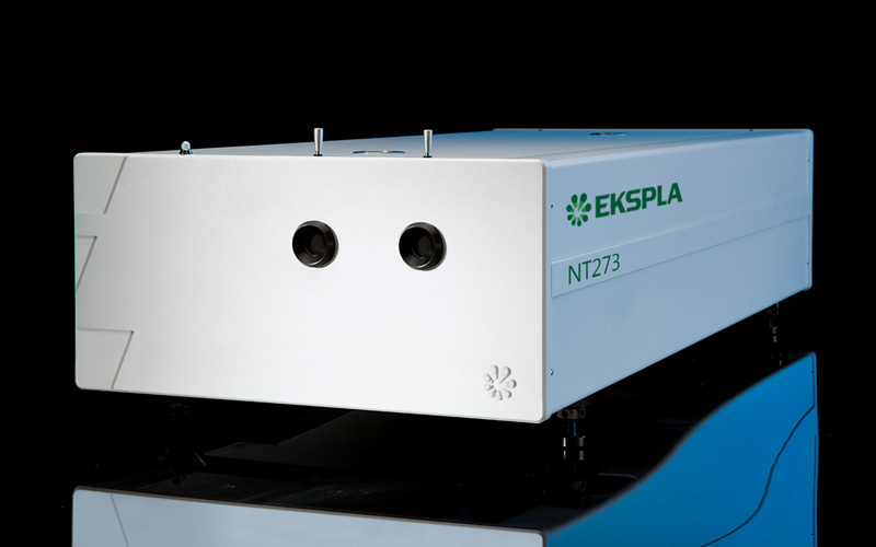
北京欧兰科技发展有限公司为您提供《褐黑色素中光解检测方案(激光产品)》,该方案主要用于其他中光解检测,参考标准《暂无》,《褐黑色素中光解检测方案(激光产品)》用到的仪器有Ekspla NT200 红外波段可调谐激光器。
我要纠错


相关方案
 咨询
咨询