

方案详情文
智能文字提取功能测试中
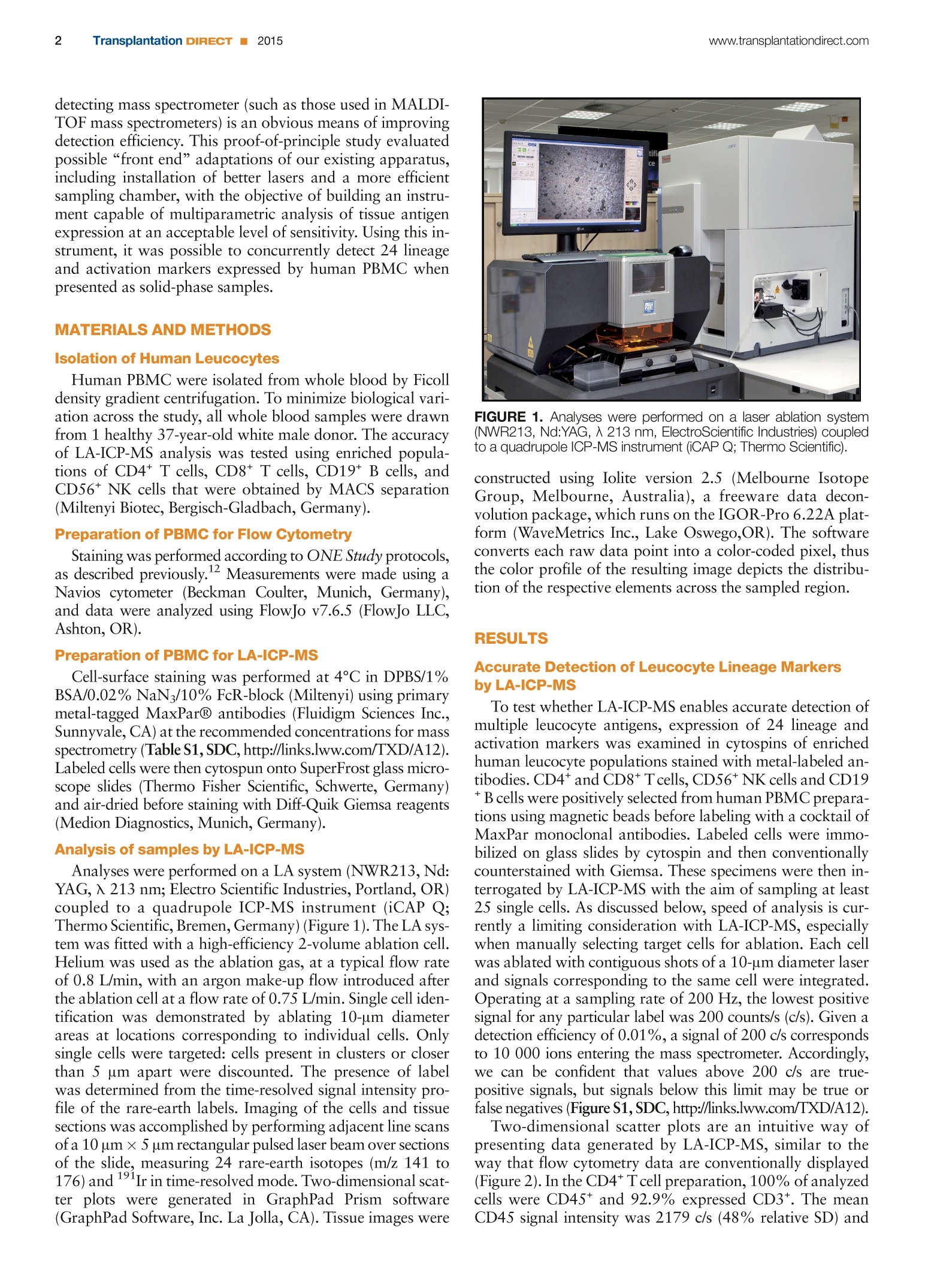
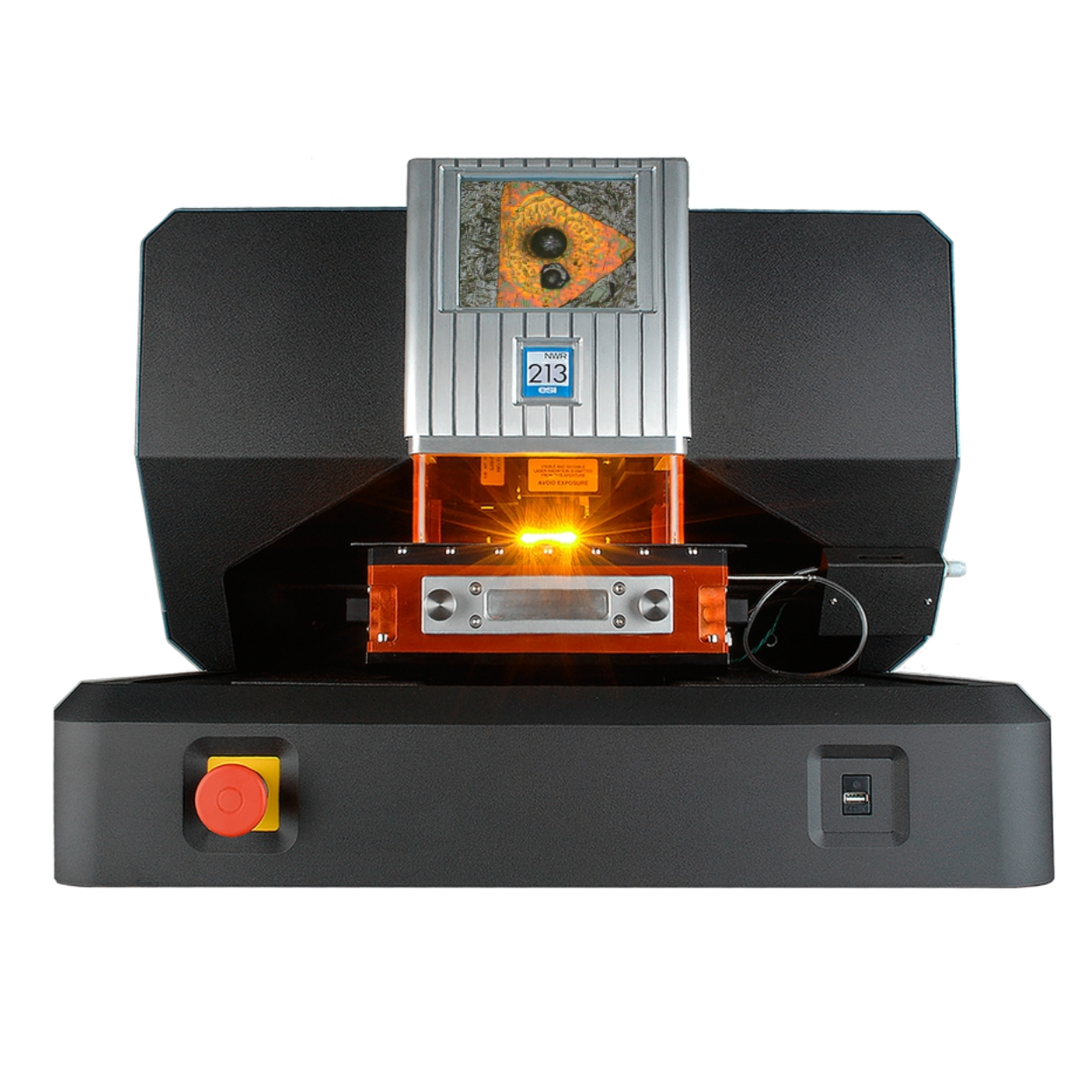
Original Basic Science 2www.transplantationdirect.com: Transplantation DIRECT 2015 www.transplantationdirect.comTransplantation DIR1 DIRECT 2015 Laser Ablation Inductively Coupled Plasma MassSpectrometry An Emerging Technology for Multiparametric Analysis of Tissue Antigens Robert W. Hutchinson, PhD, Katherine M. McLachlin, MSc, Paloma Riquelme, PhD,4 Jan Haarer, PhD,4Christiane Broichhausen, MD,4 Uwe Ritter, PhD,3 Edward K. Geissler, PhD,4 James A. Hutchinson,MBBChir, PhD4 ABSTRACT: New analytical techniques for multiparametric characterisation of individual cells are likely to reveal important infor-mation about the heterogeneity of immunological responses at the single-cell level. In this proof-of-principle study, laser ablationinductively coupled plasma mass spectrometry (LA-ICP-MS) was applied to the problem of concurrently detecting 24 lineageand activation markers expressed by human leucocytes.This approach was sufficiently sensitive and specific to identify subpop-ulations of isolated T, B, and natural killer cells. Leucocyte subsets were also accurately detected within unfractionated peripheralblood mononuclear cells preparations. Accordingly, we judge LA-ICP-MS to be a suitable method for assessing expression of mul-tiple tissue antigens in solid-phase biological specimens, such as tissue sections, cytospins, or cells grown on slides. These resultsaugur well for future development of LA-ICP-MS-based bioimaging instruments for general users. (Transplantation 2015;1:e32; doi: 10.1097/TXD.0000000000000541. Published online 25 September 2015.) mmunologists are only now beginning to appreciate theheterogeneity of immune1 responses at the level of singlecells. Developing novel technologies to measure multiplephenotypic properties of single cells in solid-phase specimenscould be a key to a more sophisticated understanding ofthe diverse behaviors of individual cells.’ Single-cell tran-scriptomics,’single-cell genomics4,5 and mass cytometry6-8 Received 15 May 2015. Revision requested 25 June 2015. IAccepted 31 July 2015. 'Electro Scientific Industries, Huntingdon, Cambridgeshire, United Kingdom. Electro Scientific Industries Inc., Bozeman, MT. Institute for lmmunology, University of Regensburg, Regensburg, Germany. “Division of Experimental Surgery, Department of Surgery, University Hospital Re-gensburg, Regensburg, Germany. Grant support: The authors gratefully acknowledge the support of the EuropeanUnion 7th Framework Programme through The ONE Study initiative (award260687). The authors declare no conflicts of interest. Corresponding: James Alexander Hutchinson, MBBChir, PhD, Department ofSurgery University Hospital Regensburg Franz-Josef-Strauss-Allee 11 93053Regensburg, Germany. (ames.hutchinson@ukr.de). Supplemental digital content (SDC) is available for this article. Direct URL citationsappear in the printed text, links to the digital files are provided in the HTML text ofthis article on the journal’s Web site (www.transplantationdirect.com). Copyright C 2015 The Authors. Transplantation Direct. Published by WoltersKluwer Health, Inc. This is an open-access article distributed under the termsof the Creative Commons Attribution-NonCommercial-NoDerivatives 3.0 Li-cense, where it is permissible to download and share the work provided it isproperly cited. The work cannot be changed in any way or used commerciallyhttp://creativecommons.org/licenses/by-nc-nd/3.0. /SSN:2373-8731 DOI: 10.1097/TXD.0000000000000541 have already revealed unexpected levels of complexity to pre-viously well-studied immune reactions. Important new in-sights into the function of immune cells within their nativemicroenvironments would undoubtedly follow if an equiva-lent, highly multiplexed instrument were available to visualizesingle cells in tissues.’ To this end, our group has adapted alaser ablation inductively coupled mass spectrometry (LA-ICP-MS) system as a tool for imaging biological specimens. The LA-ICP-MS is an analytical technique commonly usedin many fields, including material and earth sciences, that in-volves the vaporization of small parts of a solid sample with afocused pulse of high-irradiance laser energy. 1 The vaporizedmaterial is then analyzed by mass spectrometry. By contigu-ous sampling across a specimen, a data set relating spatialinformation and elemental composition is generated. Thisdata set can then be represented by 2-dimensional scatterplots or used to construct a marker distribution map. Re-cently, our group has explored the use of LA-ICP-MS as amethod of tracking gold-labeled cells in vitro and in vivo.This work demonstrated the feasiblility of detecting very in-frequent metal-labeled cells in tissue sections. Elemental tagging of tissue antigens before LA-ICP-MSusing metal-labeled antibodies, such as those currently usedfor mass cytometry, allows for detection of specific tissue an-tigens. As many as 80 isotopes could be used as labels fordetection by LA-ICP-MS; however,sensitivity is compromisedby detecting multiple elements when using currently availableinstruments with quadrupole mass analyzers. Two comple-mentary solutions to this problem exist, namely,“front end”adaptations that improve sampling efficiency and “back end”solutions that improve detection efficiency. Replacing a se-quentially detecting mass spectrometer with a simultaneously detecting mass spectrometer (such as those used in MALDI-TOF mass spectrometers) is an obvious means of improvingdetection efficiency. This proof-of-principle study evaluatedpossible “front end”adaptations of our existing apparatus,including installation of better lasers and a more efficientsampling chamber, with the objective of building an instru-ment capable of multiparametric analysis of tissue antigenexpression at an acceptable level of sensitivity. Using this in-strument, it was possible to concurrently detect 24 lineageand activation markers expressed by human PBMC whenpresented as solid-phase samples. MATERIALS AND METHODS Isolation of Human Leucocytes Human PBMC were isolated from whole blood by Ficolldensity gradient centrifugation. To minimize biological vari-ation across the study, all whole blood samples were drawnfrom 1 healthy 37-year-old white male donor. The accuracyof LA-ICP-MS analysis was tested using enriched popula-tions of CD4* T cells, CD8+T cells, CD19* B cells, andCD56* NK cells that were obtained by MACS separation(Miltenyi Biotec, Bergisch-Gladbach, Germany). Preparation of PBMC for Flow Cytometry Staining was performed according to ONE Study protocols,as described previously.Measurements were made using aNavios cytometer (Beckman Coulter, Munich, Germany),and data were analyzed using FlowJo v7.6.5 (FlowJo LLC,Ashton, OR). Preparation of PBMC for LA-ICP-MS Cell-surface staining was performed at 4°C in DPBS/1%BSA/0.02% NaN3/10%FcR-block (Miltenyi) using primarymetal-tagged MaxPar@ antibodies (Fluidigm Sciences Inc.,Sunnyvale, CA) at the recommended concentrations for massspectrometry (Table S1, SDC, http://links.lww.com/TXD/A12).Labeled cells were then cytospun onto SuperFrost glass micro-scope slides (Thermo Fisher Scientific, Schwerte, Germany)and air-dried before staining with Diff-Quik Giemsa reagents(Medion Diagnostics, Munich, Germany). Analysis of samples by LA-ICP-MS Analyses were performed on a LA system (NWR213, Nd:YAG, 入 213 nm; Electro Scientific Industries, Portland, OR)coupled to a quadrupole ICP-MS instrument (iCAP Q;Thermo Scientific, Bremen, Germany)(Figure 1). The LA sys-tem was fitted with a high-efficiency 2-volume ablation cell.Helium was used as the ablation gas, at a typical flow rateof 0.8 L/min, with an argon make-up flow introduced afterthe ablation cell at a flow rate of 0.75 L/min. Single cell iden-tification was demonstrated by ablating 10-um diameterareas at locations corresponding to individual cells. Onlysingle cells were targeted: cells present in clusters or closerthan 5 um apart were discounted. The presence of labelwas determined from the time-resolved signal intensity pro-file of the rare-earth labels. Imaging of the cells and tissuesections was accomplished by performing adjacent line scansof a 10 um × 5 um rectangular pulsed laser beam over sectionsof the slide, measuring 24 rare-earth isotopes (m/z 141 to176)and 191Ir in time-resolved mode. Two-dimensional scat-ter plots were generated in GraphPad Prism software(GraphPad Software, Inc. La Jolla, CA). Tissue images were FIGURE 1. Analyses were performed on a laser ablation system(NWR213, Nd:YAG,入 213 nm, ElectroScientific Industries) coupledto a quadrupole ICP-MS instrument (iCAP Q; Thermo Scientific). constructed using Iolite version 2.5 (Melbourne IsotopeGroup, Melbourne, Australia), a freeware data decon-volution package, which runs on the IGOR-Pro 6.22A plat-form (WaveMetrics Inc., Lake Oswego,OR). The softwareconverts each raw data point into a color-coded pixel, thusthe color profile of the resulting image depicts the distribu-tion of the respective elements across the sampled region. RESULTS Accurate Detection of Leucocyte Lineage Markersby LA-ICP-MS To test whether LA-ICP-MS enables accurate detection ofmultiple leucocyte antigens, expression of 24 lineage andactivation markers was examined in cytospins of enrichedhuman leucocyte populations stained with metal-labeled an-tibodies.CD4*and CD8* Tcells, CD56* NK cells and CD19B cells were positively selected from human PBMC prepara-tions using magnetic beads before labeling with a cocktail ofMaxPar monoclonal antibodies. Labeled cells were immo-bilized on glass slides by cytospin and then conventionallycounterstained with Giemsa. These specimens were then in-terrogated by LA-ICP-MS with the aim of sampling at least25 single cells. As discussed below, speed of analysis is cur-rently a limiting consideration with LA-ICP-MS, especiallywhen manually selecting target cells for ablation. Each cellwas ablated with contiguous shots of a 10-um diameter laserand signals corresponding to the same cell were integrated.Operating at a sampling rate of 200 Hz, the lowest positivesignal for any particular label was 200 counts/s (c/s). Given adetection efficiency of 0.01%, a signal of 200 c/s correspondsto 10 000 ions entering the mass spectrometer. Accordingly,we can be confident that values above 200 c/s are true-positive signals, but signals below this limit may be true orfalse negatives (Figure S1, SDC, http://links.lww.com/TXD/A12). Two-dimensional scatter plots are an intuitive way ofpresenting data generated by LA-ICP-MS, similar to theway that flow cytometry data are conventionally displayed(Figure 2). In the CD4*Tcell preparation, 100% of analyzedcells were CD45* and 92.9% expressed CD3*. The meanCD45 signal intensity was 2179 c/s (48% relative SD) and FIGURE 2. Assessing the accuracy of cell-surface marker detection by LA-ICP-MS. Magnetic beads were used to isolate human CD4*Tcells,CD8*Tcells, CD56* NK cells, and CD19* B cells. These cell preparations were labeled with a cocktail of metal-tagged monoclonal antibodies/Ith a co(against a panel of common human leucocyte lineage and activation markers. Labeled cells were cytospun onto glass microscope slides andcounterstained with Giemsa. Samples were then interrogated by LA-ICP-MS. The resultant data sets are presented as 2-dimensional scatterplots showing signal intensities as counts per second. the mean CD3 signal intensity for CD3* cells was 754 c/s(62% relative SD), so detection of both antigens readilyexceeded the negative cutoff. After “gating”on CD45*CD3* events, these T cells could be classified according toCD4 and CD8 expression, and then further subdividedaccording to CD62L and CD25 expression. About 84.6%of CD45* CD3* cells in the CD4* preparation expressedCD4, whereas only 3.9% was CD4- CD8*. In the CD8*T-cell preparation, 100% of analyzed cells were CD45* witha mean CD45 signal intensity of 2059 c/s (38.9% relativeSD) which was not significantly different to the level of signalmeasured in CD4*T cells (p=0.64). About 92.6% of cells inthe CD8+ T-cell preparations were associated with positiveCD3 signals, which had a mean signal intensity for CD3*cells of 624 c/s (54.8% relative SD) which was not signifi-cantly different to the signal measured in CD4* T cells (P=0.27). About 96.0% of CD45* CD3* cells in the CD8+ prepa-ration expressed CD8, whereas 0% was CD4* CD8. Hence,the frequencies of CD4* and CD8* cells in the respectiveCD4*T cell and CD8+ T cell preparations demonstrate theaccuracy of CD4 and CD8 detection by LA-ICP-MS. All cells in a near-pure preparation of CD56* NK cells wereCD45* (mean signal intensity of 1664 c/s; 72.2% relative SD) (Figure 2). However, only 29.2% of events were associatedwith CD56 signal (mean signal intensity of 200 c/s), indicatingdetection sensitivity was inadequate. Likewise, in a near-pureCD19* B-cell preparation, 100% of cells expressed CD45(mean signal intensity of 1980 c/s; 58.7%relative SD); how-ever, only 32.1% detectably expressed CD19 (mean signal in-tensity of 267 c/s; 53.0% relative SD). Within the B-cellpreparation, 42.9% cells expressed surface IgM (mean sig-nal intensity of 517 c/s; 83.3% relative SD), 46.4% cellsexpressed IgD (mean signal intensity of 385 c/s; 61.7% rela-tive SD), 85.7% cells expressed HLA-DR (mean signal inten-sity of 858 c/s; 117% relative SD), and 53.6% cells expressedCD21 (mean signal intensity of 293 c/s; 43.6% relative SD). Having detected leucocyte antigen expression in enrichedlymphocyte subsets, we next asked whether the same panelof antigens could be detected in unfractionated humanPBMC and whether a reasonable estimate of leucocytesubset frequencies could be obtained by LA-ICP-MS.Unfractionated PBMC were prepared for LA-ICP-MS analy-sis using the same protocol used for fractionated PBMC. In parallel, unfractionated PBMC were stained with fluorescentantibodies for conventional immunophenotyping by flowcytometry (Figure 3). Notably, the 24 lineage markers concur-rently detected by LA-ICP-MS had to be distributed into 6 sep-arate panels for analysis by flow cytometry. Using LA-ICP-MS,it was possible to detect CD11c* myeloid cells, CD66a*granulocytes, CD235a*erythrocytes, CD4* and CD8+ T cells, CD19* B cells and CD56* NK cells at frequencies that werebroadly similar to those measured by flow cytometry giventhe small number of sampled cells. Discrepancies in the fre-quencies of NK cells and myeloid cells determined by the 2methods is likely due to low sensitivity of detection ofCD56 signals and operator-bias in visually selecting cellsfor analysis. The precision of LA-ICP-MS will be improved oo FIGURE 3. Analysis of cell-surface marker expression in unfractionated human PBMC immobilised on a glass surface by LA-ICP-MS com-pared to conventional flow cytometry. Unfractionated human PBMC were labeled with a cocktail of metal-tagged monoclonal antibodiesagainst a panel of common human leucocyte lineage and activation markers. Labeled cells were then cytospun onto glass microscope slidesbefore counterstaining with Giemsa. Samples were interrogated by LA-ICP-MS. The resultant data sets are presented as 2-dimensional scatterplots showing signal intensities as counts per second. Forcomparison, the same PBMC were also analyzed by conventional flow cytometryusing fluorescence-labeled antibodies. by increasing the sampling speed and automation, which willallow the analysis of a greater number of cells, and increasingdetection sensitivity using an alternative mass spectrometer. Construction of Antigen Expression Maps Because each laser shot is made in a defined position, it ispossible to use LA-ICP-MS to reconstruct maps of the ana-lyzed sample that use color intensity to represent signal in-tensity. By this approach, we can visualize the distribution ofdetected antigens within a sample, creating images similarto conventional immunohistochemical pictures. Accordingly,LA-ICP-MS generates a data set that can be viewed like amicroscope image, but one that can be easily interconvertedwith numerical representations of antigen expression. To il-lustrate this concept, T-cell marker expression data obtainedfrom an unfractionated PBMC preparation is displayed as in-tensity images (Figure 4). As with conventional fluorescencemicroscopy, it is possible to create “overlay” images withLA-ICP-MS data; however, LA-ICP-MS could also be used CD45+ events CD45+ CD3+ events 100 um FIGURE 4. Representation of cell-surface marker expression inunfractionated human PBMC preparations as signal intensity maps.Unfractionated human PBMC were labeled with a cocktail of metal-tagged monoclonal antibodies against a panel of common humanleucocyte lineage and activation markers. Labeled cells were thencytospun onto glass microscope slides before counterstaining withGiemsa. Samples were interrogated by LA-ICP-MS. The resultantdata sets are represented as pictures that relate spatial positionand signal intensity. By “gating”the data shown, the complexity ofeach image can be usefully reduced. to “gate”cell populations for display within an image, whichis a useful way of reducing the high-dimensionality data setsproduced by LA-ICP-MS. DISCUSSION New technologies enabling multiparametric investiga-tion of single cells are already beginning to reveal unsus-pected levels of complexity to immunological responses.Using LA-ICP-MS, our work aims to bring the same levelof analytical power to immunohistochemical studies oftissue samples. Results presented here demonstrate thatcommercially available LA components coupled with a se-quential ICP-MS allowed detection of cell-surface antigenslabeled with metal-tagged antibodies at a adequate level fora 24-parameter phenotyping of PBMC. It was anticipatedthat use ofa sequential mass spectrometer to determine sucha large number of rare-earth antibody labels could compro-mise detection sensitivity; however, the antigens chosen forthis study appear to be expressed at sufficient density that,by allowing the mass scanning device (quadrupole) to run athigher speeds than normal, positive detection of all markerswas possible. It is anticipated that further improvements insensitivity can be achieved using of simultaneous detectors(eg, ICP-TOF-MS) in future studies. Handling multiparametric data sets presents researcherswith many unfamiliar problems, not least with their statis-tical interpretation. Here, we demonstrate 2 qualitativemethods of representing LA-ICP-MS data, namely, as imagesor scatter plots. Our reconstructed images of cytospun PBMCshow that antigen distribution data can be plotted for inter-pretation in the same way as conventional micrographs;furthermore, these images show that conditional plottingof these data (eg CD45+ → CD3*→ CD8*) can reducethe image to show particular cell types without the needto reanalyze the sample. Alternatively, 2-dimensional scat-ter plots can be used to interrogate the phenotype of indi-vidual cells, in much the same way that conventional flowcytometry data are analyzed. The sort of high dimensionalitydata produced by LA-ICP-MS could also be analyzed usingmore sophisticated computational methods. Standard statis-tical approaches for reduction of multidimensional data sets,such as principal component analysis, could be applied toLA-ICP-MS results to reveal relationships between cell pheno-types or specimens; there are also specialized algorithms forinterrogating complex phenotyping data, such as SPADEor viSNE.14 Unfortunately, speed of sampling (currently~15 cells/min) is a major limitation on the number of cellsthat can be analyzed with our current instrumentation, socollecting enough data for robust statistical analysis remainsa practical challenge. Possible solutions for improving sam-pling speed are now being investigated. At a spatial resolution of 10 um, it was possible to identifysingle cells; however, some applications may demand finerresolution. There are 2 possible solutions to this problem,namely, reducing the dimensions of the laser beam or com-bining LA-ICP-MS technology with conventional fluores-cence microscopy. Reducing the beam width is possible, buthas several technical drawbacks. Most importantly, becauseeach laser shot vaporizes a cylindrical core of material witha diameter equivalent to the beam diameter, the volume ofablated material changes with the cross-sectional area of the beam. Preliminary experiments have shown that ablationwith a 1-um laser diameter is possible; however, the samplevolume is then reduced by a factor of 100 compared to a10-um laser diameter. Accordingly, reducing the laser beamwidth compromises detection sensitivity. A complementaryapproach is to combine LA-ICP-MS detection with conven-tional fluorescence microscopy, so that antigens that mustbe visualized in finest detail could be detected using fluores-cent probes, whereas other markers could be detected by massspectrometry. Both strategies are now being developed. In conclusion, concurrent detection of 24 leucocytemarkers represents a substantial technological advance, asprevious instruments were limited to two signals. By cou-pling this efficient LA apparatus to a simultaneous detector,it should be possible to further improve the instrument's sen-sitivity. In turn, this should allow the use of a narrower laserbeam, so generating finer-resolution images. The technologi-cal advances presented in this article are important steps to-ward the ultimate goal of developing an LA-ICP-MS-basedbioimaging instruments for general users that combine thefunctionality of a microscope and a mass cytometer. REFERENCES 1. Chattopadhyay PK, Gierahn TM, Roederer M, et al. Single-cell technolo-gies for monitoring immune systems. Nat Immunol.2014;15:128-35. 2. Hoppe PS, Coutu DL, Schroeder T. Single-cell technologies sharpen upmammalian stem cell research. Nat Cell Biol.2014;16:919-27. 3. Shalek AK, Satija R, Shuga J, et al. Single-cell RNA-seq reveals dynamicparacrine control of cellular variation. Nature.2014;510:363-9. 4. Fan HC,Wang J, Potanina A, et al. Whole-genome molecular haplotypingof single cells. Nat Biotechnol. 2011;29:51-7. 5. Dey SS, Kester L, Spanjaard B, et al. Integrated genome and tran-scriptome sequencing of the same cell. Nat Biotechnol. 2015;33:285-9. 6. Becher B, Schlitzer A, Chen J, et al. High-dimensional analysis of themurine myeloid cell system. Nat Immunol. 2014;15:1181-9. 7. Bendall SC, Davis KL, Amir el-AD, et al. Single-cell trajectory detection un-covers progression and regulatory coordination in human B cell develop-ment. Cell. 2014;157:714-25. 8. Newell EW, Sigal N, Bendall SC, et al. Cytometry by time-of-flight showscombinatorial cytokine expression and virus-specific cell niches within acontinuum of CD8+Tcell phenotypes.Immunity. 2012;36:142-52. 9. Giesen C, Wang HA, Schapiro D, et al. Highly multiplexed imaging oftumor tissues with subcellular resolution by mass cytometry. NatMethods. 2014;11:417-22. 10. Managh AJ, SL Edwards, Bushell A, et al. Single cell tracking of gadolin-ium labeled CD4+ Tcells by laser ablation inductively coupled plasmamass spectrometry.Anal Chem.2013;85:10627-34. 11. Hutchinson RW, Cox AG,McLeod CW, et al. Imaging and spatial distribu-tion of beta-amyloid peptide and metal ions in Alzheimer's plaques by laserablation-inductively coupled plasma-mass spectrometry.Anal Biochem.2005:346:225-33. 12. Streitz M, Miloud T, Kapinsky M, et al. Standardization of whole bloodimmune phenotype monitoring for clinical trials: panels and methodsfrom the ONE study. Transplant Res. 2013;2:17. 13. Qiu P, Simonds EF, Bendall SC, et al. Extracting a cellular hierarchy fromhigh-dimensional cytometry data with SPADE. Nat Biotechnol.2011;29:886-9. 14. Amir el-AD, Davis KL, Tadmor MD, et al. viSNE enables visualization ofhigh dimensional single-cell data and reveals phenotypic heterogeneityof leukemia. Nat Biotechnol. 2013;31:545-52. 多参数表征单个细胞的全新分析技术可能揭示有关单细胞水平免疫反应异质性的重要信息。本原理验证研究采用激光剥蚀电感耦合等离子体质谱(LA-ICP-MS)方法,同时检测人白细胞表达的24系及活化标志物。这种方法足够敏感,可以准确识别分离的T、B和自然杀伤细胞的亚群。白细胞亚群在未分离的外周血单核细胞制剂中也得到了准确的检测。因此,我们认为LA-ICP-MS是一种适合于评估多种组织抗原在固相生物标本(如组织切片、细胞自旋或载玻片上生长的细胞)中表达的方法。这些结果预示着面向普通用户的基于LA-ICP-MS的生物成像仪器的未来发展。
关闭-
1/6

-
2/6
还剩4页未读,是否继续阅读?
继续免费阅读全文产品配置单
上海凯来仪器有限公司为您提供《单个细胞中免疫反应异质性检测方案(激光剥蚀进样)》,该方案主要用于其他中免疫反应异质性检测,参考标准《暂无》,《单个细胞中免疫反应异质性检测方案(激光剥蚀进样)》用到的仪器有ESL213 灵活的激光剥蚀系统。
我要纠错
推荐专场
相关方案
 咨询
咨询