

方案详情文
智能文字提取功能测试中
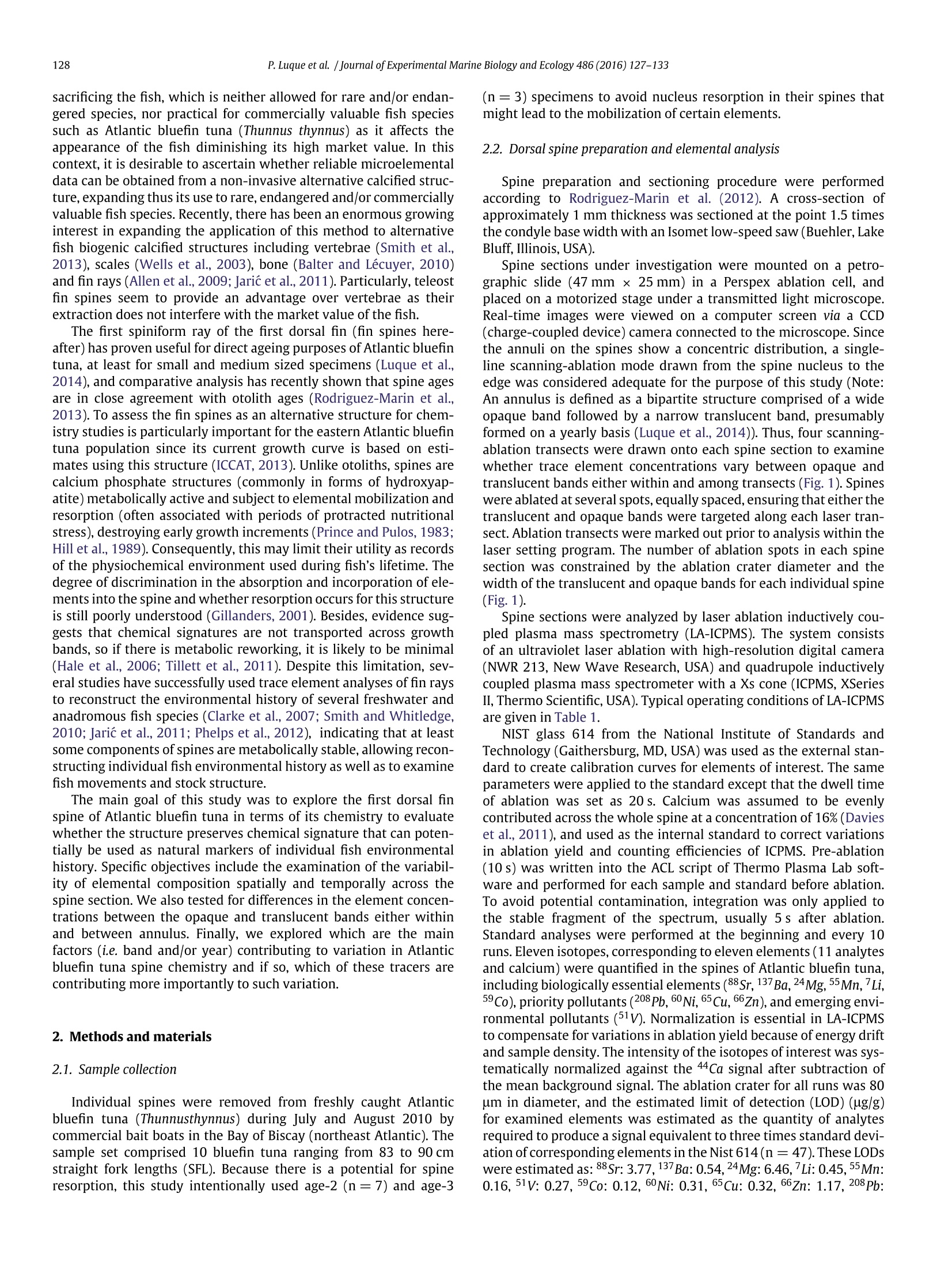
Journal of Experimental Marine Biology and Ecology 486 (2016)127-133Contents lists available at ScienceDirectJournal of Experimental Marine Biology and Ecology 128P. Luque et al. /Journal of Experimental Marine Biology and Ecology 486 (2016)127-133 EXPI TENTALMARINE BIOLOGYAND ECOLOGY ELSEVIER journal homepage: www.elsevier.com/locate/jembe Dorsal fin spines as a non-invasive alternative calcified structure formicroelemental studies in Atlantic bluefin tuna CrossMark Patricia L. Luque a,*, Saijin Zhangb, Jay R. Rookerb, Gorka Bidegain , Enrique Rodriguez-Marin Gulf Coast Research Laboratory, University of Southern Mississippi, 703 East Beach Drive, Ocean Springs, MS 39564, USA "Texas A and M University, Department of Marine Biology, 5007 Avenue U, Galveston, TX 77551, USA ‘Spanish Institute of Oceanography, Santander Oceanographic Centre, Santander 39080, Spain 1. Introduction Over the past 20 years, chemistry of fish calcified structures (i.e.otoliths, scales, vertebrae and fin ray spines) has been applied fordetermining environmental histories of fish in a diverse range ofaquatic environments including marine, estuarine and freshwatersystems (Kerr and Campana, 2014). As such, it represents one of themost powerful tools to address fundamental questions in fish ecol-ogy and fisheries science, including stock structure, site fidelity, natalorigin, and migration pathway over ecological time scale (Campanaet al.,2000;Rooker et al., 2008; Smith and Whitledge,2011; Waltherand Limburg,2012). The premise of this approach is that calcifiedstructures in fishes generally form by the periodic deposition of dailyand annual increments as the fish grows, which allow to determineages and life history parameters (e.g. growth rate) of individual fish ( * Corresponding author. ) and fish populations (Campana and Thorrold, 2001). As these bio-genic structures grow, trace amounts of elements (including heavymetals) are naturally incorporated into their mineral phase from thesurrounding environment experienced by the fish (Miller et al., 2005;Lin et al., 2007). Coupling structure bio chronology with chemicalrecord of fish's life (i.e. trace elements and/or isotopic composition)enables a retrospective description of individual fish environmentahistory including fish movements, life history traits, and ontogeneticdevelopment (Dufour et al., 2005; Whitledge et al., 2006;Whitledge,2009). The most widespread and expanding application of elementalmarkers in calcified structures has occurred using fish otoliths(Campana and Thorrold, 2001; Elsdon et al., 2008). Beside theirchronological properties, otoliths have been the preferred bonystructure to use in chemistry studies due to the fact that theyare metabolically inert calcium carbonate structures (i.e. the newlyl deposited material is neither resorbed nor reworked after depo-sition) (Campana and Neilson, 1985), and hence, only ontogeneticand environmental factors should cause changes to their chemicalcomposition (Campana, 1999). The extraction of otoliths requires sacrificing the fish, which is neither allowed for rare and/or endan-gered species, nor practical for commercially valuable fish speciessuch as Atlantic bluefin tuna (Thunnus thynnus) as it affects theappearance of the fish diminishing its high market value. In thiscontext, it is desirable to ascertain whether reliable microelementaldata can be obtained from a non-invasive alternative calcified struc-ture, expanding thus its use to rare, endangered and/or commerciallyvaluable fish species. Recently, there has been an enormous growinginterest in expanding the application of this method to alternativefish biogenic calcified structures including vertebrae (Smith et al.,2013), scales (Wells et al., 2003), bone (Balter and Lécuyer, 2010)and fin rays (Allen et al., 2009; Jaric et al., 2011). Particularly, teleostfin spines seem to provide an advantage over vertebrae as theirextraction does not interfere with the market value of the fish. The first spiniform ray of the first dorsal fin (fin spines here-after) has proven useful for direct ageing purposes of Atlantic bluefintuna, at least for small and medium sized specimens (Luque et al.,2014), and comparative analysis has recently shown that spine agesare in close agreement with otolith ages (Rodriguez-Marin et al.,2013). To assess the fin spines as an alternative structure for chem-istry studies is particularly important for the eastern Atlantic bluefintuna population since its current growth curve is based on esti-mates using this structure (ICCAT,2013). Unlike otoliths, spines arecalcium phosphate structures (commonly in forms of hydroxyap-atite) metabolically active and subject to elemental mobilization andresorption (often associated with periods of protracted nutritionalstress), destroying early growth increments (Prince and Pulos, 1983;Hill et al., 1989). Consequently, this may limit their utility as recordsof the physiochemical environment used during fish's lifetime. Thedegree of discrimination in the absorption and incorporation of ele-ments into the spine and whether resorption occurs for this structureis still poorly understood (Gillanders, 2001). Besides, evidence sug-gests that chemical signatures are not transported across growthbands, so if there is metabolic reworking, it is likely to be minimal(Hale et al., 2006; Tillett et al., 2011). Despite this limitation, sev-eral studies have successfully used trace element analyses of fin raysto reconstruct the environmental history of several freshwater andanadromous fish species (Clarke et al., 2007; Smith and Whitledge,2010; Jaric et al., 2011; Phelps et al., 2012), indicating that at leastsome components of spines are metabolically stable, allowing recon-structing individual fish environmental history as well as to examinefish movements and stock structure. The main goal of this study was to explore the first dorsal finspine of Atlantic bluefin tuna in terms of its chemistry to evaluatewhether the structure preserves chemical signature that can poten-tially be used as natural markers of individual fish environmentalhistory. Specific objectives include the examination of the variabil-ity of elemental composition spatially and temporally across thespine section. We also tested for differences in the element concen-trations between the opaque and translucent bands either withinand between annulus. Finally, we explored which are the mainfactors (i.e. band and/or year) contributing to variation in Atlanticbluefin tuna spine chemistry and if so, which of these tracers arecontributing more importantly to such variation. 2. Methods and materials 2.1. Sample collection Individual spines were removed from freshly caught Atlanticbluefin tuna (Thunnusthynnus) during July and August 2010 bycommercial bait boats in the Bay of Biscay (northeast Atlantic). Thesample set comprised 10 bluefin tuna ranging from 83 to 90 cmstraight fork lengths (SFL). Because there is a potential for spineresorption, this study intentionally used age-2 (n=7) and age-3 (n=3) specimens to avoid nucleus resorption in their spines thatmight lead to the mobilization of certain elements. 2.2. Dorsal spine preparation and elemental analysis Spine preparation and sectioning procedure were performedaccording to Rodriguez-Marin et al. (2012). A cross-section ofapproximately 1 mm thickness was sectioned at the point 1.5 timesthe condyle base width with an Isometlow-speed saw (Buehler, LakeBluff, Illinois, USA). Spine sections under investigation were mounted on a petro-graphic slide (47 mm x 25 mm) in a Perspex ablation cell, andplaced on a motorized stage under a transmitted light microscope.Real-time images were viewed on a computer screen via a CCD(charge-coupled device) camera connected to the microscope.Sincethe annuli on the spines show a concentric distribution, a single-line scanning-ablation mode drawn from the spine nucleus to theedge was considered adequate for the purpose of this study (Note:An annulus is defined as a bipartite structure comprised of a wideopaque band followed by a narrow translucent band, presumablyformed on a yearly basis (Luque et al., 2014)). Thus, four scanning-ablation transects were drawn onto each spine section to examinewhether trace element concentrations vary between opaque andtranslucent bands either within and among transects (Fig.1). Spineswere ablated at several spots, equally spaced, ensuring that either thetranslucent and opaque bands were targeted along each laser tran-sect.Ablation transects were marked out prior to analysis within thelaser setting program. The number of ablation spots in each spinesection was constrained by the ablation crater diameter and thewidth ofthe translucent and opaque bands for each individual spine(Fig. 1). Spine sections were analyzed by laser ablation inductively cou-pled plasma mass spectrometry (LA-ICPMS). The system consistsof an ultraviolet laser ablation with high-resolution digital camera(NWR 213, New Wave Research, USA) and quadrupole inductivelycoupled plasma mass spectrometer with a Xs cone (ICPMS, XSeriesII, Thermo Scientific, USA). Typical operating conditions of LA-ICPMSare given in Table 1. NIST glass 614 from the National Institute of Standards andTechnology (Gaithersburg, MD, USA) was used as the external stan-dard to create calibration curves for elements of interest. The sameparameters were applied to the standard except that the dwell timeof ablation was set as 20 s. Calcium was assumed to be evenlvcontributed across the whole spine at a concentration of 16% (Davieset al., 2011), and used as the internal standard to correct variationsin ablation yield and counting efficiencies of ICPMS. Pre-ablation(10 s) was written into the ACL script of Thermo Plasma Lab soft-ware and performed for each sample and standard before ablation.To avoid potential contamination, integration was only applied tothe stable fragment of the spectrum, usually 5s after ablation.Standard analyses were performed at the beginning and every 10runs. Eleven isotopes, corresponding to eleven elements (11 analytesand calcium) were quantified in the spines of Atlantic bluefin tuna,including biologically essential elements (88Sr, 137Ba,24Mg,55Mn,Li,59Co), priority pollutants (208Pb, 60Ni, 65 Cu,66Zn), and emerging envi-ronmental pollutants (51V). Normalization is essential in LA-ICPMSto compensate for variations in ablation yield because of energy driftand sample density. The intensity of the isotopes of interest was sys-tematically normalized against the 44Ca signal after subtraction ofthe mean background signal. The ablation crater for all runs was 80um in diameter, and the estimated limit of detection (LOD)(ug/g)for examined elements was estimated as the quantity of analytesrequired to produce a signal equivalent to three times standard devi-ation of corresponding elements in the Nist 614(n=47).These LODswere estimated as: 88Sr:3.77,137Ba:0.54,24Mg: 6.46,7Li:0.45,55Mn:0.16, 51V: 0.27, 59Co: 0.12, 60Ni: 0.31, 65 Cu: 0.32, 66Zn: 1.17, 208Pb: Fig. 1. A cross-spine section showing the line scanning-ablation mode used across the spine section. A, B, C, D: Ablation line transects drawn from the nucleus to the edge of thespine. Zoom of ablation line transect 'B'showing five ablation spots targeting the opaque and translucent bands of each annulus. 2.3. Statistical analysis Before statistical testing, residuals were examined for normal-ity (Shapiro-Wilkinson test) and homogeneity of variance (Lev-ene's test) among factor levels. When it was needed, Lambda fromBoxplots was used to meet parametric assumptions. Multivariateanalysis of variance (MANOVA) was used to examine the effect ofscanning-ablation transect (fixed factor) on the response variable(elemental composition) in each spine section. Pillai's trace statis-tic was used for multivariate test because it is considered the mostrobust test (Wilkinson et al., 1996). If no significant difference inspine chemistry was detected within and between the four scanning-ablation transects, we deemed it appropriate to pool these data toexamine the effect of band and year (fixed factors) on the spinechemistry using two-way MANOVA. Morevover, univariate analy-sis (two-way ANOVA) was used to explore which of these tracerscontributed to the differences in spine chemistry for the two studied Table 1 Settings Laser ablation system Energy 60% Repetition rate 10 HZ Ablation diameter 80pm Ablation pattern Spot Ablation dwell time 12 s ICPMS Uptake delay Os Washout delay 20s Acquisition dwell time 20 ms Duration time 70s Acquisition resolution 44Ca Internal standard 44Ca, 8.570% for NIST614,16% for spines factors (i.e. band and year). All statistical analyses were run using thesoftware R 2.12.2 (The R Foundation for Statistical Computing). 3. Results 3.1. Elemental concentrations A total of eleven trace elements were detected in Atlantic bluefirable tuna spine samples above the limit of detection: 88sr, 1337SEr, 137 Ba, 24Mg,55Mn and 66 Zn were detected in 100% of samples whereas 7Li, and65Cu appeared in 60% of the samples. Other elements quantifiedsuch as 59Co, 60Ni, and 208Pb, were found below detection limitand were not included for further analysis. Magnesium (Mg) andStrontium (Sr) showed the highest concentrations in Atlantic bluefintuna spines with values ranging from 1582 to 5074 (ug/g) and 384.5to 547.2 (ug/g), respectively. In contrast, Co, Ni and Pb showed thelowest concentration values (Table 2). Table 2 Summary of trace element concentrations (Mean±SD) in bluefin tuna spines showingthe limit of detection (LODs) for each tracer. Elements n Mean Std dev Min Max Limit of detection (ug/g) (ug/g) (ug/g) (LODs)(ug/g) Sr 168 452.60 33.90 384.52 547.21 3.77 Mg 168 2357.71 660.02 1582 5074 6.46 Ba 164 1.21 0.37 0.66 2.34 0.54 Mn 168 3.18 0.84 1.62 5.48 0.16 Li 168 0.43 0.08 0.28 0.70 0.45 Zn 163 31.51 6.17 19.52 47.63 1.17 Cu 166 0.85 1.91 0.09 11.01 0.32 Co 167 0.03 0.01 0.02 0.06 0.12 Ni 165 0.01 0.04 0.03 0.25 0.31 v 164 0.15 0.07 0.05 0.40 0.27 Pb 164 0.01 0.01 0.01 0.12 0.35 3.2. The spatial and temporal variability ofelemental composition inAtlantic bluefin tuna spine sections. Factor/s that contribute tovariations in spine chemistry Results of MANOVA indicated that elemental signatures mea-sured in all spine sections did not vary significantly among the fourscanning-ablation transects. Consequently, the effect of the band andyear was examined using different transect data as replicates. Assuch, Pillai’s test showed that the two fixed factors, i.e. band and yearand their interaction have a significant effect in the overall spineelemental composition (p≤0.001) (Table 3). The factorialANOVA results for each element presented in Table 4showed that year has a significant effect on the concentration of alltracers except for Zn, whereas band has a significant effect on theconcentration of Sr, Ba, Mn, Zn and Cu. Moreover, an effect of theinteraction between these two factors was found significant in Sr,Ba, Li and Cu. In addition, for those tracers that both factors havea significant effect, the factor that contributed more importantly tothe overall variance was year for Mn, band for Sr, while for Ba andCu, both factors contributed similarly. Box plots showed that con-centration of Sr, Ba and Li displayed similar pattern with the highestconcentration in the translucent band of the annulus 2. Mg andmore slighly Co showed an increasing pattern from annulus 1 tothe spine edge, with a Mg concentration particularly high in the lastdeposited opaque band. In contrast,Mn showed a decreasing patternby annulus, with the lowest concentration at the edge of the spine.A certain annual periodicity was displayed by V and less clear for Zn,with regardless the annulus, higher concentrations in the translu-cent and opaque bands, respectively. Finally, Ni, Cu and Pb, wereirregular, with unclear annual pattern, which made it more difficultto associate to the transitions between different annuli (Fig. 2). 4. Discussion Several studies have demonstrated that trace elements and sta-ble isotopic composition of otoliths can serve as natural markersof environmental history for individual fishes in either freshwateror marine environments (Kerr and Campana, 2014, for a review).Such research for dorsal fin spines is limited and to our knowledgethere has not been any investigation that examined the suitabil-ity of spines as an alternative structure for Atlantic bluefin tuna. Inthis study, two-year-old specimens were used to limit the poten-tial for spine nucleus resorption (Luque et al., 2014). The presentstudy explored whether the elemental markers are sufficiently sta-ble spatially and temporally across the bipartite annular structureof the spine. The chemical profile was examined across sectionedspines at four scanning-ablation transects to test for spatial variabil-ity in tracer concentration. Thus, seven (Sr, Ba,Mg, Mn, Zn, Li and Cu)out of eleven tracers initially analyzed appeared consistently abovethe LODs in Atlantic bluefin tuna spines. Moreover, all elementalsignatures measured in all spine sections did not vary significantlyamong the four scanning-ablation transect providing an important Table 3 Sumary of MANOVA test. Effects of the three fixed factors and their interactionon bluefin tuna spine tracers concentration. The effect was considered statisticallysignificant at ps0.001. Factor df Pillai's F df df error p trace (den df) Band 1 0.54 23.45 7 146 *** Year 1 0.68 46.31 7 146 *** Ablation site 1 0.08 1.74 7 146 0.11 BandxYear 1 0.43 15.78 7 146 *** BandxAblation site 1 0.03 0.82 7 146 0.57 YearxAblation site 1 0.06 1.36 7 146 0.22 BandxYearxAblation site 1 0.02 0.46 7 146 0.86 Table 4Sumary of factorial ANOVA results for each tracer. n.s (non-significant). *** Statistically significant at p<0.001. ** Statistically significant at p<0.01. * Statistically significant at p<0.05. insight regarding the stability of some tracer concentrations acrossthe spine. This indicates that some tracer signatures are certainlyincorporated into the mineral fraction of the spines,and can be likelyvaluable elemental markers to reconstruct the environmental his-tory of Atlantic bluefin tuna from spine chemistry. Although it isearly to draw any conclusion based only on such a young fish sam-ple, results support the evidence (Hale et al., 2006; Tillett et al., 2011)that certain elements are metabolically stable and might not be nottransported across growth bands. In light of this, the physiologicalreworking of the soft calcium phosphate ("hydroxyapatite') whichconstitute the corpus calcareum of the spines is likely to be minimal.Results of this study also coincide with previous studies (Clarke et al.,2007; Smith and Whitledge, 2010; Jaric et al., 2011; Phelps et al.,2012) that have successfully used fin rays to reconstruct the envi-ronmental history of several freshwater and anadromous fish speciesand therefore support the potential use of fin spines chemistry toreconstruct the environmental history of Atlantic bluefin tuna. Ingeneral, elements are naturallyacquired throughrespiratoryanddietary pathways and assimilated into actively calcifying structuresincluding scales, shells, and otoliths (Campana, 1999). But elemen-tal composition can be further modified by temperature, that has astrong influenceontheratesofchemical and metabolicprocesses(BathMartin and Thorrold, 2005), salinity, and ambient elemental concen-tration (i.e. water chemistry) (Elsdon and Gillanders, 2002, 2004). In addition, physiological regulation of internal elemental compositioncan result in active competition or facilitation uptake of elements,modifyingthus relationships withenvironmentalconditions.The abil-ity to reconstruct the environmental history of fish and migratorypatterns from spine chemistry relies on the concentration ofcertainelements to change in predictable manners with the environmentalvariables (Elsdon and Gillanders,2002,2004).Juvenile Atlantic bluefintuna individuals produced in the Mediterranean Sea appear to staywithin this basin (including the Strait ofGibraltar) during the first yearof life and they may carry out migrations to foraging areas out of theMediterranean Sea such as the Bay of Biscay (Fraile et al., 2016).Nev-ertheless, the Atlantic bluefin tuna migration patterns in the easternAtlantic and Mediterranean Sea remain poorly known (Fromentin andLopuszanski,2014). The concentration of Strontium (Sr) in Atlantic bluefin tuna spineswas significantly higher in the translucent band, particularly in the second annulus that corresponds with the second winter growth(Luque et al., 2014). This, together with the belief that sources andpathways of elements to fin spines are similar to that in vertebrae(Gillanders, 2001), suggests a negative relationship of Sr with tem-perature as detected in fish otoliths (Elsdon and Gillanders, 2002;Secor et al., 1995). The observed increasing of Sr from ages 1 to 2may reflect a change in water temperature, perhaps indicating themovement of juvenile Atlantic bluefin tuna from epipelagic waternursery areas in the Mediterranean Sea to mesopelagic waters in theBay of Biscay (Abascal et al., 2016;Fraile et al., 2016). Regardingsalinity, the level of Sr is positively correlated with water salinity(Secor et al., 1995;Limburg et al., 2001; Zimmerman,2005). Allenet al. (2009) and Jaric et al.(2011) found changes of such magni-tude in pectoral fin rays of green sturgeon (Acipenser medirostris)and Danube sturgeon species, supporting the fact that fin rays reflectcertain changes in ambient salinity conditions and could indicate habitat transitions. Here, the concentration of Sr was slightly higherin the opaque band of the first annulus that correspond with thefirst summer of fish’life, very likely in the Mediterranean sea. Incontrast, in the second annulus, the concentration of Sr was sig-nificantly higher in the translucent band which corresponds withthe second winter of fish' life in the Atlantic Ocean-Bay of Biscay(with lower salinity). This pattern seems to be inconsistent with thepositive relationship of Sr with water salinity but may be explainedby the relative and likely interactive effects of temperature, salinity,and ambient concentration of Sr on the resulting concentration ofthis tracer in the spine Elsdon and Gillanders (2004). These authorsconclude for black bream (Acanthopagrus butcheri) otoliths that theinteraction between salinity and the ambient Sr concentration hasmore effect than salinity alone on Sr concentrations. Hence, it seemsplausible that the effect of ambient Sr outweighs that of salinity ininfluencing otolith chemistry. There is a further need of experimen-tation to ascertain whether the use of spine Sr and Sr:Ca ratios canbe an appropriate tool for describing migration across significanttemperature and salinity gradients. Barium (Ba) pattern was similar to Sr (Fig. 2), with higherconcentrations in the translucent bands, and significantly higherconcentration in the second annulus corresponding to the secondwinter growth. Ba increasingly incorporates into fin spines acrossyears with higher concentrations during winter time representinga negative relationship with temperature as reported in previousstudies e.g.Balter and Lécuyer (2010), Smith et al. (2013). Bariumincorporation into the hydroxyapatite of fish scales, bone, and teethhas also revealed either positive or no relationship with temperature(Miller, 2009). These discrepancies suggest that Ba concentration islikely to represent interactive effects between environmental vari-ables and species-specific responses (Elsdon and Gillanders, 2004).This could confound interpretations of field data particularly in studyareas with remarkable gradients in both temperature and Ba:Ca inwater. Barium is known to show nutrient-like distributions, i.e. verylow concentrations in surface waters and considerably higher val-ues in deeper waters (Chan et al.,1977). As it was suggested forcephalopods (Loligo gahi) (Arkhipkin et al., 2004), the pattern of Bain bluefin tuna spines across the annular structure may be an indica-tor for vertical movements of bluefin tuna, from surface waters (Balower concentrations) at the larval and juvenile stage to mesopelagicwaters (Tudela et al., 2011; Abascal et al., 2016) when they arejuveniles and adults (Ba high concentrations). The concentration of Magnesium (Mg) showed an increasingtrend across the spine section albeit no significant differencesbetween opaque and translucent bands. The increasing pattern ofMg observed in Bluefin tuna spines may indicate an increasingaccumulation rate with age associated with a physiological regula-tion that would be reflected in a comparatively consistent patternof incorporation, as was observed in vertebrae of round stingrays(Smith et al., 2013). Experimental studies of Mg incorporation intothe otoliths of several fish species including red drum (Sciaenops ocel-latus) (Hoff and Fuiman, 1995), gray snapper (Lutjanus griseus) (BathMartin and Wuenschel,2006) and Pacific cod (Gadus macrocephalus)(DiMaria et al., 2010) concluded that Mg:Ca was not affected neitherby temperature or salinity. Manganese (Mn) concentration decreased across annulus andwas significantly higher in the opaque bands compared to translu-cent bands regardless of the annuli. The concentration of Mn wassignificantly higher in the first opaque band which corresponds withthe first summer of fish’s life right after spawning (i.e. spawningin the western Mediterranean occurs from mid-June to mid-July(Rooker et al.,2007)). This result suggests that the concentration ofMn in spines could be also used as a potential indicator of natalorigin of Atlantic bluefin tuna as in otoliths (Brophy et al., 2003;Rooker et al., 2003, 2001). Considering the annual periodicity in theformation of opaque bands in bluefin tuna spines during summer months (Luque et al., 2014), higher Mn concentration in opaquebands may coincide with a positive relationship of Mn with temper-ature (Smith et al., 2013). Nevertheless, further controlled labora-tory studies are needed to assess the factors (e.g., water chemistry,spine structure) controlling Mn incorporation into spines and clar-ify the utility of this element as an indicator of fish' environmentalhistory. Zinc (Zn) concentration was higher in opaque bands insinuat-ing a positive influence of temperature as it has been suggestedin round stingrays vertebrae (Smith et al.,2013). Zinc is an essen-tial micronutrient that is involved in various metabolic pathways(Watanabe et al., 1997). In synthetic hydroxyapatites, the majorityof Zn seems to incorporate through inclusion into interstitial spaces(Tang et al., 2009) and is being representative of environmentalconditions (Campana, 1999; Bath Martin and Thorrold, 2005). In bio-genic hydroxyapatites such as spines, Zn is bound within the proteinmatrix and unlikely to be a reliable proxy of ambient environmen-tal conditions (Watanabe et al., 1997). In addition, there is evidencethat trace of Zn in seawater becomes toxic at high levels (Watanabeet al., 1997) and consequently prone to contamination (Gosnell et al.,2012). To date there has not been any investigation referring to theutility of dorsal spines as environmental pollution proxy and furtherresearch is needed to verify this since it may simply correspond tolife history changes in fish. Copper (Cu) and Lithium (Li) showed a conspicuous increasealong the spine section. The sudden increase of Cu may be explainedby atmospheric (deposition of dust and rainwater) and riverineinputs such as the Loire and the Gironde that enrich shelf watersof the Gulf of Biscay (OSPAR, 2000). Copper is known to occur atlower concentrations in surface waters, where larvae and very youngjuveniles swim (Boyle et al., 1985), which is consistent with low con-centrations along almost the entire longitudinal section of the spine.The more gradual increase of Li may reflect an increase in seawaterconcentrations combined with a high degree of physiological mod-ulation (Baumann et al., 2015). Similar patterns for Cu and Li werefound along the longitudinal section of the otolith in young Pacificbluefin tuna (Baumann et al., 2015). In conclusion, findings here provide an important insight regard-ing the metabolic stability of some tracer concentrations acrossthe growth bands of the spine. Physiological reworking of the softcalcium phosphate (‘hidroxyapatite') which constitutes the corpuscalcareum of the spines is likely to be minimal, strengthening its useas a non-invasive alternative structure in elemental markers for thisspecies. Using tracers in spine chemistry as a proxy of environmen-tal conditions (e.g. temperature, salinity and elemental concentra-tions), and consequently for migration patterns studies, will dependon a more robust seawater chemistry characterization and furtherresearch on the elements incorporation mechanisms into the hardstructure. Hence, it is crucially important to do more experimen-tation under controlled conditions to explore and test how tracersare bound within spine hydroxyapatite. Further research should alsoinclude older specimens where resorption has already taken placeand the resorption effect on these trace elements. Acknowledgments The authors would like to thank the Spanish Ministry of Scienceand Innovation (ACI2008-0824) for supporting this work and by theSpanish data collection program within the EU Fisheries Data Collec-tion Regulation Framework.The authors would like to thank to MartaRuiz and Pablo Quelle for their assistance in the preparation of spinesection samples. The authors would also like to thank to the largenumber of people who collected the biological samples and data thatunderpin the analyses of this paper. [SS] Abascal, F.J.,Medina, A., De La Serna,J.M., Godoy, D., Aranda, G., 2016. Tracking bluefintuna reproductive migration into the Mediterranean Sea with electronic pop-upsatellite archival tags using two tagging procedures. Fish.Oceanogr. 25(1),54-66. Allen, P.J., Hobbs, J.A., Cech, J.J., Jr, Van Eenennaam, J.P., Doroshov, S.I., 2009. Usingtrace elements in pectoral fin rays to assess life history movements in sturgeon:estimating age at initial seawater entry in Klamath River green sturgeon. Trans.Am. Fish. Soc. 138 (2),240-250. Arkhipkin, A.I., Campana, S.E., FitzGerald,J., Thorrold, S.R., 2004. Spatial and temporalvariation in elemental signatures of statoliths from the patagonian longfin squid(Loligo gahi). Can. J. Fish. Aquat. Sci. 61(7), 1212-1224. Balter, V., Lécuyer, C., 2010. Determination of Sr and Ba partition coefficients betweenapatite from fish (Sparus aurata) and seawater: the influence of temperature.Geochim. Cosmochim. Acta 74 (12),3449-3458. Bath Martin, G., Thorrold, S.R., 2005. Temperature and salinity effects on magnesium,manganese, and barium incorporation in otoliths of larval and early juvenile spotLeiostomus xanthurus. Mar. Ecol. Prog. Ser. 293, 223-232. Bath Martin, G., Wuenschel, M.J., 2006. Effect of temperature and salinity on otolithelement incorporation in juvenile gray snapper Lutjanus griseus. Mar. Ecol. Prog.Ser. 324, 229-239. Baumann, H., Wells, R., Rooker, J.R., Zhang, S., Baumann, Z., Madigan, D.J., Dewar, H.,Snodgrass, O.E., Fisher, N.S., 2015. Combining otolith microstructure and traceelemental analyses to infer the arrival of juvenile Pacific bluefin tuna in theCalifornia current ecosystem. ICES J. Mar. Sci.:J. Conseil.fsv062. Boyle, E., Chapnick, S., Bai, X., Spivack, A., 1985. Trace metal enrichments in theMediterranean Sea. Earth Planet. Sci. Lett. 74(4), 405-419. Brophy,D.,Danilowicz, B.,Jeffries,T., 2003. The detection of elements in larval otoliths from Atlantic herring using laser ablation ICP-MS. J. Fish Biol. 63 (4), 990-1007.Campana, S., Chouinard, G., Hanson, J., Frechet, A., Brattey,J., 2000. Otolith elemental fingerprints as biological tracers of fish stocks. Fish. Res. 46 (1-3), 343-357.Campana, S.E., 1999. Chemistry and composition of fish otoliths: pathways, mecha-nisms and applications. Mar. Ecol.Prog. Ser. 188, 263-297. Campana, S.E., Neilson, J.D., 1985. Microstructure of fish otoliths. Can. J. Fish. Aquat.Sci. 42(5), 1014-1032. Campana, S.E., Thorrold,S.R., 2001. Otoliths, increments, and elements: keys to a com-prehensive understanding of fish populations? Can. J. Fish. Aquat. Sci. 58 (1),30-38. Chan, L., Drummond, D., Edmond, J., Grant, B., 1977. On the barium data from theatlantic geosecs expedition.Deep-Sea Res.24(7),613-649. Clarke, A.D., Telmer, K.H., Mark Shrimpton, J.,2007. Elemental analysis of otoliths,1mifin rays and scales: a comparison of bony structures to provide population andlife-history information for the arctic grayling (Thymallus arcticus). Ecol. Freshw.Fish 16(3),354-361. Davies, C., Brophy, D., Jeffries, T., Gosling, E., 2011. Trace elements in the otolithsand dorsal spines of albacore tuna (Thunnus alalunga, bonnaterre, 1788): anassessment of the effectiveness of cleaning procedures at removing postmortemcontamination. J. Exp. Mar. Biol. Ecol. 396 (2), 162-170. DiMaria, R., Miller, J., Hurst, T., 2010. Temperature and growth effects on otolith ele-mental chemistry of larval pacific cod, Gadus macrocephalus. Environ. Biol. Fish 89(3-4),453-462. Dufour, E., Patterson, W.P., Hook, T.O., Rutherford, E.S., 2005. Early life history ofLake Michigan alewives (Alosa pseudoharengus) inferred from intra-otolith stableisotope ratios.Can. J. Fish. Aquat. Sci. 62(10),2362-2370. Elsdon, T.S., Gillanders, B.M., 2002. Interactive effects of temperature and salinity onotolith chemistry: challenges for determining environmental histories of fish.Can. I. Fish. Aquat. Sci. 59 (11),1796-1808.a 6-1802 Elsdon, T.S., Gillanders, B.M., 2004. Fish otolith chemistry influenced by exposure tomultiple environmental variables. J. Exp. Mar. Biol. Ecol. 313 (2), 269-284. Elsdon, T.S., Wells, B.K., Campana, S.E., Gillanders, B.M., Jones, C.M., Limburg, K.E., Secor, D.H., Thorrold, S.R., Walther, B.D., 2008. Otolith chemistry to describemovements and life-history parameters of fishes: hypotheses, assumptions, lim-itations and inferences.Oceanogr.Mar. Biol. Annu. Rev. 46(1), 297-330. Fraile, I., Arrizabalaga, H., Groeneveld, J., Kolling, M.,Santos, M.N., Macias, D., Addis, P.,Dettman, D.L., Karakulak, S.,Deguara, S., et al. 2016. The imprint of anthropogenicCO2 emissions on Atlantic bluefin tuna. J. Mar. Syst. 158, 26-33. Fromentin, J.-M., Lopuszanski, D., 2014. Migration, residency, and homing of bluefintuna in the western Mediterranean Sea. ICES J. Mar. Sci. 71 (3),510-518. Gillanders, B., 2001. Trace metals in four structures of fish and their use for estimatesof stock structure. Fish.Bull. 99 (3), 410-419. Gosnell, K.J., Landing, W.M., Milne, A., 2012. Fluorometric detection of total dissolvedzinc in the southern Indian Ocean. Mar.Chem.132,68-76. Hale, L.F., Dudgeon, J.V., Mason, A.Z., Lowe, C.G., 2006. Elemental signatures in thevertebral cartilage of the round stingray, Urobatis halleri, from seal beach, cali-fornia.. Special Issue: Age and Growth of Chondrichthyan Fishes: New Methods,Techniques and AnalysisSpringer.,pp.317-325. Hill, K.T., Cailliet, G.M., Radtke, R.L., 1989. A comparative analysis of growth zonesin four calcified structures of pacific blue marlin, Makaim. Fish. Bull. 87 (4),829-843. Hoff, G.R., Fuiman,L.A., 1995. Environmentally induced variation in elemental compo-sition of red drum (Sciaenops ocellatus) otoliths. Bull. Mar. Sci. 56 (2), 578-591. ICCAT, 2013. Report of the 2012 atlantic bluefin tuna stock assessment session. Coll.Vol. Sci. Pap.69, 1-198. Jaric, I., Lenhardt, M., Pallon, J., Elfman, M., Kalauzi, A., Suciu, R., Cvijanovic, G.,Ebenhard, T., 2011. Insight into Danube sturgeon life history: trace elementassessment in pectoral fin rays. Environ. Biol. Fish 90 (2), 171-181. Kerr, L.A., Campana, S.E., 2014. Chapter eleven - chemical composition of fish hardparts as a natural marker of fish stocks. In: Cadrin, S.X., Kerr, L.A., Mariani, S.(Eds.), Stock Identification Methods (Second Edition). Academic Press,San Diego,pp.205-234. Limburg, K.E., Landergren, P., Westin, L., Elfman, M., Kristiansson, P., 2001. Flexiblemodes of anadromy in baltic sea trout: making the most of marginal spawningstreams. J. Fish Biol. 59 (3), 682-695. Lin,S.-H., Chang, C.-W., lizuka, Y.,Tzeng, W.-N., 2007. Salinities, not diets, affect stron-tium/calcium ratios in otoliths of Anguilla japonica. J. Exp. Mar. Biol. Ecol.341(2),254-263. Luque,P.,Rodriguez-Marin, E., Landa,J.,Ruiz, M., Quelle, P., Macias, D., Ortiz De Urbina,J., 2014. Direct ageing of Thunnus thynnus from the eastern Atlantic Ocean andwestern Mediterranean Sea using dorsal fin spines. J. Fish Biol.84(6),1876-1903. Miller, J., 2009. The effects of temperature and water concentration on the otolithincorporation of barium and manganese in black rockfish Sebastes melanops. J.Fish Biol. 75(1), 39-60. Miller, J., Banks, M., Gomez-Uchida,D., Shanks, A.,2005. A comparison of populationstructure in black rockfish (Sebastes melanops) as determined with otolith micro-chemistry and microsatellite DNA. Can. J. Fish. Aquat. Sci. 62 (10), 2189-2198. OSPAR, 2000. Quality Status Report 2000: Region ⅣV: Bay of Biscay and Iberian Coast.Commission for the Protection of the Marine Environment of the North-EastAtlantic. Phelps, Q.E., Whitledge, G.W., Tripp, S.J., Smith, K.T., Garvey, J.E., Herzog, D.P., Osten-dorf, D.E.,Ridings, J.W., Crites, J.W., Hrabik, R.A., et al. 2012. Identifying river oforigin for age-0 Scaphirhynchus sturgeons in the Missouri and Mississippi riversusing fin ray microchemistry. Can. J. Fish. Aquat.Sci. 69 (5),930-941. Prince, E., Pulos, L. (Eds.), 1983. Proceedings of the International Workshop on AgeDetermination of Oceanic Pelagic Fishes: Tunas,Billfishes, and Sharks. 1982. Vol.8. National Oceanic and Atmospheric Administration, National Marine FisheriesService, Florida. Rodriguez-Marin, E., Luque, P.L., Busawon, D., Campana, S., Golet, W., Koob, E., Neil-son, J., Quelle, P., Ruiz, M., 2013. An attempt of validation of Atlantic bluefin tuna(Thunnus thynnus) ageing using dorsal fin spines. Rodriguez-Marin, E., Luque, P.L., Ruiz, M., Quelle, P., Landa,J., 2012. Protocol for sam-pling, preparing and age interpreting criteria of Atlantic bluefin tuna (Thunnusthynnus) first dorsal fin spine sections. Coll. Vol. Sci. Pap.68, 240-253. Rooker, J.R., Alvarado Bremer, J.R., Block, B.A., Dewar, H., De Metrio, G., Corriero, A.,Kraus, R.T., Prince, E.D., Rodriguez-Marin, E., Secor, D.H., 2007. Life history andstock structure of Atlantic bluefin tuna (Thunnus thynnus). Rev. Fish. Sci. 15 (4),265-310. Rooker, J.R., Secor, D.H., DeMetrio, G., Kaufman, A.J., Rios, A.B., Ticina, V., 2008. Evi-dence of trans-atlantic movement and natal homing of bluefin tuna from stableisotopes in otoliths. Mar.Ecol. Prog. Ser. 368, 231-239. Rooker,J.R., Secor, D.H., Zdanowicz, V.S., De Metrio, G.,Relini, L.O., 2003. Identificationof Atlantic bluefin tuna (Thunnus thynnus) stocks from putative nurseries usingotolith chemistry. Fish.Oceanogr. 12(2), 75-84. Rooker, J.R., Secor, D.H., Zdanowicz, V.S., Itoh, T., 2001. Discrimination of northernbluefin tuna from nursery areas in the Pacific Ocean using otolith chemistry.Mar.Ecol.Prog. Ser.218,275-282. Secor, D.H., Henderson-Arzapalo, A., Piccoli, P., 1995. Can otolith microchemistry chartpatterns of migration and habitat utilization in anadromous fishes? J. Exp. Mar.Biol. Ecol. 192(1),15-33. Smith, K.T., Whitledge, G.W., 2010. Fin ray chemistry as a potential natural tag forSmallmouth Bass in northern Ilinois rivers. J. Freshw.Ecol.25(4), 627-635. Smith, K.T., Whitledge, G.W., 2011.Evaluation of a stable-isotope labelling techniquefor mass marking finrays ofage-Olake sturgeon.Fish.Manag.Ecol.18(2),168-175. Smith, W.D., Miller, J.A., Heppell, S.S., 2013. Elemental markers in elasmobranchs:effects of environmental history and growth on vertebral chemistry. PloS One 8(e62423). Tang, Y., Chappell, H.F., Dove, M.T.,Reeder, R.J.,Lee, Y.J., 2009. Zinc incorporation intohydroxylapatite. Biomaterials 30 (15), 2864-2872. Tillett, B.J., Meekan, M.G., Parry, D., Munksgaard,N., Field, L.C., Thorburn, D.,Bradshaw,C.J.A., et al. 2011. Decoding fingerprints: elemental composition of vertebrae cor-relates to age-related habitat use in two morphologically similar sharks. Mar.Ecol. Prog. Ser. 434, 133-142. Tudela,S.,Sainz-Trapaga, S.,Cermeno, P., Hidas, E.,Graupera,E.,Quilez-Badia, G., 2011.Bluefin tuna migratory behavior in the western and central Mediterranean Searevealed by electronic tags. Collect. Vol.Sci. Pap. ICCAT 66(3),1157-1169. Walther, B.D., Limburg, K.E., 2012. The use of otolith chemistry to characterizediadromous migrations. J. Fish Biol.81(2),796-825. Satoh, Watanabe, T., Kiron, V., 1997.Trace minerals in fish nutrition. Aquaculture 151(1), 185-207. Wells, B.K., Rieman, B.E., Clayton, J.L., Horan, D.L., Jones, C.M., 2003. Relationshipsbetween water, otolith, and scale chemistries of westslope cutthroat trout fromthe Coeur d'Alene river, Idaho: the potential application of hard-part chemistryto describe movements in freshwater. Trans. Am. Fish. Soc. 132(3), 409-424. Whitledge, G.W., 2009. Otolith microchemistry and isotopic composition as poten-tial indicators of fish movement between the Illinois River drainage and LakeMichigan. J. Great Lakes Res. 35(1), 101-106. Whitledge, G.W., Johnson, B.M., Martinez, P.J., 2006. Stable hydrogen isotopic compo-sition of fishes reflects that of their environment. Can. J. Fish. Aquat. Sci. 63 (8),1746-1751. Wilkinson, L., Blank, G., Gruber, C., 1996. Desktop Data Analysis SYSTAT. 1st Edition,Prentice Hall PTR, Upper Saddle River, NJ, USA. Zimmerman, C.E., 2005. Relationship of otolith strontium-to-calcium ratios and salin-ity: experimental validation for juvenile salmonids. Can. J. Fish. Aquat. Sci. 62 (1),88-97. 鱼类钙化结构中的化学特征代表了环境化学和物理特征的自然标记。研究了大西洋蓝鳍金枪鱼非侵入性结构背脊替代耳石的适用性。首次通过比斯开湾的蓝鳍金枪鱼鳍刺年增长环带(即半透明和不透明带)研究了微量元素在全年增长过程中随空间和时间的变化。利用LA-ICP-MS在不同鳍刺截面做四条线扫分析,以研究微量元素的变化。首先,结果证实了微量元素在背鳍刺中空间稳定性问题。其次,大多数分析元素 88Sr, 137Ba, 24Mg, 55Mn, 7Li(生命必需元素),66Zn 和 65Cu(污染元素)在检出限之上。锶和钡在整个环带中表现出相似的模式,第二环的半透明带中的浓度显著升高。镁的浓度呈环状增加的趋势,半透明带和不透明带(第二个冬季)之间没有差别。相反,锰在整个环层中的浓度呈下降趋势,在不透明带(即夏季带)中的浓度显著高,而不是整个截面含量高。Li,Cu,Zn分布模式不清楚,尽管Zn与生长过程同步沉积。研究结果表明,某些生物必需元素的化学特征在脊椎骨中保持稳定,加强了它作为大西洋蓝鳍金枪鱼化学研究的非侵入性替代结构的应用。
关闭-
1/7

-
2/7
还剩5页未读,是否继续阅读?
继续免费阅读全文产品配置单
上海凯来仪器有限公司为您提供《大西洋蓝鳍金枪鱼中微量元素检测方案(激光剥蚀进样)》,该方案主要用于其他中微量元素检测,参考标准《暂无》,《大西洋蓝鳍金枪鱼中微量元素检测方案(激光剥蚀进样)》用到的仪器有ESL213 灵活的激光剥蚀系统。
我要纠错
推荐专场
相关方案
 咨询
咨询