

方案详情文
智能文字提取功能测试中
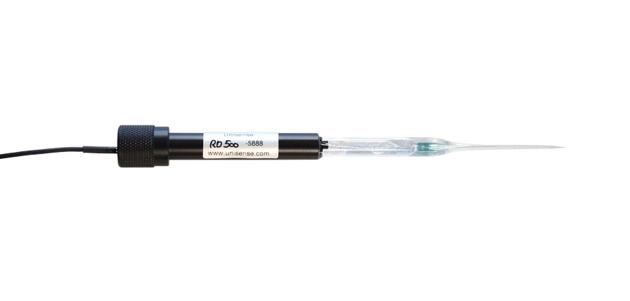
Acta Biomaterialia 73(2018)559-566 560D. Zhao et al./Acta Biomaterialia 73 (2018) 559-566 https://doi.org/10.1016/j.actbio.2018.04.0321742-7061/@2018 Published by Elsevier Ltd on behalf of Acta Materialia Inc. Contents lists available at ScienceDirect Acta Biomaterialia ELSEVIER journal homepage: www.els evier.com/locate/actabiomat Full length article In vivo quantification of hydrogen gas concentration in bone marrowsurrounding magnesium fracture fixation hardware using anelectrochemical hydrogen gas sensor Daoli Zhaoa1, Andrew Brownab.c.d,1, Tingting Wang, Sayuri Yoshizawab., Charles Sfeir b.c.d.e.*,William R. Heinemana,* Department of Chemistry, University of Cincinnati, 301 Clifton Court, Cincinnati, OH 45221-0172, USA "Department of Periodontics and Preventative Dentistry, University of Pittsburgh, 3501 Terrace Street, Pittsburgh, PA 15261, USA The Center for Craniofacial Regeneration, University of Pittsburgh, 335 Sutherland Drive, Pittsburgh, PA 15261, USA Department of Bioengineering, University of Pittsburgh, 3700 O'Hara Street, Pittsburgh, PA 15261, USA The McGowan Institute for Regenerative Medicine, 450 Technology Drive, University of Pittsburgh, Pittsburgh, PA 15219, USA Article history: Received 2 November 2017 Received in revised form 9 April 2018 Accepted 18 April2018 Available online 20 April 2018 Keywords: Magnesium MicroCT Fixation devices Fracture fixation Amperometric hydrogen sensor ABSTRACT Magnesium (Mg) medical devices are currently being marketed for orthopedic applications and have acomplex degradation process which includes the evolution of hydrogen gas (H2). The effect of H2 expo-sure on relevant cell types has not been studied; and the concentration surrounding degrading Mgdevices has not been quantified to enable such mechanistic studies. A simple and effective method tomeasure the concentration of H2 in varying microenvironments surrounding Mg implants is the first stepto understanding the biological impact of H2 on these cells. Here, the in vivo measurement of H2 sur-rounding fracture fixation devices implanted in vivo is demonstrated. An electrochemical H2 microsensordetected increased levels of H2 at three anatomical sites with a response time of about 30 s. The sensorshowed the H2 concentration in the bone marrow at 1 week post-implantation (1460±320pM) to bemuch higher than measured in the subcutaneous tissue (550±210uM) and at the skin surface (120±50 uM). Additionally, the H2 concentrations measured in the bone marrow exceeded the concentrationin a H2 saturated water solution (~800uM). These results suggest that H2 emanating from Mg implantsin bone during degradation pass through the bone marrow and become at least partially trapped becauseof slow permeation through the bone. This study is the first to identify H2 concentrations in the bonemarrow environment and will enable in vitro experiments to be executed at clinically relevant H2 concen-trations to explore possible biological effects of H2 exposure. Statement of Significance An electrochemical H2 sensor was used to monitor the degradation of a Mg fracture fixation system in alapine ulna fracture model. Interestingly, the H2 concentration in the bone marrow is 82% higher than H2saturated water solution. This suggests H2 generated in situ is trapped in the bone marrow and bone isless permeable than the surrounding tissues. The detectable H2 at the rabbit skin also demonstrates a H2sensor’s ability to monitor the degradation process under thin layers of tissue. H2sensing shows promiseas a tool for monitoring the degradation of Mg alloy in vivo and creating in vitro test beds to more mech-anistically evaluate the effects of varying H2 concentrations on cell types relevant to osteogenesis.@ 2018 Published by Elsevier Ltd on behalf of Acta Materialia Inc. ( * Corresponding authors at: D epartment of Periodontics and P reventative Den- tistry, University of Pittsburgh, 3501 Terrace S t reet, P i ttsburgh, P A 15 2 61, USA(C. Sfeir) . Department of Chemistry, U n iversity of Cincinnati, 3 0 1 Cl i fton Court, C i ncinnati, OH 45221-0172, USA (W.R . Heineman) ) ( E-mail addresses: csfeir @ pitt.ed u (C . Sfeir ), William . Heineman@uc.e d u (W.R. Heineman). ) ( T h ese au t hors contributed equally to this work. ) Biodegradable implants for fracture fixation offer many advan-tages over the permanent stainless steel and titanium deviceswidely used today [1-10]. Importantly, implants based onbiodegradable materials provide mechanical support during bone It is known that Mgand its alloys degrade in aqueous environ-ments, generating magnesium (Mg*) and metallic alloying ele-ment ions, hydroxide (OH-) ions, as well as hydrogen (H2) gas. Mg and its alloys have been used in pre-clinical animal studiesevaluating fracture fixation and ACL graft fixation with differentdevices such as plates, screws, and pins [19-26]. These studiesused a combination of radiographic (micro-computed tomographyand X-ray imaging) and histological methods to assess the degra-dation of the Mg devices and their impact on surrounding tissuesand cells. Some of these studies describe accumulation of gas pock-ets surrounding the Mg implants. However, the generation of H2gas was not measured nor was its distribution throughout the var-ious tissues surrounding the implants assessed. Additionally,Bartsch et al. measured pH changes surrounding Mg devicesimplanted in vivo by fluorescence imaging, but this methodrequired the injection of fluorescent dye seminaphthorhodafluor[27]. Several research groups have extrapolated Mg device degra-dation rates to Mg*concentrations observed by cells in vivo tobegin exploringthe molecular mechanisms;Lunderlying theenhanced bone regeneration observed around Mg devices whichhighlights the importance of understanding and identifying thepresence of the alloys’ various degradation products within thehealing environment [25,28,29]. However, no studies exist thatassess the impact of H2 on the behavior of cells relevant to fracturehealing (osteoblasts, osteoclasts, bone marrow stromal cells, etc.). Since one molecule of H2 generated is liberated for one metallicMg atom that degrades, measuring the evolved H2 allows a directmeasurement reflecting the degradation of Mg [30]. In additionto enabling definition of Mg alloys' degradation products withinthe biological environment for mechanistic studies, direct andnon-invasive measurement of Mg device degradation whileimplanted in the body would enable monitoring to ensure thatdevice degradation was proceeding appropriately given the relatedbone regeneration. Monitoring the degradation of a Mg device isfrequently done in vivo using radiographic measures such asmicrocomputed tomography (MicroCT) and X-ray imaging, how-ever, these techniques require major equipment and exposure toX-ray absorption and their sensitivities are limited. MicroCT canbe used for in vivo imaging, but typically only in small animalmodels. Recently, we have demonstrated that the measurement of H2generated by degradation of Mg and its alloys implanted subcuta-neously using an electrochemical H2 sensor in vivo is a simple andeffective non-invasive method for monitoring H2 concentrationstransdermally in small animal models [31-35]. The H2 concentra-tion measured at the skin surface above the implanted alloy wasfound to be proportional to the weight loss of explanted implants.The faster the degradation rate of a Mg alloy, the higher the H2 con-centration that was detected. This method employed a highly sen-sitive H2 electrochemical sensor and relied on tlthe highpermeability of H2 through skin. While effective at translatingobserved H2 concentration into degradation rate, these studies did not evaluate functional devices implanted in bony environ-ments where complex tissues’ varying H2 diffusion rates likelyimpact the accumulation of H2. This study aims to monitor the degradation of a Mg fracture fix-ation system in a lapine ulna fracture model using electrochemicalsensors for H2 gas. A more comprehensive and quantitative assess-ment of H2 distribution within varying tissue environments sur-rounding implants could lead to improved understanding of themechanisms underlying the enhanced bone regeneration observedsurrounding a Mg device, as well as the factors contributing to theaccumulation versus dispersion of H2 degradation byproducts.Completion of this study would serve as the first step in establish-ing a test bed for evaluating the impact of H2 exposure, at clinicallyrelevant H2 concentrations, on cell populations exposed to degrad-ing Mg devices. 2. Experimental 2.1. Device design and fabrication The device fabrication process follows our earlier procedure[19].Mg rod stock (99.9% Mg, Goodfellow, PA) was subjected toCNC machining to produce fixation plates (4.5×20 mm) with fourholes for fixation screw placement and screws that were 7 mmlong with a shaft outer diameter of 1.75 mm. Fixation plates as-machined were then polished with 400, 600,800 and 1200 grit dia-mond films. Both fixation plates as-polished, and screws as-machined were then acid etched in a solution of glycerol, aceticacid and nitric acid for 10-15 s to homogenize surface characteris-tics. Prior to implantation the fixation plates and screws were sub-jected to gamma sterilization with a dose of 20 kGy at a dose rateof 23.5 Gy/min (Mark I 68, JL Shepherd and Associates, San Fer-nando, CA). 2.2.Surgical implantation A rabbit ulna fracture model was used to assess Mg devicedegradation in a functional orthopedic application as it is well-established in the literature [36] and has been previously usedby our group [19,20]. All the animal experiments and procedureswere approved by the University of Pittsburgh Institutional AnimalCare and Use Committee and carried out in accordance with theNIH Guide for Care and Use of Laboratory Animals. A total of 9New Zealand White rabbits (12 weeks old, Charles River Laborato-ries, Wilmington,MA) were used in the study. Right or left ulnaefrom forearms of eight of the nine animals were subjected to sur-gical implantation of the fixation plate and screw following osteot-omy creation and one rabbit was used as a sham surgical controlwith no osteotomy created or fixation plates and screws implanted(Fig.1). Priorto surgery, rabbits were sedated using ketamine and xyla-zine, intubated and anesthetized using isoflurane, and forearmswere shaved and disinfected. The skin and muscle overlying theulnae were dissected with a scalpel and the overlying musclewas elevated and retracted to enable surgical manipulation ofthe ulnae. In each experimental rabbit, an osteotomy was per-formed at the midpoint of the ulna using a rotating burr, resultingin a defect approximately 1 mm in proximal-distal length andthrough the full width and height of the ulna. A plate and screwset was then implanted by drilling 4 pilot holes (2 on each sideof the osteotomy site) using a hand-held drill, placing the plateacross the osteotomy site, and securing in place with 4 screws suchthat all screws engaged cortical bone on both sides of the ulnae asshown in Fig. 2a and b. A 2 mm burr hole was then created throughthe anterior cortical wall, using the handheld drill, both proximal -1 sham control - 1 sham control -1 sham control Fig. 1. Experimental timeline and sample size at each assessment. The study began with 9 total rabbits, 8 assigned to the experimental arm receiving an osteotomy and repairand 1 sham control who did not receive an osteotomy or repair. On the third postoperative day one rabbit expired and was removed from H2 monitoring procedures and the4 week endpoint. Fig. 2. (a) Schematic drawing showing placement of Mg fixation plate attached to the fractured ulna bone with four Mg screws that penetrate through the bone marrowinterior. The osteotomy site (fracture) in the ulna is marked by the vertical dashed line. Sensor measurements were made by inserting the probe tip through the tissue andinto the predrilled hole in the bone shown on the right (The scheme is not to scale). (b) Photograph of Mg device for fracture fixation during the implantation stage. Themeasuring hole for H2 sensing is to the right of the fixation plate (marked with blue arrow). (c) H2 microsensor assembled on a micromanipulator for measuring H2 from Mgalloy implanted in a rabbit ulna. (For interpretation of the references to colour in this figure legend, the reader is referred to the web version of this article.) and distal to the Mg plate to allow monitoring of H2 evolution withthe sensor. The overlying soft tissue was then closed in layers usingresorbable Vicryl sutures and the location of the H2 monitoringport was marked on the skin with a tattoo. A sham surgery wasperformed on one rabbit such that the osteotomy and subsequentplate and screw fixation were not performed, but a burr hole for H2monitoring was created. All rabbits received post-operative Ketophen for analgesia twicea day for 3 days and Baytril for antibiotic coverage twice a day for3 days. Rabbits also received e-collars post-operatively to preventchewing of sutures until incision sites had healed (approximately10 days post-op). Rabbits were monitored for general behavior,movement, and food and water intake, with standard diet andstandard enrichment beginning in the immediate post-operativeperiod. Rabbits were monitored daily for signs of lameness,lethargy, wound dehiscence and infection. This post-operative careprocedure was carried out following the surgical implantation, aswell as H2 monitoring procedures. 2.3. H2 measurement by electrochemical H2 sensor H2 measurements were made with two electrochemical H2 sen-sors (Unisense, Aarhus, Denmark) that operate in the amperomet-ric mode by applying a voltage to the working electrode andmeasuring the current resulting from oxidation of H2 [31-35,37].One sensor was outfitted with a needle sensing tip (1.6 mm tipdiameter,H2-NP) and was used for measurements where the rabbit skin was penetrated, such as inside the bone marrow and the sub-cutaneous tissue, and for some transdermal measurements bypressing or just gently touching the tip to the rabbit skin. The othersensor was outfitted with a flat glass capillary tip (50 um tip diam-eter, H2-50) and was used for measurements made transdermallyby pressing or just gently touching the tip to the rabbit skin. Theamperometric microsensors were connected to a multimeter formeasurements (Unisense, Aarhus, Denmark). The H2 sensors werecalibrated and the multimeter readout converted from microsen-sor current to H2 concentration using a calibration curve generatedfrom standards prepared by dilution of H2 saturated water follow-ing our method reported earlier [31-35,37]. H2 concentrationsgreater than those in saturated water were determined based onextrapolation of the calibration curve to higher concentrations. For in vivo measurements, the H2 sensors were sanitized by sub-merging in 70% ethanol for 5 min between animals. Rabbits weresedated using ketamine and xylazine and anesthetized usingisoflurane (delivered via mask). Rabbits were maintained on aheated table where the microsensor tip was positioned for mea-surement with a micromanipulator as shown in Fig. 2c. Controlmeasurements for H2 sensing were made by inserting the needlesensor tip inside the bone marrow (using the monitoring port cre-ated proximal to the plate and screws) and the subcutaneous tissueor just touching the flat capillary sensor tip on the skin of the sur-gical sham control rabbit without an osteotomy or Mg implant. Allmeasurements were conducted on all rabbits at both 1 week and 2weeks post-surgical implantation. Measurements in the bone marrow were made by penetratingthe skin at the tattoo mark with the H2 needle sensor and thengently pushing it through the tissue and into the hole predrilledin the bone before the implantation of the Mg plate and screwsshown in Fig. 2a and b. Measurements were then made in subcuta-neous tissue by withdrawing the sensor tip out of the bone andunderlying tissue so that it was in contact with the subcutaneousspace. Finally, H2 was measured non-invasively on skin above theimplant by pressing the H2 sensor tip (either needle or flat capil-lary) against the skin covering the hole. This last measurement pro-cedure is similar to that described for non-invasive transdermalmeasurements in our earlier reports [32,33]. Two techniques wereused for the transdermal measurements: pressing the sensor tip tothe skin to ensure a good contact with the skin and just gentlytouching the sensor tip to the skin. Although each measurementrequired only about 30s for a steady state signal to be achieved,more stable H2 signals with better precision were obtained bywaiting for 90 s or longer. 2.4. Microcomputed tomography characterization Four weeks (3 rabbits) or eight weeks (4 rabbits) post-osteotomy and fixation device implantation, rabbits were eutha-nized and the radius/ulna complex was explanted and fixed in for-malin for 3 days. The sham control rabbit was not euthanized.Micro-computed tomography characterization was performedsimilar to methods described in Chaya, et. al. [19,20]. Microcom-puted tomography scanning was performed at 10 um voxel size,60 kVp beam energy, 400 ms exposure, 10 frames averaged perview and 360 degrees angular range of scan (Skyscan 1172,Bruker-Skyscan, Belgium). Raw scan files were then reconstructedwith Recon and analyzed using CTAn (Skyscan). Device volumewas measured by manually defining regions of interest aroundall screws and plates and then setting upper and lower thresholdlevels for binarisation that separated the devices from the sur-rounding soft and hard tissues. The lower threshold levels weredetermined by selecting the inflection point between the maxi-mum value of the soft tissue and background histogram curveand the maximum value of the Mg device histogram curve (aver-age 58±4 a.u.). Three-dimensional reconstruction and cross-sectional views were also obtained for qualitative characterization. 2.5. Statistical analysis All data are expressed as the mean±SD (standard deviation)unless indicated otherwise. For statistical analysis of H2 generatedby the Mg alloy corrosion, measurements were made on 7 experi-mental rabbit forearms, and one sham rabbit forearm. The statisti-cal analysis was performed using GraphPad Prism 3 software(GraphPad Prism Software, Inc.). Statistical significance of differ-ences between the same location at different time points and themeasurement by different sensors was assessed by the student t-test. P values less than 0.05were considered statisticallvsignificant. 3. Results and discussion The overall goal of this research was to characterize the H2 con-centrations in the environment surrounding a functional Mgimplant system in a clinically relevant animal model. Specifically,this study determined that H2 accumulates at high concentrationswithin the bone marrow and then decreases in the surroundingsubcutaneous space and decreases further on the cutaneous sur-face in the setting of the bone fracture fixation model. Further-more, the concentrations of H2 measured in this model were significantly higher than the concentrations of H2 within a satu-rated solution of water. Surgical implantation of plates and screws was uneventful forall rabbits. However, one rabbit was found expired in its cage onpost-operative day 3 due to unknown causes. This rabbit was des-ignated for the four-week endpoint and was thus excluded fromfurther assessments. All other rabbits had uneventful healingcourses and exhibited no signs of distress or infection with theaforementioned analgesia and antibiotic plan. The H2 sensor responses to positioning the tip in bone marrow,in subcutaneous tissue, pressing skin and gently touching the skinon the rabbit after one week of implantation are shown in Fig. 3.Fig. 3a shows a representative sensor response signal for one mea-surement with the H2needle sensor on a single rabbit. The sensorresponse signal is current from the oxidation of H2 versus time asthe sensor is positioned at the four measurement sites. The signalwas initially a very small (ca. 40 pA), flat background as the sensorwas held in air for about 40 s; the sensor was then slowly pushedthrough the skin and into the tissue above the hole in the bone dur-ing which time the signal increased gradually to about 250 pA as itwas pushed deeper into the tissue, indicating that the H2 concen-tration was increasing as the hole in the bone was approached;the signal then exhibited an abrupt increase as it entered the holewhere it was then held in position; and, finally, the signal leveledoff to a steady value of ca. 1400 pA that reflects a much higher con-centration of H2 in the marrow. After about 200 s the sensor waswithdrawn from the port,resulting in the sharp drop in sensor sig-nal, and positioned in the subcutaneous space above the portwhere the signal leveled off to a steady value (ca.300 pA) that issimilar to that obtained just before the sensor was pushed downinto the port. At about 300 s the sensor was withdrawn from therabbit subcutaneous tissue causing the signal to drop down tobackground. Then, the tip was pressed firmly against the skinabove the burr hole for a measurement, the signal increases frombackground as the sensor is in position and levels off to a valueabout half (ca. 150 pA) that when it was in the subcutaneous tis-sue. Lastly, at about 400 s the sensor was just gently touched tothe skin and the signal dropped off to a low value comparable tothe initial background signal. This same pattern of signal responsewas obtained with the other rabbits. Fig. 3b is a bar graph summarizing the combined results formeasurements at 4 different locations (in bone marrow, in subcu-taneous tissue, and transdermally by pressing skin and gentlytouching the skin, from left to right) on all seven experimental rab-bits with the H2 needle sensor. For this graph, the raw sensor cur-rent signals were converted to H2 concentrations by means of acalibration plot. a) Fig. 3. H2 measurements on anesthetized rabbits with Mg device. (a) Sensor current signal versus time for H2 concentrations measured in bone marrow, in subcutaneoustissue, and transdermally by pressing skin and gently touching the skin (from left to right) on a single representative rabbit after 1 week implantation.(b) Average H2concentrations after 1 week of implantation measured for all seven rabbits at these four points as determined from a calibration curve.(c) Average H2 concentration for allseven rabbits with Mg devices after 2 weeks implantation. Error bars are standard deviations for measurements made on seven rabbits (n=7). By comparison, the H2 concentration in the subcutaneous spaceadjacent to the bone above the port was 550±210 uM, or about 3xlower. To obtain these results the sensor tip was withdrawn fromthe access hole back into the subcutaneous space above the holeso the probe tip was approximately equidistant from the holeand the end of the fixation plate. The H2 detected in this regioncan originate from three sources: H2 escaping from the bone mar-row through the hole, H2 formed by degradation of the plate endthat is closest to the probe tip above the hole and is diffusing tothe probe tip, and H2 from corrosion within the bone marrow thatis permeating through the bone and overlying muscle. The mostunlikely of these is permeation through the bone, which seemsmore impermeable to H2 given the high concentration found inthe marrow. The H2 concentration dropped to 120±50 uM when the electro-chemical sensor was withdrawn and then pressed against the rab-bit skin, non-invasively directly above tthe implant. Thismeasurement indicates that some H2 permeates from the implant through the subcutaneous tissue and the rabbit skin to the surfaceand that the path length for permeation of the H2 to the skin sur-face has a significant effect on the concentration of H2 measured.For transdermal measurements at the surface with the needleprobe we found that the degree of pressure exerted on the probesignificantly affected the H2 signal. The measured H2 concentrationdropped to only 12±5 pM when the probe was just gently touch-ing the rabbit skin covering the predrilled hole. Although the mea-surement was at the same position, the apparent concentrationdecreased about 10 times compared to pressing the rabbit skin.We attribute this to the slanted surface of the opening in the nee-dle probe tip, which makes it difficult to ensure complete contactwith the skin surface so that H2 cannot escape through any gapbetween the probe and the skin. The pressure on the H2 sensoraffects the H2 measurements; with more pressure, more H2 is cap-tured by the H2 sensor giving a higher signal. Thus, the larger con-centration measured with the sensor tip pressed against the skin ismore representative of the true concentration of H2 at the skinsurface. Our previously reported transdermal measurements were all onthe subcutaneous implant mouse model where the path length forH2 diffusion to the skin surface was only through the very thin skinon the back of the mouse [31-35]. Here we have shown that someH2 permeates through subcutaneous tissue that includes the thinmuscle covering the ulna for measurement at the surface. ThisH2 level at the skin surface is much lower compared to H2 gener-ated by Mg alloys implanted subcutaneously in the mouse model,where H2 only has to permeate the thin mouse skin to reach thesurface [32-35]. We attribute the lower concentration to the lowerpermeability of the rabbit skin compared to the mouse skin, whichis probably due to differences in skin thickness. The thickness ofthe mouse whole skin is about 0.7 mm, whereas it is about 2 mmfor rabbit [42,43], or about 3x thicker. Moreover, the Mg alloy Table 1Volume changes of plates and screws. Device Plates Screws Volume (mm’) Decrease in Volume (%) Volume (mm’) Decrease in Volume (%) As-machined 57.5 9.7±0.4 4 week Endpoint 57.1±0.6 0.7 8.6±0.3 11.3 8 week Endpoint 54.3±4.0 5.6 7.8±0.4 19.6 was implanted deeper compared to the subcutaneous implant inthe mouse model. Consequently, the blood flow through the mus-cle overlaying the implant would be expected to carry away someof the H2 as it diffuses through tissue, further lowering the H2 con-centration at the skin surface. As the control, H2 measurements were made on the rabbit bonewithout a Mg implant. The H2 concentration was 6±3 uM and 5±2 pM in the bone marrow and subcutaneous tissue, respectively.The H2 concentration is 2±1 pM when pressing the rabbit skin,whereas no H2 was detected when only gently touching the rabbitskin.This rabbit skin background level in the control rabbit is sim-ilar to the background level in control experiments for mouse skinof 1.0-7.3 uM [33]. Since the needle point for the needle sensor is slanted, it cannotcapture all the H2 permeating through the rabbit skin at the touch-ing point unless it is pressed to the skin at an angle so that theslanted surface is flush with the skin. Therefore, the electrochemi-cal H2 glass capillary sensor (H2-50) was also used to measure theH2 permeability through the rabbit skin above the hole. The sensortip of the H2 glass capillary sensor is flat and about 50 um in diam-eter, which is much smaller compared to the H2 needle sensor (1.6mm). Due to the small, flat tip of the glass sensor, it can tightly con-tact with the skin and capture more H2 per unit area. Holding thesensor vertically to the skin and pressing just enough to slightlydepress the skin gave very reproducible readings, which we alsofound in our earlier studies on mice [33].Indeed, the H2 concentra-tions measured with the glass sensor were much higher. The H2concentrations were 62±10 uM when only gently touching therabbit skin covering the predrilled holes, which is ~5 times higherthan with the needle probe under the same condition as men-tioned earlier (12±5pM)(p=0.0001). The concentration of H2 willbe even higher if the glass sensor is pressed against the rabbit skin.However, we did not press against the skin with the glassy capil-lary sensor due to its fragility. The glass capillary sensor can easilydetect the low concentration of H2 by simply touching the skin.Moreover, the glass sensor probe gives a more accurate readingof the concentration of H2 at the skin surface since a considerableamount of H2 escapes measurement with the needle probe becauseof poor contact with the skin. Thus, to get a better evaluation of thedegradation of Mg alloys by transdermal low H2 concentrationmeasurement, the H2 glass sensor is recommended. Fig. 3c shows results from H2 needle sensor measurements onseven rabbits after 2 weeks implantation. The H2 concentrationhad dropped to 1058±158 pM in the bone marrow. The H2 con-centrations are 187±96 pM and 94±48 pM for the subcutaneoustissue and pressing the rabbit skin, respectively. The H2 concentra-tion is 5±3 pM for gently touching the skin.The H2 concentrationsfrom all these different spots are lower compared to the first weekof measurement. Compared to the first week of measurement, theH2 concentration is significantly less in the bone marrow (p=0.0115), subcutaneous (p=0.0013), and gently touching the skin(p=0.0080). However, there is no significant difference in H2 con-centration for gently pressing the rabbit skin (p=0.3406) after 1week and 2 weeks implantation. The lower H2 concentration inthe bone marrow,subcutaneous tissue and pressing the rabbit skinsuggests a lower degradation rate of the Mg alloy in the second week. The faster degradation behavior of the screw plate systemin the first week than afterward was also observed for LAE442 alloyin an in vitro bone model [41]. This decreased generation of H2 isexpected due to the slowing corrosion caused by formation of anincreasingly thick corrosion layer of Mg(OH)2 and MgCOs thathas been shown to coat the implant surface [32,35]. Although thegeneration of H2 decreases at 2 weeks implantation compared tothe first week, the H2 concentration in the bone marrow is stillhigher than an H2 saturated water solution. MicroCT analysis revealed plate volume at the 4 week endpointdecreased from 57.5 mm’as machined to 57.1±0.6 mm’at the 4week endpoint representing a 1% average decrease in total volume(n=3) (Table 1). Screw volume decreased from 9.7±0.4 mm asmachined (n=8) to 8.6±0.6 mm’at the 4 week endpoint repre-senting an 11% average decrease in total volume (n=12). Due tothe low deviations measured across the devices, only one plateand four screws as machined were scanned prior to the implanta-tion. At the 8 week endpoint, plate volume was 54.3±4.0 mm(n=4) and screw volume was 7.8±0.4 mm(n=15). Due to a techni-cal error, one screw from the eight week timepoint could not beanalyzed. The screws displayed increased volume loss comparedto the plates, which is similar to observations in Chaya et al.[19]. This is likely due to the increased surface area/volume ratioof the screws compared to the plate, particularly within thethreaded shaft region and could also be due to the increased fluidflow within the marrow space (where the screw resides) versus theunderlying muscle space (where the plate resides). The increasedvolume loss observed in the screws also supports the increasedH2 concentrations measured in the marrow space versus the sub-cutaneous tissue. Qualitative assessment of microCT samples correlated with thequantitative evaluation (Fig. 4). The site of the osteotomy (o)remains visible 4 weeks post-surgical creation, as does the holecreated for insertion of the H2 sensor into the marrow space (h).However, by 4 weeks post-surgical creation, the H2 sensor holehas been infiltrated with mineralized tissue, which would preventmeasurement at this time point and beyond, an important consid-eration for future studies. Marked pitting corrosion is noted at sev-eral locations on the plate (solid arrows) while more uniformcorrosion is noted on the screws, particularly loss of the threadedportion and distal ends of the screw shaft (dashed arrows). Callousformation and bone overgrowth(c) is noted and has been observedin other studies using this surgical model [19,20,44]. Limitations of this study include the inability to extrapolate H2concentrations to specific degradation rates that would likely beuseful long-term to enable clinically relevant information to affectdecision making. The H2 monitoring technique, as performed,isinvasive and requires anesthesia to perform bone marrow and sub-cutaneous measurements. Future developments could focus oncreating less invasive methods to perform more comprehensivecharacterizations of H2 concentration in different tissue environ-ments. The effect of varying concentrations of H2 gas on the variousaffected cell populations is also not well understood. Future studiesmay involve in vitro work to understand mechanisms affected byvarying H2 concentrations,as well as the evaluation of Mg alloyswith varying degradation rates in order to determine the ability Fig. 4. Representative microCT cross-section of an ulnae explanted 4 weeks post-osteotomy and fixation with Mg plates and screws. The osteotomy site (o) remainsvisible however callous formation is also visible (c). The H2 monitoring hole (h) isnoted to now be infilled with bone. Device degradation is noted on both the plate(solid arrow) and screws (dashed arrows) (n=3). of the described H2 monitoring technique to determine differencesin degradation rate. In addition, due to the low cost, the X-ray anal-ysis in the future experiments will be considered. More intermedi-ate measurement time points will be performed to provide anadditional clinical information on the device degradation, boneregeneration and H2 gas cavity development. 4. Conclusion The degradation of a Mg alloy in the bone fracture fixationmodel in rabbit ulnae was investigated by an electrochemical H2sensor. The H2 concentration in the bone marrow was much higherthan previous in vivo studies where H2 was measured transder-mally with Mg alloys with the subcutaneous mouse model. Theseresults suggest that H2 generated from degradation of the Mgscrew segment inside the marrow was trapped in the bonemarrow. The H2 concentration was found to be significant, but sub-stantially lower in the subcutaneous tissue above the plate.Although the H2 concentrations obtained by non-invasive trans-dermal measurements on the skin surface above the implant arelower, the electrochemical H2 sensor can adequately detect H2 atthese levels when pressed against the skin. These results confirmthat Mg in the marrow of the bone corrodes at an easily measur-able rate and that the bone is less permeable to H2 than the sur-rounding tissue, which leads to a buildup of H2 in the boneinterior. H2 sensing shows more promise as an effective meansfor monitoring the degradation of Mg alloys in vivo with larger testanimals than mice and with human patients. Based on these datamany biological questions arise, such as whether there is a H2effect on any of the components ofthe healing environment. DoesH2 gas accumulation affect the extracellular matrix, cells,cell sig-naling? Or is it an innocuous element? Further studies on the bio-logical effect of H2 gas are key to further establishing the biologicaleffects of implantable Mg devices. Acknowledgements The authors thank the National Science Foundation (NSF ERC0812348) for financial support. The surgical support of Dr. SamerZaky, as well as microCT assistance provided by Dr. Kostas Verdelisand Rong Chong is appreciated. References [2] S. Zhang, X. Zhang, C.Zhao, J. Li, Y. Song,C. Xie, H. Tao, Y. Zhang, Y. He, Y. Jiang,Y. Bian, Research on an Mg-Zn alloy as a degradable biomaterial, ActaBiomater. 6(2010) 626-640. [3]Y. Chen, Z. Xu, C. Smith, J.Sankar, Recent advances on the development ofmagnesium alloys for biodegradable implants, Acta Biomater. 10 (2014)4561-4573. [4] D. Chou, D. Hong, P.Saha, J. Ferrero, B. Lee, Z. Tan, Z. Dong, P.N. Kumta, In vitroand in vivo corrosion, cytocompatibility and mechanical properties ofbiodegradable Mg-Y-Ca-Zr alloys as implant materials, Acta Biomater. 9(2013)8518-8533. [5]S.E. Henderson, K. Verdelis,S. Maiti, S. Pal, W.L. Chung, D.T. Chou, P.N. Kumta,A.J. Almarza, Magnesium alloys as a biomaterial for degradable craniofacialscrews, Acta Biomater. 10 (2014)2323-2332. [6] E. Poinern, S. Brundavanam, D. Fawcett, Biomedical magnesium alloys: Areview of material properties, surface modifications and potential as abiodegradable orthopaedic implant, Am.J. Biomed. Eng. 2 (2012) 218-240. [7] F. Witte, J. Fischer,J. Nellesen, H.A. Crostack,V. Kaese, A. Pisch, F. Beckmann, H.Windhagen, In vitro and in vivo corrosion measurements of magnesium alloys,Biomaterials 27 (2006) 1013-1018. [8] F. Witte, V. Kaese, H. Haferkamp, E. Switzer, A. Meyer-Lindenberg, C.J.Wirth, H.Windhagen, In vivo corrosion of four magnesium alloys and the associatedbone response,Biomaterials 26 (2005) 3557-3563. 9Y.H. Yun, Z. Dong, N. Lee, Y. Liu, D. Xue, X. Guo, j. Kuhlmann, A. Doepke, H.B.Halsall, W. Heineman, S. Sundaramurthy, M.J. Schulz, Z. Yin, V. Shanov, D.Hurd, P. Nagy, W. Li, C. Fox, Revolutionizing biodegradable metals, Mater.Today 12 (2009)22-32. [10]D. Hong, P. Saha, D. Chou, B. Lee, B.E. Collins, Z. Tan, Z. Dong, P.N. Kumta, Invitro degradation and cytotoxicity response of Mg-4% Zn-0.5% Zr (ZK40) alloyas a potential biodegradable material, Acta Biomater. 9(2013)8534-8547. [11] M.P. Staiger, A.M. Pietak, J. Huadmai, G. Dias, Magnesium and its alloys asorthopedic biomaterials: a review, Biomaterials 27 (2006) 1728-1734. [12]F. Witte, J. Fischer, J. Nellesen, C. Vogt, J. Vogt, T. Donath, F.Beckmann, In vivocorrosion and corrosion protection of magnesium alloy LAE442, Acta Biomater.6(2010)1792-1799. ( [13] F . Witte, N . H ort , C . Vogt, S . Cohen, K.U. K aine r , R . W illu m eit, F . F eyerabend, Degrada b le biomaterials based on m a gnes i um cor r os i on, Curr. O p i n. Solid St. M. 1 2 (2008)63 - 72 . ) ( [14] R . Z eng, W. Di e tzel, F. W i tte, N. H o rt, C. Blawert, P r o gress and chal l enge for m agnesium alloys as biomater i als, Adv. E n g . Mater. 1 0 (2008) B3-B1 4 . ) [151]F. Feyerabend, J. Fischer, J. Holtz, F. Witte,R. Willumeit, H. Drucker, C. Vogt, N.Hort, Evaluation of short-term effects of rare earth and other elements used inmagnesium alloys on primary cells and cell lines, Acta Biomater. 6 (2010)1834-1842. ( [16] S .H. By un, H.K. Lim, S.M . Ki m , S .M. Lee,H.E . Kim , J.H. L ee , The bioresorptio n and guided b one r egeneration of absorbable hy d roxyapatite-coated magnesium m es h 1 Cran i n far Sure 2 8 ( 2017)518-5 2 3 b fac . Surg . 28 ( 2 ) ( [171 J . W. Lee, H.S. Han, K . J . H an , J. P ark , H. J e o n, M.R . Ok, H.K. Seok,J . P . Ahn, K.E. L e e , D .H. Lee,S.J. Yang, S.Y. Cho, P.R. Cha, H. Kwon,T . H. N am, J.H. L o Ha n , H . J. Rho, K. S . Lee, Y.C. Kim, D. Mantovani, Long-term cli n ical study and mu l t i sc a l e a nalysi s o f i n vivo b iodegr a dation m ec h anism of Mg alloy, Proc.Na t l. Ac ad. S c i. U . S .A. 1 1 3 (20 1 6)716-721. ) ( [18] H . W indhage n , K. R a dtke , A . W e izbauer, J. D i ekmann, Y. N o ll, U. K reimeyer,R. S chavan, C. Stuk en borg-Co l sman, H. Waiz y , Biodegradable magnesium-based s crew clin ic ally equivalent to titanium sc r ew in h all u x v algus s urgery: s h o rt term r esults o f t he f i rst p r o spective, r an d o m ized, co n trolled cl i nical p i l o t s t u d y , Biome d. E n g. Onli n e 1 2 (2 0 13)62. ) ( [19] A . C haya, S . Y o shizawa,K. V e r d elis, N. Mye r s, B.J. Cos t ello,D.T. Chou, S. Pal, S. M a i ti, P.N. Kumt a , C . S feir, In vivo study of magnesium pl a te a n d sc r ew d eg ra d ation and b o ne frac t ur e he ali ng , A c ta Bioma t e r . 18 (2 0 15)262-269. ) ( [201 A . Chaya,S. Yoshiza w a, K. Ver d elis, S. Noo r ani, B.J . Co s tello, C. Sfeir, Fractur e h ealing using degradable magnesium fixa t ion plates and screws,J. Oral. Maxil. S urg. 73 (20 1 5) 295- 3 05. ) ( [21 ] B. Schaller, N. Saulacic, T. Imwin k elried, S . B eck, E .W.Y. L i u , J . G rall a , K . N akahara, W. Hofstetter, T. I i zuka, In vivo degradation o f m agnesium pla t e/ screw osteos yn thesi s i m pl ant s ys tems: S oft a nd hard t issue r esponse in a c alvarial mod e l i n m i n iature pigs, J . C r anio. M ax ill . Su rg . 44 ( 2016) 309- 317 . ) ( [22] P . C heng, P. H a n, C . Z hao, S. Zhang,H. Wu, J. H . N i , P. H o u, Y. Z hang,J. L i u, H . Xu, S . L i u, X . Zha n g, Y. Zheng, Y . Ch ai , High-purit y magnesium i nt e rference screws p romote f i brocartila g inous e ntheses r e g en eration i n t h e a n terior c r uciate ligament reconstruc ti on rabbi t model vi a a c cumu l atio n o f BMP-2 and V E GF, Biomat e r i a ls 81 (2016) 1 4 - 2 6. ) ( [ 23] D . Zhao,S. H u ang, F. L u, B . Wang,L. Y a ng, L. Qin, K. Y a ng, Y. L i , W. L i , W. Wang, S .Ti a n, X. Z hang, W. Gao, Z. W a ng, Y . Zh a n g , X. Xie, J. W a ng,J. Li, Vascular i zed bone g ra fti ng f i xed by b iodegradable magnesium s crew f o r r treating o steonecrosis o f the fem o ra l head, Biomaterials 81 (2016)84-92. ) ( [241 B . S ch a ller, N. S aulacic, S. Beck, T . Imwinkelried, B. T. G oh, K . N a ka h ara, W. H o f stetter,T. lizu k a, I n vivo degradation o f a new concep t o f magn e s i um-ba s ed r ivet-scr e ws i n t h e m inipig ma n dibu l ar bon e , Mat. Sci. Eng. C-Ma t er.69 ( 2016) 247-254. ) ( [ 25] Y .Zhang, J . K .Xu , Y. R u an, M. Yu , M.O' L a ughlin, H . Wis e , D. C hen, L. T ia n , D. S h i, J . Wang , S. C hen,J. F e ng , D.H.K. Chow, X. Xi e , L. Zheng, L. Hu a ng, S. H uang , K. L e un g , N . L u , L . Z hao, H . F . L i , D . Z hao, X. G u o, K. C h an, F. W i t te, H. Chan , Y. Z h eng, L.Qin, Implant- de rived magnesium induces local neu r ona l pro d uction o f C GRP t o improve bone-fra ct ure he a ling in ra t s, Nat. M ed. 2 2 (2016) 1 1 60- 1 1 69. ) ( [26] P . H an, P. Cheng, S. Zhang, C . Zhao,J. Ni, Y . Z h ang, W.Zhong , P . Hou, X. Zhan g , Y .Z hen g , Y. Chai, In vitro and in viv o stu d ie s on the degr a dation of high-purity Mg VO ST ( 9 9.9 9 wt. % ) s crew w i th fe m oral in t r a co ndylar fr a ct u r e d ra b bit mo d el, Biomaterials 64 (2015)57- 6 9. ) [27] I. Bartsch, E. Willbold, B. Rosenhahn, F. Witte, Non-invasive pH determinationadjacent to degradable biomaterials in vivo, Acta Biomater.10 (2014)34-39. [28]S.Yoshizawa, A. Brown, A. Barchowsky, C. Sfeir, Magnesium ion stimulation ofbone marrow stromal cells enhances osteogenic activity, simulating the effectof magnesium alloy degradation, Acta Biomater. 10 (2014) 2834-2842. [29] S. Minardi, B. Corradetti, F. Taraballi, M. Sandri, J. Van Eps, F.J. Cabrera, B.K.Weiner, A. Tampieri, E. Tasciotti, Evaluation of the osteoinductive potential ofa bio-inspired scaffold mimicking the osteogenic niche for bone augmentation,Biomaterials 62 (2015)128-137. [30] A. Atrens, G. Song, M. Liu, Z. Shi, F. Cao, M.S. Dargusch, Review of recentdevelopments in the field of magnesium corrosion, Adv. Eng. Mater. 17 (2015)400-453. [31] D. Zhao, T. Wang,X. Guo,J. Kuhlmann, A. Doepke, Z. Dong, V.N. Shanov, W.R.Heineman, Monitoring biodegradation of magnesium implants with sensors,JOM-J.Min. Met. S. 68 (2016) 1204-1208. ( [ 3 2] J . Ku h l m ann, I . Bartsch,E . W i llbold, S . Schuchardt, O. Holz , N. Hort , D. Ho c h e , W .R. H e ineman, F. Witte, Fast e scap e of h ydrogen f r om g a s c a viti e s a round corroding magnesium i mp l ants, Acta Biomater. 9 (2013)8714-8 7 21. ) [33] D. Zhao, T. Wang,J. Kuhlmann, Z. Dong, S. Chen, M. Joshi, P. Salunke,V.N.Shanov, D. Hong, P.N. Kumta, W.R. Heineman, In vivo monitoring thebiodegradation of magnesium alloys with an electrochemical H2 sensor, ActaBiomater. 36 (2016) 361-368. [34]D. Zhao,T. Wang, B. Hoagland,D. Benson, Z.Dong, S. Chen, D. Chou,D. Hong,J.Wu, P.N. Kumta, W.R.R.Heineman, Visual H2 sensorr ffor monitoringbiodegradation of magnesium implants in vivo, Acta Biomater. 45 (2016)399-409. [35] D. Zhao, T.Wang, K. Nahan, X. Guo, Z. Zhang, Z. Dong, S. Chen, D. Chou, D.Hong,P.N.Kumta, W.R. Heineman, In vivo characterization of magnesium alloybiodegradation using electrochemical H2 monitoring, ICP-MS, and XPS, ActaBiomater. 50 (2017) 556-565. ( [36] L .A. M i lls, A .H . R.W. S i mpson, I n v i vo m od els of bo ne repair, J . Bo n e Joint Su r g. B r. 94b (2012)865-874. ) [37] D. Zhao, T. Wang, W.R. Heineman, Advances in H2 sensors for bioanalyticalapplications, Trac-Trend Anal. Chem. 79 (2016) 269-275. [38]T. Seo, R. Kurokawa, B. Sato, A convenient method for determining theconcentration of hydrogen in water: use of methylene blue with colloidalplatinum, Med. Gas Res. 2 (2012). [39]F. Witte, A. Eliezer, S. Cohen, The history, challenges and the future ofbiodegradable metal implants, Adv. Mat. Res. 95 (2010) 3-7. [40] T.A. Huehnerschulte, N. Angrisani, D. Rittershaus, D. Bormann, H. Windhagen,A. Meyer-Lindenberg, In vivo corrosion of two novel magnesium alloys ZEK100and AX30 and their mechanical suitability as biodegradable implants,Materials 4 (2011) 1144-1167. ( [41] L 1 . W olters, S . B e sdo, N . A n grisa n i , P . W r iggers , B . H e r i ng , J .M . S e it z , J . Reifenrath, Degradation behaviour of L AE44 2 - b ased p l a te-screw-syst e ms i n a n i n v itro bone m odel, M at. S ci. Eng. C - M ater. 4 9 (2015)30 5 -31 5 . ) [42]E.C. Jung, H.I. Maibach, Animal models for percutaneous absorption, J. Appl.Toxicol. 35 (2015)1-10. [43]G.D. Thorburn, B.H. Casey, G.S. Molyneux, Distribution of blood flow within theskin of the rabbit with particular reference to hair growth, Circ. Res. 18 (1966)650-659. [44] J. Reifenrath, A.K. Marten, N. Angrisani, R. Eifler, A. Weizbauer, In vitro andin vivo corrosion of the novel magnesium alloy Mg-La-Nd-Zr: influence of themeasurement technique and in vivo implant location, Biomed. Mater. 10(2015). 镁植入材料(Mg)是市场上骨科领域的一种很常规的骨折固定的材料,这种材料有一个复杂的降解过程,包括氢气(H2)的演化。而H2暴露的影响有关细胞类型的研究尚未见报道以及Mg周围的氢气浓度设备还没有被量化,难以提出相关的作用机制。一种简单而有效的方法来被用来测量Mg植入物周围不同微环境中H2的浓度,是了解H2对这些细胞生物学影响的第一步。研究人员研究了动物体内植入镁材料作为骨折固定装置周围的H2浓度的体内测量。电化学H2微传感器在三个解剖位置检测到H2浓度的增加,其中该电极对氢气响应时间约为30秒。微电极传感器显示植入镁植入材料后1周对应的骨髓中H2浓度(1460±320 uM)远高于皮下组织(550±210 uM )和皮肤表面(120±50 uM)。此外,在镁植入材料周围的骨髓中测量的H2浓度超过了在饱和H2水溶液(800 uM)中的浓度。这些结果表明,从Mg植入体降解过程中释放出的H2通过骨髓缓慢渗透到骨中,至少部分遗留在骨头中(图1所示)。这项研究首次确定了骨中H2的浓度,并将使体外实验能够在与临床相关的H2浓度下顺利进行,来探索H2暴露可能存在的生物学效应。图1、兔子骨髓中的氢气浓度的原位在线监测研究人员应用unisense氢气微电极传感器,实现了对兔子尺骨骨折模型中监测骨折固定系统的Mg植入体降解情况的全面了解。发现了兔子骨髓中的H2浓度比H2饱和水溶液高82%。这表明原位生成的H2被困在骨髓中,骨的渗透性低于周围组织。在兔皮肤上也检测到H2证明了H2传感器能够监测薄层组织下Mg合金植入体材料的降解过程。H2传感技术有望作为一种监测镁合金体内降解的工具,并能更系统地建立体外实验临床评价不同H2浓度对成骨相关细胞类型的影响。 这项研究首次提出了一种原位测试技术(unisense微电极研究系统)确定了动物骨头中H2的浓度,并将使体外实验能够在与临床相关的H2浓度下顺利进行,来探索H2暴露可能存在的生物学效应。这也说明H2传感技术(unisense微电极研究系统)有望作为一种监测镁合金体内降解的工具,并能更系统地建立体外实验临床评价不同H2浓度对成骨相关细胞类型的影响。
关闭-
1/8
-
2/8
还剩6页未读,是否继续阅读?
继续免费阅读全文产品配置单
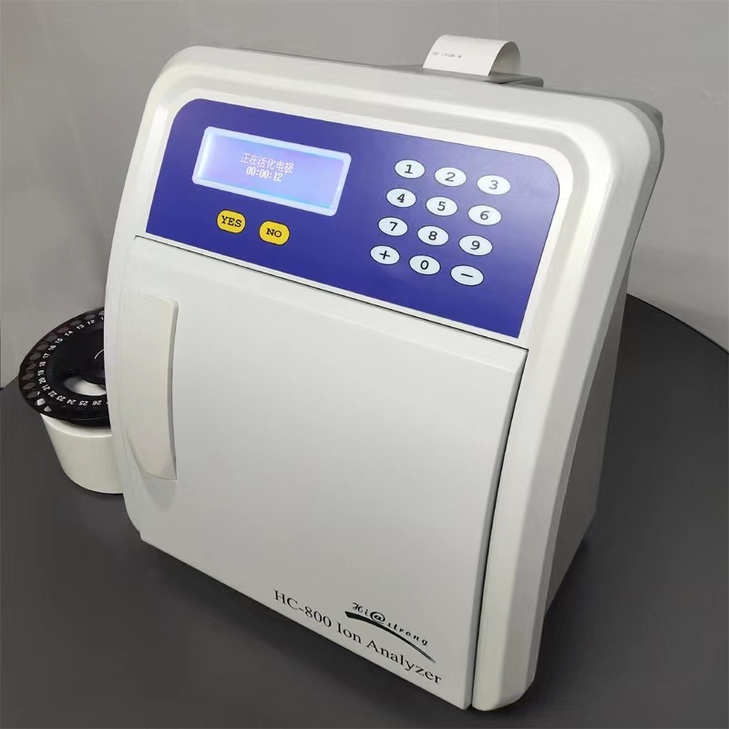
上海谓载科技有限公司为您提供《动物骨头组织中氢气检测方案(其他电化学仪)》,该方案主要用于其他中氢气检测,参考标准《暂无》,《动物骨头组织中氢气检测方案(其他电化学仪)》用到的仪器有丹麦Unisense氢气测量仪。
我要纠错
相关方案
 咨询
咨询